. 2016 Mar 7:3:7.
doi: 10.3389/fnut.2016.00007. eCollection 2016.

Dietary Plant Lectins Appear to Be Transported from the Gut to Gain Access to and Alter Dopaminergic Neurons of Caenorhabditis elegans, a Potential Etiology of Parkinson's Disease

Affiliations

Dietary Plant Lectins Appear to Be Transported from the Gut to Gain Access to and Alter Dopaminergic Neurons of Caenorhabditis elegans, a Potential Etiology of Parkinson's Disease

Jolene Zheng et al. Front Nutr. .

Abstract

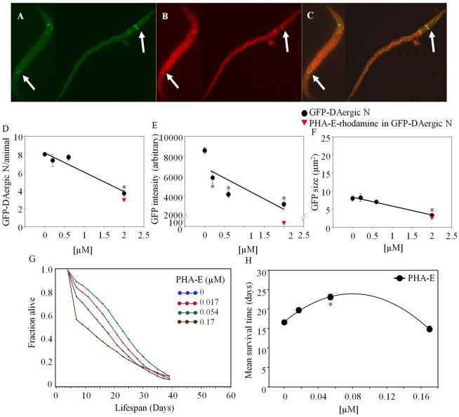
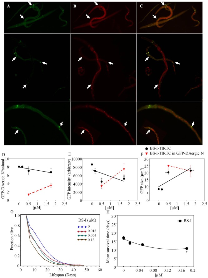
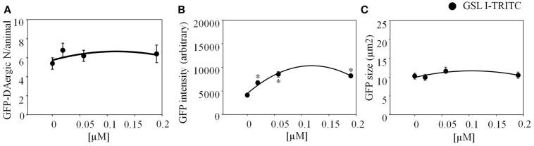
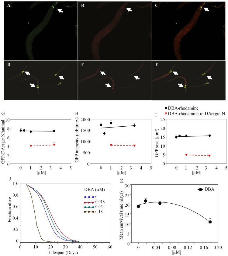
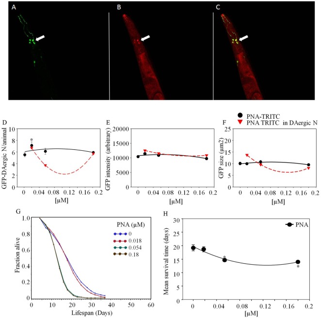
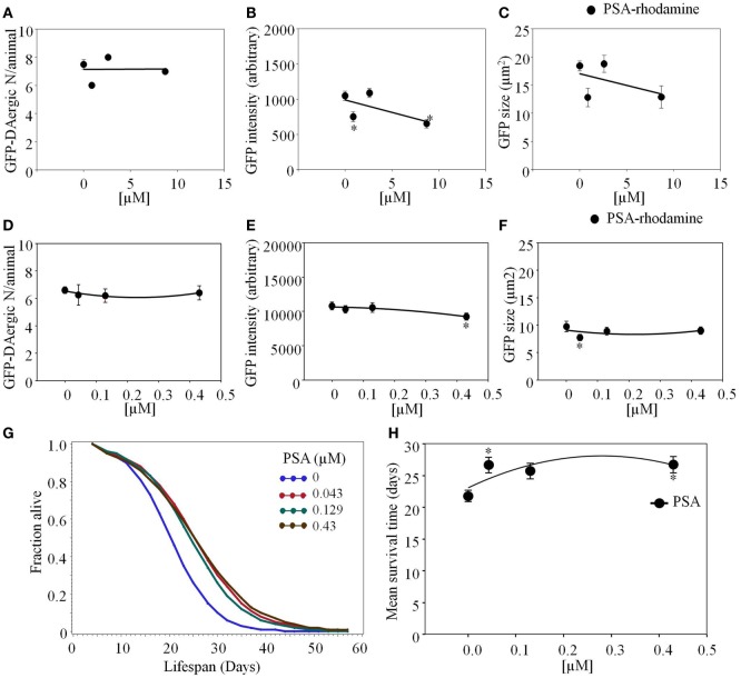
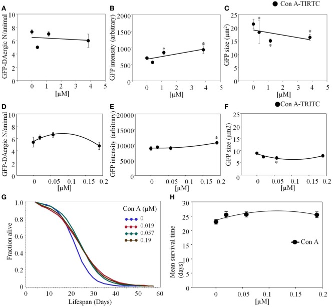
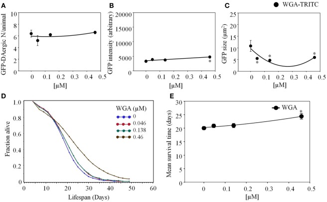
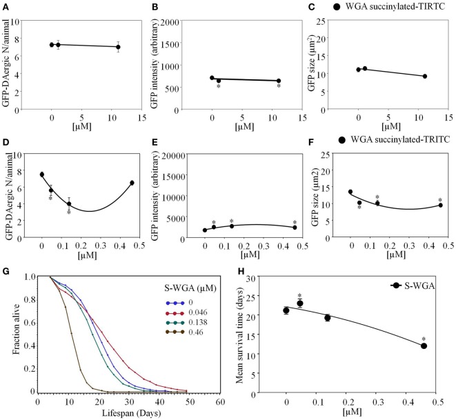
Lectins from dietary plants have been shown to enhance drug absorption in the gastrointestinal tract of rats, be transported trans-synaptically as shown by tracing of axonal and dendritic paths, and enhance gene delivery. Other carbohydrate-binding protein toxins are known to traverse the gut intact in dogs. Post-feeding rhodamine- or TRITC-tagged dietary lectins, the lectins were tracked from gut to dopaminergic neurons (DAergic-N) in transgenic Caenorhabditis elegans (C. elegans) [egIs1(Pdat-1:GFP)] where the mutant has the green fluorescent protein (GFP) gene fused to a dopamine transport protein gene labeling DAergic-N. The lectins were supplemented along with the food organism Escherichia coli (OP50). Among nine tested rhodamine/TRITC-tagged lectins, four, including Phaseolus vulgaris erythroagglutinin (PHA-E), Bandeiraea simplicifolia (BS-I), Dolichos biflorus agglutinin (DBA), and Arachis hypogaea agglutinin (PNA), appeared to be transported from gut to the GFP-DAergic-N. Griffonia Simplicifolia and PHA-E, reduced the number of GFP-DAergic-N, suggesting a toxic activity. PHA-E, BS-I, Pisum sativum (PSA), and Triticum vulgaris agglutinin (Succinylated) reduced fluorescent intensity of GFP-DAergic-N. PHA-E, PSA, Concanavalin A, and Triticum vulgaris agglutinin decreased the size of GFP-DAergic-N, while BS-I increased neuron size. These observations suggest that dietary plant lectins are transported to and affect DAergic-N in C. elegans, which support Braak and Hawkes' hypothesis, suggesting one alternate potential dietary etiology of Parkinson's disease (PD). A recent Danish study showed that vagotomy resulted in 40% lower incidence of PD over 20 years. Differences in inherited sugar structures of gut and neuronal cell surfaces may make some individuals more susceptible in this conceptual disease etiology model.

Keywords: Caenorhabditis elegans; dopamine transporter; dopaminergic neurons; fluorescence; plant lectins.


Figures

Figure 1
Figure 1
Phaseolus vulgaris (PHA-E)-rhodamine co-localized and diminished GFP-DAergic neurons in C. elegans post feeding. (A) GFP-DAergic neurons. (B) PHA-E-rhodamine in several GFP-DAergic neurons. (C) Merged (A,B) showing co-localization of the PHA-E-rhodamine with GFP-DAergic neurons. (D) Dose-dependent reduction of the number of GFP-DAergic neurons per animal: 4.4 ± 0.3 (2 × 10−3 mM, n = 3, P < 0.01). (E) Dose-dependent reduction of fluorescence intensity per GFP-DAergic neurons: 5,837 ± 556 (2 × 10−4–2 × 10−3mM, n = 3, P < 0.01). (F) Size reduction of GFP-DAergic neurons: 3.3 ± 0.4 μm2 (2 × 10−3 mM, n = 3, P < 0.01). (G) The APS was increased dose-dependently. (H) Mean survival time of each group. *indicates statistical significance.
Figure 2
Figure 2
Bandeiraea simplicifolia (BS-I)-rhodamine co-localized with GFP-DAergic neurons: (A) GFP-DAergic neurons. (B) BS-I-rhodamine in GFP-DAergic neurons (5.26 × 10−4 or 1.75 × 10−3 mM). (C) Merged (A,B) showing co-localization of the BS-I-rhodamine with GFP-DAergic neurons. (D) Dose-dependent reduction of GFP-DAergic neurons per animal: 7.0 ± 1.0 (1.75 × 10−4 mM) or 6.7 (1.75 × 10−3 mM) (n = 3, P > 0.05). (E) Reduction in fluorescence intensity per GFP-DAergic neuron (up to 60%): 4,468 ± 332 or 5,166 ± 300 (5.3 × 10−4 or 1.75 × 10−3mM, n = 3, P < 0.01). (F) The size of GFP-DAergic neurons was elevated dose-dependently from 8.0 ± 0.8 to 20.1 ± 1.7 μm2 (5.26 × 10−4) or 21.4 ± 2.5 μm2 (1.57 × 10−4) (n = 3, P < 0.01). (G) The APS was dose-dependently decreased at all doses. (H) Mean survival time of each group. *indicates statistical significance.
Figure 3
Figure 3
Griffonia simplicifolia I (GSL-I)-rhodamine appeared to affect the GFP-DAergic neurons. (A) GSL-I did not affect the number of the GFP DAergic neuron. (B) GSL-I increased fluorescence intensity of the neuron at all doses (P < 0.05). (C) GSL-I did not affect neuron size. *indicates statistical significance.
Figure 4
Figure 4
Dolichos biflorus agglutinin (DBA)-rhodamine in C. elegans co-localized with GFP-DAergic neurons of aged and young nematodes (L3). GFP-DAergic neurons in aged animals (A) or young animals (D). DBA-rhodamine in GFP-DAergic neurons in aged animals (1.0 × 10−3 or 3.33 × 10−3 mM) (B) or young animals (E). Merged (A,B) showing co-localization of the DBA-rhodamine with GFP-DAergic neurons in aged animals (C) or young animals (F). (G) The number of DAergic neurons was not altered (7.5, n = 5, P > 0.2) and 50% of them were co-localized with DBA-rb (1.0 × 10−3 or 3.33 × 10−3 mM, n = 3) in the aged animals. (H) The fluorescence intensity per GFP-DAergic neuron. (I) The size of the GFP-DAergic neurons was not changed. These observations were similarly seen in young animals. (J) The APS was increased at lower doses of DBA, and decreased at higher doses. (K) Mean survival time of each group. *indicates statistical significance.
Figure 5
Figure 5
Arachis hypogaea agglutinin (PNA)-TRITC post-feeding C. elegans co-localized with GFP-DAergic neurons (Leica, TCS SP5, Germany). (A) GFP-DAergic neurons (green), (B) PNA-TRITC in the neuron (red), (C) Co-localization of the GFP-DAergic neurons in merged (A,B) (yellow). (D) Number of GFP-DAergic neurons was increased at low dose (P < 0.05). (E) The intensity of GFP-DAergic neurons was not altered. (F) The size of GFP-DAergic neurons was not altered. (G) APS was dose-dependently reduced at all doses. (H) Mean survival time of each group. *indicates statistical significance.
Figure 6
Figure 6
Pisum sativum (PSA)-rhodamine affected the GFP-DAergic neurons in C. elegans. (A) The number of GFP-DAergic neurons per animal was not reduced overall (8.70 × 10−4–8.7 × 10−3 mM, n = 6, P > 0.05). (B) The fluorescence intensity of GFP-DAergic neurons was decreased at high dose (651 ± 61, 8.7 × 10−3 mM). (C) The size of GFP-DAergic neurons appeared to be reduced at high dose from 18.4 ± 0.9 μm2 (control) to 12.9 μm2 (8.70 × 10−4 mM, n = 3, P > 0.05) in a dose-dependent trend. Direct co-localization of PSA-rhodamine with GFP-DAergic neurons was not detected. (D) PSA alone did not affect the number of the neurons. (E) PSA decreased the intensity of DAergic neurons at the highest dose (P < 0.05). (F) PSA decreased the size of the DAergic neurons at lowest dose (P < 0.05), which was mitigated at the higher dose (P > 0.05). (G) The APS was increased at all doses. (H) Mean survival time of each group. *indicates statistical significance.
Figure 7
Figure 7
Concanavalin A (Con A)-TRITC in C. elegans appeared to have a mild effect on the GFP-DAergic neurons. (A) The number of GFP-DAergic neurons per animal was slightly reduced at the high dose (7 ± 0.4, P > 0.05). (B) The fluorescence intensity per GFP-DAergic neuron was dose-dependently increased from 691 ± 45 (control, n = 3) to 942 ± 118 (3.84 × 10−3 mM, n = 4, P < 0.05). (C) The size of GFP-DAergic neurons was significantly reduced from 21.4 ± 1.8 μm2 (control) to 15.1 ± 0.7 μm2 (1.15 × 10−3 mM, n = 4, P < 0.05). Direct co-localization of Con A-TRITC with GFP-DAergic neurons was not detected. In liquid culture, Con A-TRITC in C. elegans appeared to have a mild effect on the GFP-DAergic neurons. (D) The number of the GFP-DAergic neuron was not affected (P > 0.05). (E) The fluorescent intensity of the DAergic neuron was increased at the highest dose (P < 0.05). (F) The area of GFP-DAergic neurons was decreased at middle dose (P < 0.05). (G) The APS was increased at all doses. (H) Mean survival time of each group. *indicates statistical significance.
Figure 8
Figure 8
Triticum vulgaris (WGA)-rhodamine affected the intensity and area of DAergic neurons in liquid culture. (A) WGA did not affect the number of DAergic neurons (B) WGA increased the intensity of the GFP-DAergic neurons. (C) The area of the DAergic neurons was reduced at all doses (P < 0.05). (D) The APS was increased at all doses. (E) Mean survival time of each group. *indicates statistical significance.
Figure 9
Figure 9
Triticum vulgaris (Succinylated) S-WGA-TRITC affected C. elegans GFP-DAergic neurons. (A) The number of GFP-DAergic neurons per animal was similar to the effect of WGA-TRITC (7.3–7.0, n = 4, P > 0.5). (B) The fluorescence intensity was reduced from 735 ± 26 (control) to 667 ± 25 (1.11 × 10−2 and 11.12 × 10−3, n = 4, P < 0.05). (C) The size of GFP-DAergic neurons were slightly reduced at the high dose only to 9.2 ± 0.6 μm2 (11.12 × 10−2, n = 4, P = 0.4). Direct co-localization of WGA-TRITC with GFP-DAergic neurons was not detected. S-WGA-TRITC affected the GFP-DAergic neurons in liquid culture. (D) The number of the GFP-DAergic neurons was decrease at low doses. (E) The fluorescence intensity of the GFP-DAergic neurons was increased. (F) The size of the GFP-DAergic neurons was decreased. (G) The APS was increased at a low dose, and decreased does-dependently at higher doses. (H) Mean survival time of each group. * indicates statistical significance.

Similar articles

Cited by

References

    1. Svensson E, Horvath-Puho E, Thomsen RW, Djurhuus JC, Pedersen L, Borghammer P, et al. Vagotomy and subsequent risk of Parkinson’s disease. Ann Neurol (2015) 78:522–9.10.1002/ana.24448 - DOI - PubMed
    1. Nachbar MS, Oppenheim JD. Lectins in the United States diet: a survey of lectins in commonly consumed foods and a review of the literature. Am J Clin Nutr (1980) 33:2338–45. - PubMed
    1. Ho SC, Woo J, Lee CM. Epidemiologic study of Parkinson’s disease in Hong Kong. Neurology (1989) 39:1314–8.10.1212/WNL.39.10.1314 - DOI - PubMed
    1. Sharon N, Lis H. History of lectins: from hemagglutinins to biological recognition molecules. Glycobiology (2004) 14:53R–62R.10.1093/glycob/cwh122 - DOI - PubMed
    1. Bruylants M, Vennemann M. Le Jequirity. Bull Acad R Med Belg (1884) 3:147