. 2016 Sep;68(9):2300-13.
doi: 10.1002/art.39694.

Monocyte MicroRNA Expression in Active Systemic Juvenile Idiopathic Arthritis Implicates MicroRNA-125a-5p in Polarized Monocyte Phenotypes

Affiliations

Monocyte MicroRNA Expression in Active Systemic Juvenile Idiopathic Arthritis Implicates MicroRNA-125a-5p in Polarized Monocyte Phenotypes

Grant S Schulert et al. Arthritis Rheumatol. 2016 Sep.

Abstract

Objective: Systemic juvenile idiopathic arthritis (JIA) is an inflammatory disease of childhood in which cells of the monomyelocytoid lineage are thought to be key effector cells. Monocytes from patients with systemic JIA have a distinct phenotype, with features of both M1 and M2 alternative activation. MicroRNAs are critical regulators of monocyte polarization and function, but cellular microRNAs in systemic JIA have not been examined systematically.

Methods: MicroRNA TaqMan arrays were used to determine the expression profiles of monocytes from children with systemic JIA. Expression of microRNA-125a-5p (miR-125a-5p) and its contribution to monocyte polarization were examined using in vitro-polarized THP-1 cells and primary human monocytes.

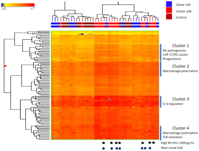
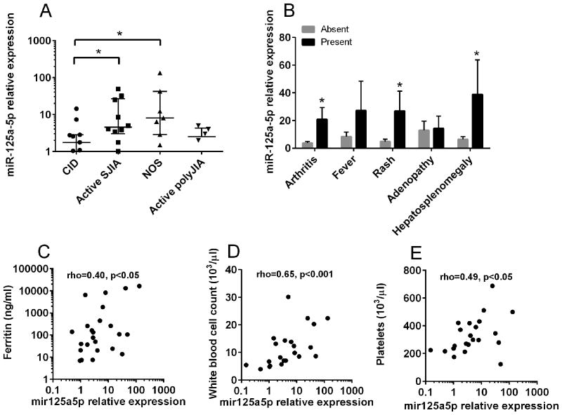
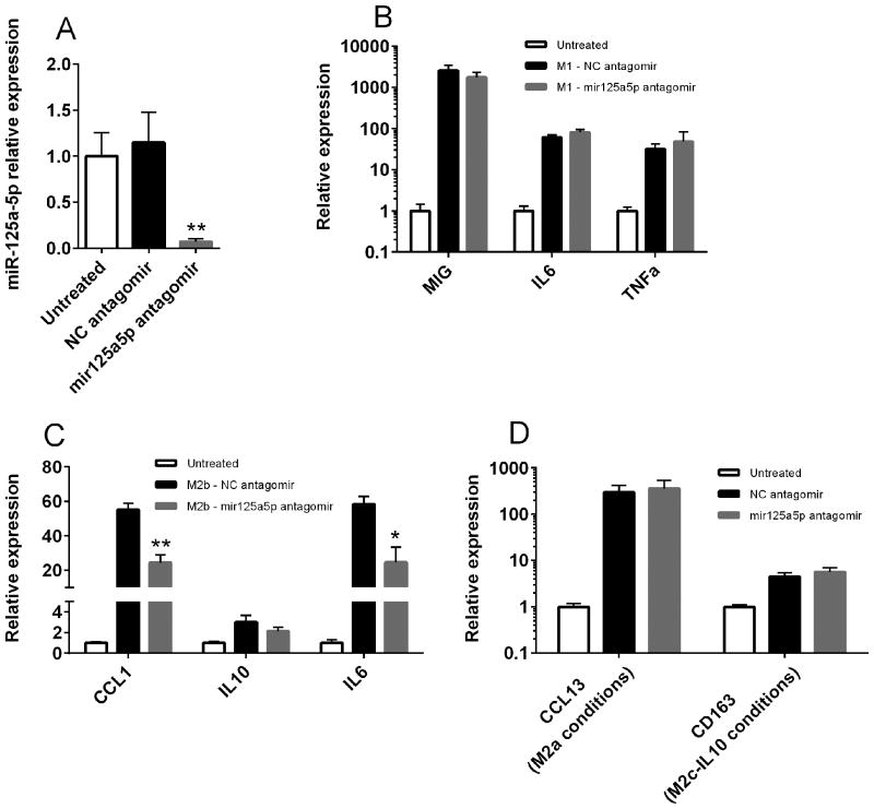
Results: A total of 110 microRNAs were found to be differentially expressed in monocytes from patients with active systemic JIA, including molecules implicated in rheumatoid arthritis pathogenesis, cytokine production, and monocyte polarization. MicroRNA-125a-5p was identified as being highly up-regulated in monocytes from children with active systemic JIA, as compared to those from children with clinically inactive JIA or those with active polyarticular JIA, and correlated with systemic features of the disease. In vitro, monocyte miR125a-5p expression was increased after polarization under M2b or M2c conditions. Inhibition of miR-125a-5p showed that this microRNA contributed to full polarization of M2b regulatory macrophages. In contrast, miR-125a-5p overexpression enhanced M2b polarization and altered other polarized populations, including increasing the production of M2 markers. Indeed, in vitro overexpression of this microRNA altered the macrophage phenotype toward that observed in systemic JIA.

Conclusion: Children with active systemic JIA have profound alterations in the expression of microRNAs that are implicated in monocyte function and polarization. One of these microRNAs, miR-125a-5p, is also a regulator of immunoregulatory M2b macrophages.


Figures

Figure 1
Figure 1
Clustering analysis of differentially expressed microRNAs between patients with active SJIA and those with CID and healthy controls. The list of differentially expressed genes was generated using a three-way ANOVA comparing patients with active SJIA to those with CID and controls, with Benjamini and Hochberg corrected p<0.05. In this tree, each row represents a separate microRNA and each column represents a separate sample. The normalized expression levels for each microRNA in each sample is indicated by color. The colored line above the tree indicates patients with CID (blue), active SJIA (red) or controls (brown). Patients with high serum ferritin >209ng/ml are indicated with stars. Patients with new-onset (untreated) SJIA are indicated with dark circles.
Figure 2
Figure 2
Elevated levels of miR-125a-5p in monocytes from patients with active SJIA and associated with features of systemic disease. Expression levels of miR-125a-5p were determined by Taqman qRT-PCR and normalized against RNU48. A, Expression of miR-125a-5p in patients with active SJIA, new-onset disease (NOS), active polyarticular JIA, and CID. Lines indicate median and error bars indicate SEM. B, Expression of miR-125a-5p in patients with SJIA with presence (black bars) or absence (gray bars) of clinical features. *=p<0.05, Mann-Whitney U-test. C-E, Correlation of miR-125a-5p level with serum ferritin (C), white blood cell count (D), and platelet count (E) as determined by Spearman rank correlation method.
Figure 3
Figure 3
miR-125a-5p is required for M2b macrophage polarization. A, THP-1 monocytic cells were differentiated with PMA and left untreated (white bar) or treated with negative control (black bar) or specific miR-125a-5p antagomir (gray bar) as described. miR-125a-5p levels were determined by individual Taqman assays and normalized to RNU48. Data are representative of three independent experiments performed in triplicate. B-D, THP-1 monocytic cells were differentiated with PMA and left untreated or treated with negative control or miR-125a-5p antagomir as described. Cells were then polarized under M1 (B), M2b (C), M2a or M2c-IL10 (D) conditions for 24 hours as described. Target gene expression was determined using qRT-PCR and normalized to GAPDH. Data are pooled from three independent experiments performed in triplicate. **=p<0.01; *=p<0.05 as determined by student's t-test.
Figure 4
Figure 4
Overexpression of miR-125a-5p enhances expression of regulatory macrophage-associated genes. A, THP-1 monocytic cells were differentiated with PMA and transfected with negative control (black bar) or miR-125a-5p mimic (white bar). miR-125a-5p expression was determined by individual Taqman assays and normalized to RNU48. Data is representative of three independent experiments performed in triplicate. B-E, THP-1 monocytic cells were differentiated with PMA, transfected as described, and polarized under M1 (B), M2b (C), M2a (D), or M2c-IL10 (E) conditions for 24 hours. Target gene expression was determined using qRT-PCR and normalized to GAPDH. Data are pooled from three independent experiments performed in triplicate. *=p<0.01 as determined by student's t-test.

References

    1. Petty RE, Southwood TR, Manners P, Baum J, Glass DN, Goldenberg J, et al. International League of Associations for Rheumatology classification of juvenile idiopathic arthritis: second revision, Edmonton, 2001. J Rheumatol. 2004;31:390–2. - PubMed
    1. Ravelli A, Grom AA, Behrens EM, Cron RQ. Macrophage activation syndrome as part of systemic juvenile idiopathic arthritis: diagnosis, genetics, pathophysiology and treatment. Genes Immun. 2012;13:289–298. - PubMed
    1. Mellins ED, Macaubas C, Grom AA. Pathogenesis of systemic juvenile idiopathic arthritis: some answers, more questions. Nat Rev Rheumatol. 2011;7:416–426. - PMC - PubMed
    1. Fall N, Barnes M, Thornton S, Luyrink L, Olson J, Ilowite NT, et al. Gene expression profiling of peripheral blood from patients with untreated new-onset systemic juvenile idiopathic arthritis reveals molecular heterogeneity that may predict macrophage activation syndrome. Arthritis Rheum. 2007;56:3793–3804. - PubMed
    1. Pascual V, Allantaz F, Arce E, Punaro M, Banchereau J. Role of interleukin-1 (IL-1) in the pathogenesis of systemic onset juvenile idiopathic arthritis and clinical response to IL-1 blockade. J Exp Med. 2005;201:1479–1486. - PMC - PubMed

Publication types