Observational Study
. 2016 Mar;95(12):e3109.
doi: 10.1097/MD.0000000000003109.

Anatomic and Functional Evaluation of Central Lymphatics With Noninvasive Magnetic Resonance Lymphangiography

Affiliations
Observational Study

Anatomic and Functional Evaluation of Central Lymphatics With Noninvasive Magnetic Resonance Lymphangiography

Eun Young Kim et al. Medicine (Baltimore). 2016 Mar.

Abstract

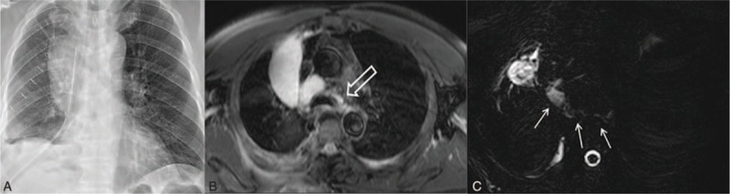
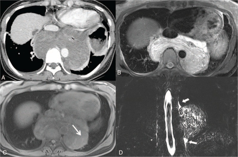
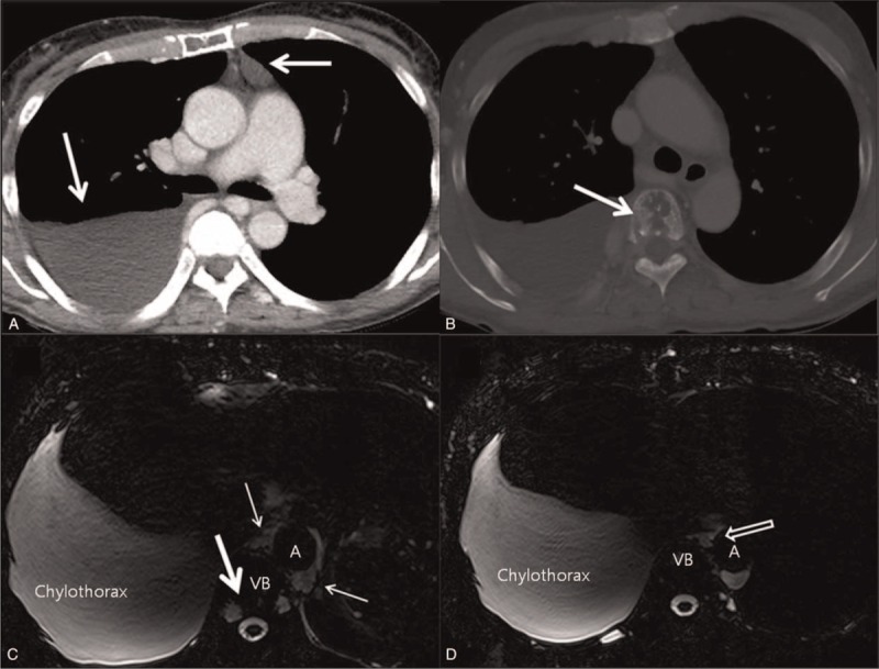
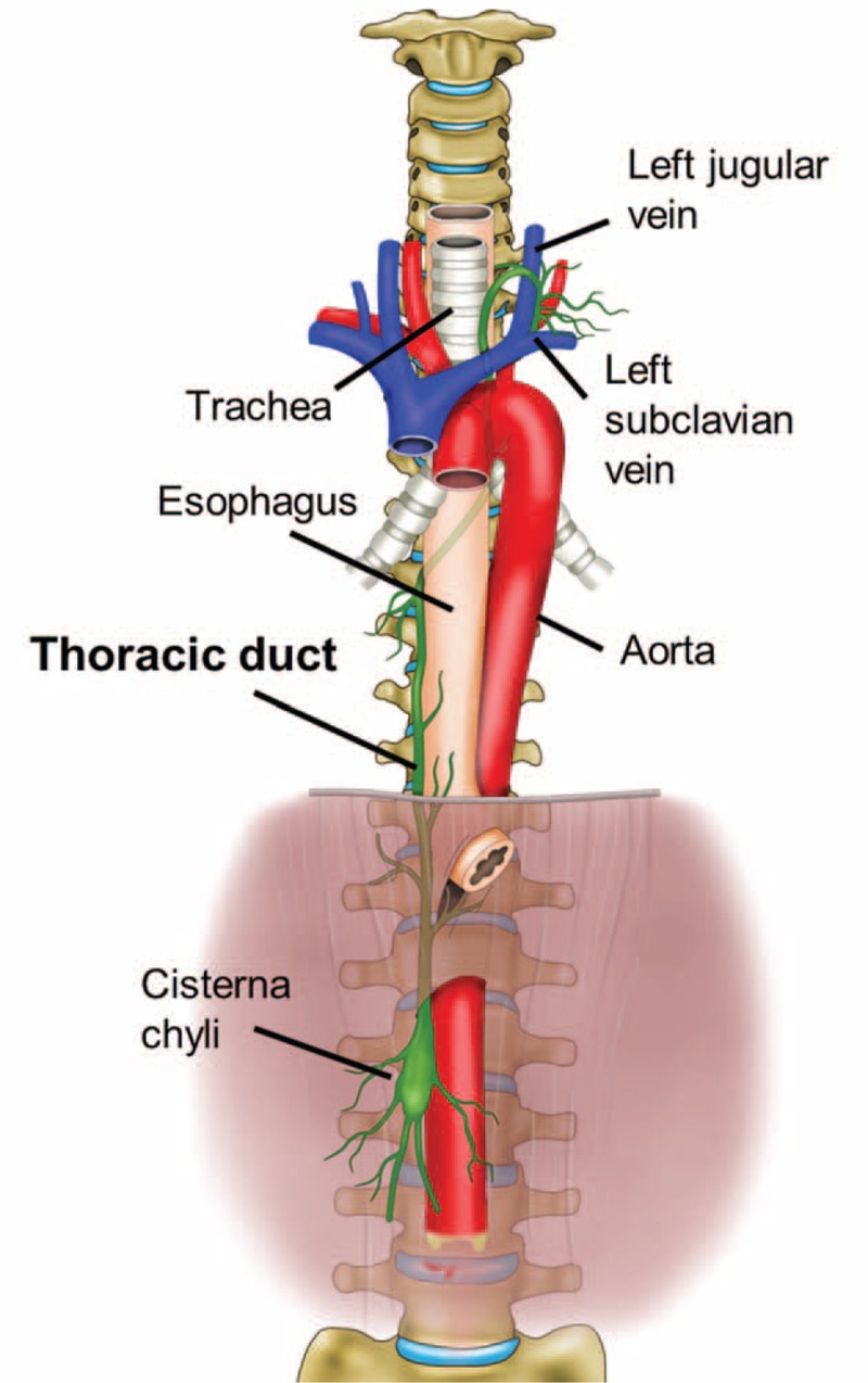
Accurate assessment of the lymphatic system has been limited due to the lack of optimal diagnostic methods. Recently, we adopted noncontrast magnetic resonance (MR) lymphangiography to evaluate the central lymphatic channel. We aimed to investigate the feasibility and the clinical usefulness of noninvasive MR lymphangiography for determining lymphatic disease.Ten patients (age range 42-72 years) with suspected chylothorax (n = 7) or lymphangioma (n = 3) who underwent MR lymphangiography were included in this prospective study. The thoracic duct was evaluated using coronal and axial images of heavily T2-weighted sequences, and reconstructed maximum intensity projection. Two radiologists documented visualization of the thoracic duct from the level of the diaphragm to the thoracic duct outlet, and also an area of dispersion around the chyloma or direct continuity between the thoracic duct and mediastinal cystic mass.The entire thoracic duct was successfully delineated in all patients. Lymphangiographic findings played a critical role in identifying leakage sites in cases of postoperative chylothorax, and contributed to differential diagnosis and confirmation of continuity with the thoracic duct in cases of lymphangioma, and also in diagnosing Gorham disease, which is a rare disorder. In patients who underwent surgery, intraoperative findings were matched with lymphangiographic imaging findings.Nonenhanced MR lymphangiography is a safe and effective method for imaging the central lymphatic system, and can contribute to differential diagnosis and appropriate preoperative evaluation of pathologic lymphatic problems.


Conflict of interest statement

The authors have no funding and conflicts of interest to disclose.

Figures

FIGURE 1
FIGURE 1
Postoperative chylothorax in a 68-year-old male who underwent right lower lobectomy due to lung cancer (T2aN0M0, stage IB). A, Posteroanterior chest radiography obtained 10 days after surgery shows large right pleural effusion despite chest tube drainage. B, Axial T2-weighted MR imaging obtained the same day demonstrates a large right pleural effusion continuous into the mediastinum between the aorta and azygos vein (white open arrow). C, Axial heavily T2-weighted MR imaging demonstrates a leak site (white arrows) in thoracic duct near the azygos arch. MR = magnetic resonance.
FIGURE 2
FIGURE 2
Cavernous lymphangioma in a 41-year-old female. A, Enhanced axial CT scan demonstrates a well-defined homogenous low attenuating mass encasing the esophagus in the posterior mediastinum. B, T2-weighted MR imaging shows high signal intensity with a multilocular septum within the mass. C, T1-weighted MR imaging shows an isosignal intensity with faint high signal intensity (white arrow), which led to suspicion of hemorrhage. D, Coronal heavily T2-weighted maximum intensity projection (MIP) image reveals continuation between the thoracic duct (arrow head) and mass. CT computed = tomography, MR = magnetic resonance.
FIGURE 3
FIGURE 3
Gorham disease in a 63-year-old female with idiopathic chylothorax. A, Enhanced axial CT with mediastinal window setting shows large right pleural effusion and abnormal fluid density lesion (white arrow) in the anterior mediastinum. B, Axial CT with bone window setting reveals a mottled osteolytic lesion (white arrow) in vertebral body. C, D, Axial heavily T2-weighted MR imaging obtained the same day demonstrates abrupt discontinuation of the thoracic duct (open arrow) with dispersion of chyle into the mediastinum (thin arrows), suggesting leakage around the cistern chyli. The mottled osteolytic lesion mentioned above showed high T2 signal intensity (thick arrow), suggesting a dilated lymphatic channel within the vertebral body. CT computed = tomography, MR = magnetic resonance.
FIGURE 4
FIGURE 4
Schematic illustration of the anatomical course of the thoracic duct. The thoracic duct is a continuation of the cisterna chyli from its abdominal segment into the thorax. Typically, the lymphatic pathway originates in the cisterna chyli and enters into the thoracic cavity thorough the aortic hiatus. In the thorax, the thoracic duct ascends along the right anterior surface of the vertebral column between the aorta and the azygos vein posterior to the esophagus. At the T5 to T6 vertebral level, it crosses left of the midline and extends posterior to the aortic arch. It has a close relationship with the trachea, ascends approximately 2 to 3 cm above the clavicle, and then crosses anterior to the subclavian and thyrocervical trunk, making an arch inferiorly. Finally, it terminates at the junction of the left subclavian vein and the internal jugular veins.

References

    1. Chen E, Itkin M. Thoracic duct embolization for chylous leaks. Semin Intervent Radiol 2011; 28:63–74. - PMC - PubMed
    1. Raman SP, Pipavath SN, Raghu G, et al. Imaging of thoracic lymphatic diseases. AJR Am J Roentgenol 2009; 193:1504–1513. - PubMed
    1. Silvestri RC, Huseby JS, Rughani I, et al. Respiratory distress syndrome from lymphangiography contrast medium. Am Rev Respir Dis 1980; 122:543–549. - PubMed
    1. Clement O, Luciani A. Imaging the lymphatic system: possibilities and clinical applications. Eur Radiol 2004; 14:1498–1507. - PubMed
    1. Matsumoto T, Yamagami T, Kato T, et al. The effectiveness of lymphangiography as a treatment method for various chyle leakages. Br J Radiol 2009; 82:286–290. - PubMed

Publication types