. 2016 Mar 25;14(3):e1002395.
doi: 10.1371/journal.pbio.1002395. eCollection 2016 Mar.

Hemotin, a Regulator of Phagocytosis Encoded by a Small ORF and Conserved across Metazoans

Affiliations

Hemotin, a Regulator of Phagocytosis Encoded by a Small ORF and Conserved across Metazoans

José I Pueyo et al. PLoS Biol. .

Abstract

Translation of hundreds of small ORFs (smORFs) of less than 100 amino acids has recently been revealed in vertebrates and Drosophila. Some of these peptides have essential and conserved cellular functions. In Drosophila, we have predicted a particular smORF class encoding ~80 aa hydrophobic peptides, which may function in membranes and cell organelles. Here, we characterise hemotin, a gene encoding an 88aa transmembrane smORF peptide localised to early endosomes in Drosophila macrophages. hemotin regulates endosomal maturation during phagocytosis by repressing the cooperation of 14-3-3ζ with specific phosphatidylinositol (PI) enzymes. hemotin mutants accumulate undigested phagocytic material inside enlarged endo-lysosomes and as a result, hemotin mutants have reduced ability to fight bacteria, and hence, have severely reduced life span and resistance to infections. We identify Stannin, a peptide involved in organometallic toxicity, as the Hemotin functional homologue in vertebrates, showing that this novel regulator of phagocytic processing is widely conserved, emphasizing the significance of smORF peptides in cell biology and disease.


Conflict of interest statement

The authors have declared that no competing interests exist.

Figures
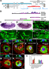
Fig 1
Fig 1. Identification and phenotypical characterisation of the hemotin gene.
(A) hemo genomic locus including the hemo, CG7691, fray, and fruitless genes (blue arrows). The hemo A4 deletion (red bar) was generated by FRT-mediated recombination using the P{RS3}fray CB-0706-3 and the P-Bac{WH}fru f02684 transposable elements (blue triangles). Transcript models are represented under their respective genes, orange boxes represent coding exons, whereas gray boxes indicate noncoding exons (untranslated regions, UTRs). hemo A4 completely removes hemo and CG7691 plus the first noncoding exons of fray and fruitless. The P{PZ}fray 07551 insertion is a lethal fray allele [25] and was used for genetic complementation experiments between hemo A4 and fray. (B) Top: Ribosomal profiling reads obtained from polyribosomes from S2 cells (Poly-Riboseq;(3)) mapped to the hemo full-length transcript (hemoFL). hemo-ORF is translated more efficiently than ORF2 (hemo-ORF RPKM: 29.4, coverage: 0.9 ORF; ORF2 RPKM: 6.6, coverage: 0.7. Note that the reads per kilobase of transcript per million mapped reads [RPKM] value of ORF2 is below the 11.8 cut-off to be considered translated [3]). Bottom: schematic representation of other constructs used in this manuscript. hemo-ORF is a minigene consisting of an mRNA fragment truncated immediately after the hemo-ORF stop codon, ORF2 consists of a mini-gene construct carrying the ORF2 sequence only, including 6 nt upstream of its start codon (to conserve its endogenous Kozak sequence). hemo-GFP (green fluorescent protein) is a hemo-ORF-GFP fusion construct in which the GFP sequence (devoid of a start codon) was cloned into the hemoFL construct, immediately downstream and in frame with hemo-ORF (devoid of a stop codon) (see Materials and Methods). (C) Pattern of expression of hemo in germ band-retracted embryos revealed by in situ hybridisation. hemo is specifically expressed in embryonic hemocytes (arrows; compare with D) in the head, amnioserosa, and dispersed along the body. (D) Spatial distribution of embryonic hemocytes at germ band retraction stage revealed by in situ hybridisation of hemocyte-specific croquemort (crq) gene, showing similar distribution in the head, amnioserosa, and along the body (arrows). (E) Cluster of early embryonic hemocytes of the cephalic region expressing the hemo transcript revealed by FISH (fluorescent in situ hybridisation). Some hemocytes show drop-shape morphologies (asterisk) and membrane projections such as filopodia (arrows). (F) Embryonic hemocytes labelled with crq-Gal4;UAS-GFP expression from the head region displaying similar cellular morphologies (arrows and asterisk) as those in E. (G–H) White prepupal thoracic hemocytes revealed by crq-Gal4;UAS-GFP expression in wild-type (G) and hemo A4 mutants (H). In hemo A4 mutants, hemocytes display enlarged vacuoles within the cytoplasm (arrowheads), with larger occupied area index (OAI). Scale bar (50 μm). (I–N) hemocytes observed ex vivo [15] showing Tubulin (green) and Actin (red) cytoskeletons and nuclei (2-(4-Amidinophenyl)-6-indolecarbamidine dihydrochloride, DAPI) with its corresponding orthogonal projection of confocal microscopy z-stacks (above inset) showing only tubulin cytoskeleton (green) and DAPI (blue) staining in the nucleus (n). Scale bar (5 μm). (I) Wild-type hemocyte. (J) hemo A4 mutant hemocyte shows large disruptions of the tubulin cytoskeleton that appear as rounded vacuoles (arrows; arrowhead in inset). (K) Knocking down the expression of hemo with a UAS-hemo-RNAi construct phenocopies the vacuolation phenotype (arrows and arrowhead in inset). (L) Expression of hemo full length transcript (UAS-hemoFL) rescues the vacuolated hemo A4 phenotype. Expression of hemo-ORF only (M) also rescues the hemo A4 mutant vacuolation. (N) Expression of ORF2 does not rescue the hemo A4 mutant vacuolated phenotype (arrows and arrowhead in inset). (O) Vacuolation measurements in ex vivo primary pre pupal hemocytes. hemo A4 mutant hemocytes show significantly higher occupied volume index (OVI) (see Materials and Methods) than wild-type. Rescue experiments show that the vacuolation phenotype is specific to the peptide encoded by hemo-ORF. All upstream activating sequence (UAS) constructs were driven by crq-Gal4. Error bars represent standard error of the mean (SEM). Statistical analysis was performed using one-way ANOVA test indicating that samples were significantly different [F(9,486) = 9.5, p < 0.0001]. A post hoc Bonferroni multicomparison test showed that hemo A4, UAS-hemo-RNAi, UAS-ORF2-hemo A4, UAS-hemoFS (expressing a hemo full-length transcript containing frameshifts in hemo-ORF and ORF2)-hemo A4 and CG7691 genomic fragment (GF)-hemo A4 were significantly different than wild-type. The UAS-hemoFL-hemo A4,UAS-hemo-ORF-hemo A4,UAS-hemoGFP-hemo A4 and fray PZ /hemo A4 were not significant to wild-type (n ≥ 24, p < 0.05). Supplemental data are shown in S1 Fig and S1 Data.
Fig 2
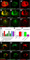
Fig 2. The Hemotin peptide is required for proper endosomal maturation in hemocytes.
(A–A”) Distribution of acidic organelles in hemo A4 mutant ex vivo hemocytes revealed by the expression of LAMP1-GFP lysosomal marker. The intracellular vacuoles that disrupt the beta-tubulin cytoskeleton (A, A”; red) accumulate LAMP1-GFP positive compartments (A, A’; green). Compare with wild-type in S2A–S2A” Fig. Scale bar (5 μm). (B–B”) Distribution of the endosomal marker FYVE (PI(3)P binding zinc finger domain, early endosomal marker, named after being found in Fab1, YOTP, Vac1, EEA1) (green) (B, B’) and Lysotracker (red) (B, B”) organelles in a hemo A4 mutant ex vivo hemocyte showing enlarged intracellular compartments coexpressing FYVE and Lysotracker. Scale bar (5 μm). (C–C”) Wild-type ex vivo hemocyte labelled as in (B), showing little overlap between early endosome-FYVE positive (green) (C,C’) and lysosomal (red) (C,C”) compartments. (D) Quantification of the FYVE OAI in ex vivo hemocytes (see Materials and Methods). hemo A4 mutants display a significantly larger FYVE area than wild-type. This phenotype is rescued by the expression of the hemo full length transcript (UAS-hemoFL) and is specific to hemo-ORF function, as it is also rescued by the expression of the hemo-ORF mini gene (UAS-hemo-ORF) or C-terminal-tagged hemo-GFP (UAS-hemo-GFP). No rescue was observed by a CG7691 genomic fragment (CG7691-GF), or with a hemo full-length transcript containing a frameshift in the hemo-ORF (UAS-hemoFS), or with the ORF2 mini gene (UAS-ORF2). All UAS constructs were driven with He-Gal4. Error bars represent SEM. One-way ANOVA analysis shows that there is a statistically significant difference between these groups [F(7,286) = 27.12, p < 0.0001]. Post hoc comparisons using Bonferroni test indicated that the mean score of hemo A4, UAS-hemo-ORF2-hemo A4, UAS-hemoFS-hemo A4, and CG7691-GF-hemo A4 did significantly differ from wild-type (p < 0.05), whereas UAS-hemoFL-hemo A4, UAS-hemoORF-hemo A4, and UAS-hemoGFP-hemo A4 did not. (E) Analysis of the overlap between FYVE-positive early endosomal and Lysotracker-positive compartments using Pearson’s correlation coefficient in wild-type and hemo A4 mutant hemocytes. In hemo A4 hemocytes, there exist significantly more intracellular compartments displaying FYVE and Lysotracker colocalisation than in the wild-type as shown by a Two-tailed Mann-Whitney test (n ≥ 17; p < 0.05). Error bars represent SEM. (F) Statistical analysis of the Pearson’s coefficient measurements of Hemotin-GFP (Hemo-GFP) with early endosomal (FYVE-cherry) and lysosomal (Lysotracker) markers. Tagged-Hemotin peptides are significantly enriched in early endosomal compartments in comparison with lysosomes as shown by a two tailed Mann-Whitney test (n ≥ 20, p < 0.05). Error bars represent SEM. (G–G”) Localisation of Hemo-GFP peptides (green)(G’) and the endosome FYVE marker (red)(G”) in hemocytes (He-Gal4;UAS-hemo-GFP). A substantial part of Hemo-GFP pattern colocalizes with FYVE-positive compartments (G) (arrows). Scale bar (5 μm). (H–H”) Distribution of Hemo-GFP peptides (green) (H’) and the lysosomal marker (lysotracker; red) (H”) in hemocytes. Only a small overlap exists between Hemo-GFP compartments and lysosomes (H) (arrow). Supplemental data are shown in S2 Fig, S6 Fig, and S1 Data.
Fig 3
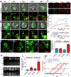
Fig 3. hemotin is involved in phagocytic processing and is necessary for optimal bacterial clearance and life span.
(A–B) Time-lapse imaging showing the phagocytic trafficking of pHrodo-labelled bacterial particles (arrowhead, red) in ex vivo hemocytes expressing the early endosomal marker FYVE-GFP (green) with the He-Gal4 driver in wild-type (A) or hemo A4 mutants (B) (see S1 and S2 Videos). (t0) represents the time when the particle docks into the cell membrane, displaying a relatively faint intensity. By t = 6 minutes (min), the pHrodo particles are in FYVE-positive early endocytic vesicles in both wild-type (A) and hemo A4 mutant hemocytes (B). By t = 22 min in the wild-type hemocyte (A), the FYVE signal around the particle is dramatically reduced, while the intensity of the pHrodo signal increases, suggesting that the vesicle has progressed into a PI3P-depeleted and acidified endolysosome. In the hemo A4 mutant hemocyte (B), the FYVE signal around the particle is still visible at t = 44 min, showing an increased prevalence of PI(3)P in this vesicle, and therefore an extended early endocytic phase. However, the intensity of the pHrodo signal is lower than in wild-type, indicating a delay in the acidification of the endocytic vesicles (See also C and D). The insets show a magnification of the specific particles. Scale bars = 5 μm. (C) Magnified raw images from the particles shown in (A) and (B), showing the region of interest (ROI) used to quantify their fluorescent intensity. The integrated intensity read-out for each time point is indicated in red. (D) Quantification of fluorescent intensity of pH-sensitive pHrodo particles undergoing phagocytosis in wild-type (blue) or hemo A4 mutant (red) hemocytes. Average integrated intensity per pixel is represented for each time point. Error bars represent SEM. A two-way ANOVA analysis with Bonferroni post test indicates that the difference between these curves is significantly different from t = 16.5 min (n = 8, p < 0.001). (E) Quantification of FYVE prevalence on pHrodo particles undergoing phagocytosis in wild-type (blue) or hemo A4 mutant (red) hemocytes. The FYVE signal remains significantly longer in hemo A4 mutants (mean = 46.06 +/− 5.3 min) than wild-type (mean = 19.87 +/− 2.1 min) as indicated by a one-tailed unpaired t test (n = 11, p < 0.0003). Error bars represent SEM. (F–I) Visualisation of bacterial up-take and processing in vivo. Dorsal vessel-associated adult hemocytes (see Materials and Methods) expressing FYVE-GFP (green) driven by He-Gal4 from wild-type (F, G) or hemo A4 mutant flies (H, I) infected with mCherry-expressing E. coli bacteria (red) (OD600 = 0.05) and dissected 20 min (F, H) or 120 min (G, I) postinjection. Wild-type and mutant flies exhibit a similar number of bacterial cells per hemocyte after 20 min; however, this number increases in mutant flies after 120 min, whereas it remains constant in wild-type flies, suggesting that mutant hemocytes accumulate undigested bacterial cells. Yellow dashed lines represent the cell body outline. (J) Quantification of bacterial uptake. hemo A4 mutant hemocytes contain a similar number of bacterial cells as wild-type after 20 min but significantly more bacterial cells after 120 min. Average number of bacterial cells per hemocyte is represented in the y-axis. (n = 30, p < 0.0001). (K) Visualisation of bacterial load from either wild-type or hemo A4 mutant 30days (d)old adult fly homogenates. Each spot represents the bacterial colonies grown from an individual male fly. Each homogenate was plated in decreasing dilution (1:1 or 1:10). Note the higher density of bacterial colonies in the spots from hemo A4 mutants compared to wild-type. (L) Quantification of bacterial colonies grown from single adult fly homogenates. hemo A4 mutants contain significantly more bacteria than wild-type, both at 10 (n = 12, p < 0.0054) or 30 d old (n = 8, p < 0.0078). Error bars represent SEM. (M) hemo A4 mutants (red) have a reduced viability over time compared to wild-type (blue), with a median life-span of 23 +/− 6 d, compared to 49 +/− 3 d for wild-type. The addition of antibiotics (penicillin-streptomycin) to the food media significantly increases the median life span of hemo A4 mutants to 38 +/− 6 d, as determined by a paired t test (p < 0.05). For each condition, five different replicates were analysed for a total of 50 flies. Error bars represent SEM. Supplemental data are shown in S3 Fig, S1 Video, S2 Video, and S1 Data.
Fig 4
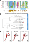
Fig 4. Conserved sequence and structure in Stannin, the Hemotin vertebrate homologue.
(A) Alignment showing Hemotin amino acid peptide sequences (see S4 File) from two insect lineages (Diptera and Hymenoptera) and vertebrate Stannin. The “Conservation” lane at the bottom reflects the conservation of physical–chemical properties in the amino acids, while the “Quality” lane scores the likelihood of observing the mutations displayed in each particular position of the alignment. Note that there exists high conservation in the transmembrane domain (blue) motif among these peptides. The N-terminal transmembrane and C-terminal alpha-helix domains are indicated below the alignment. Sequences studied in C) (see below) are highlighted with stars. (B) Cladistic guide tree showing the relationships between insect Hemotin and vertebrate Stannin peptides shown in (A). The Hemotin and Stannin sequences cluster with their respective counterparts from related species. Insect Hemotin sequences are branched into two main clusters, one represented by flies and the other by hymenopterans, with other dipterans such as mosquitoes being between these two. New Stannin homologues identified by us cluster with annotated Stannin sequences (prefixed by snn_) and show an overall correct phylogenetic position, with agnathans Lamprey and Hagfish basal to other vertebrates and closer to insect sequences (see also S4 Fig). Sequences studied in C) (see below) are highlighted with stars. (C) Threading of the peptide sequences (shown in red) from a basal vertebrate (hagfish), a basal hymenopteran (the wasp Microplitis), and D. melanogaster onto the human Stannin structure (shown in grey) (see Materials and Methods). It has been proposed that the N-terminal α-helix of the Stannin peptide transverses the membrane at an 80o angle, whereas the C-terminal α-helix lies upon the lipid bilayer at the cytoplasmic side [36]. Note that the three peptides can adopt similar tertiary structures. GDT indicates the value of the High-Accuracy Global Distance Test, which measures the average distance (in Angstroms) between the Human Stannin model and the model for each peptide [38]. As expected, the Hagfish peptide, more closely related to human Stannin, obtains a lower distance, but insect peptides display values in the same range despite their lower sequence similarity (see A) above and S4C Fig). Supplemental data are shown in S4 Fig, S3 File and S4 File.
Fig 5
Fig 5. Stannin and Hemotin replicate each other’s functions and counteract 14-3-3ζ function during endosomal maturation.
(A) Mouse macrophage-like RAW264.7 cells treated with control-scrambled siRNAs and labelled with the acidic pH-sensitive Lysotracker (Red). Scale bar (5 μm). (B) RAW264.7 cells treated with two Fluorescein isothiocyanate (FITC)-labelled snn siRNAs (green) (see S5 Fig) and stained with Lysotracker. Note the highly enlarged lysosomal compartment in snn siRNA-treated cells (see C). Scale bar (5 μm). (C) Lysosomal OAI, revealed by Lysotracker, in nontransfected or control siRNA and snn siRNA-treated RAW264.7 cells, showing that snn siRNA-treated RAW 264.7 cells have significantly larger lysosomal compartments than control samples (see S1 Data). The graph shows averages of three independent experiments. One-way ANOVA test showed that samples were significantly different [F(2,780) = 185.6, p < 0.0001]. Post hoc Bonferroni multicomparison test showed that the siRNA-snn sample was significantly different to nontransfected and siRNA-control samples (n ≥ 240, p < 0.05). On average, snn siRNA-treated cells show a reduction in snn expression of 60% relative to nontreated cells, whereas cells treated with control siRNA only show a reduction of 0.8% (see S5A Fig). Error bars represent SEM. (D–D”) Hemo-GFP and Snn-FLAG peptides expressed in hemocyte-like Drosophila Kc165 cells (20) using the Act5-Gal4 driver. Hemo-GFP (green) (D’) and Snn-FLAG (red) (D”) peptides colocalize in intracellular vesicles and punctate organelles (arrows). Scale bar (5 μm). (E) Colocalisation of Hemo-GFP peptides and Nt-tagged HA-14-3-3ζ protein in intracellular compartments in ex-vivo hemocytes. (E’) Hemo-GFP. (E”) HA-14-3-3ζ. (E”‘) Merged image. UAS constructs were driven with He-Gal4. Scale bar (10 μm). (F) Pull down of myc-14-3-3ζ with Hemo-GFP in transfected Drosophila Kc167 cells. Myc-14-3-3ζ interacts with Hemo-GFP but not with a GFP-only control. Molecular weight is indicated in kilodaltons Retention of Hemo-GFP and GFP is shown in S5C Fig. (G) Vacuole OVI measurements (see Materials and Methods and S1 Data) in primary hemocytes. Expression of human snn (UAS-snn) rescues the hemo A4 vacuolation phenotype to a similar extent as the rescue observed by the UAS-hemoFL and hemo-ORF constructs. Reducing the dosage of 14-3-3ζ (in a heterozygous null 14-3-3ζ 12BL /+ background, labelled 14-3-3ζ −/+) reduces the hemo A4 vacuolation phenotype. Conversely, overexpression of 14-3-3ζ (UAS-14-3-3ζ) in hemocytes induces the formation of larger vacuoles. The induction of vacuoles by excessive 14-3-3ζ is reversed by simultaneous overexpression of hemo full-length transcript (UAS-hemoFL) or overexpression of the human Stannin peptide (UAS-snn) but not by the expression of the control UAS-GFP construct. One-way ANOVA test showed that the means were significantly different [F(9,365) = 14.26, p < 0.0001]. Post hoc multiple comparison Bonferroni’s test showed that hemo A4, UAS-14-3-3ζ and UAS-14-3-3ζ;UAS-GFP samples were significantly different than wild-type, whereas the rest were not (n ≥ 20, p < 0.05). Error bars represent SEM. (H) Measurement of the occupied FYVE area index (OAI) in ex vivo prepupal hemocytes (see Materials and Methods and S1 Data). Overexpression of human Snn peptide (UAS-snn) rescues the hemo A4-enlarged FYVE compartments. Similarly, reducing 14-3-3ζ function by expression 14-3-3ζ-RNAi restores the size of hemo A4 mutant FYVE-organelles to wild-type. Conversely, overexpression of 14-3-3ζ (UAS-14-3-3ζ) mimics the hemo A4 mutant FYVE phenotype. The overexpression 14-3-3ζ-phenotype is reversed by coexpression with hemo full-length transcript (UAS-hemoFL). UAS constructs were driven with He-Gal4. One-way ANOVA test showed that the means of samples were significantly different [F(8,346) = 23.15, p < 0.0001]. Multiple comparison post hoc Bonferroni’s test indicated that UAS-14-3-3ζ and hemo A4 were significantly different than wild-type whereas the rest of the samples were not (n ≥ 20, p < 0.05). Error bars represent SEM. (I–N) Intracellular distribution of FYVE-positive (green) compartments in ex vivo prepupal hemocytes (see also H). Scale bar (5 μm). (I) In wild-type FYVE-positive organelles appear as small rings and punctae. (J) In hemo A4 mutant hemocytes, FYVE compartments contain larger rings than wild-type (arrowheads). (K) Expression of Hemo-ORF peptide (UAS-hemo-ORF) rescues the enlarged hemo A4 mutant FYVE compartments. (L) Expression of snn (UAS-snn) reduces the hemo A4 FYVE phenotype. (M) Overexpression of 14-3-3ζ (UAS-14-3-3ζ) produces enlarged FYVE compartments (arrowheads). (N) Reducing 14-3-3ζ function with RNAi rescues the hemo A4 mutant FYVE phenotype. UAS constructs were driven with He-Gal4. Yellow dashed lines indicate the cell body. Supplemental data are shown in S5 Fig, S6 Fig, and S1 Data.
Fig 6
Fig 6. Hemotin modulates PI(3)P formation by repressing 14-3-3ζ-mediated activation of PI(3)K68D kinase.
(A–I) Distribution of FYVE-positive compartments in ex vivo hemocytes (see also J). Yellow dashed lines indicate cell body area, and arrowheads indicate enlarged FYVE compartments. Scale bar (5 μm). (A) Wild-type hemocytes. (B) hemo A4 mutants show enlarged FYVE compartments (arrowheads), (C) this phenotype is rescued by mtm gain of function (UAS-mtm). (D) Hemocytes overexpressing 14-3-3ζ (UAS-14-3-3ζ) or (E) the PI3K68D Kinase (UAS-PI3K68D) also show enlarged FYVE compartments (arrowheads), similar to those observed in hemo A4 mutants. (F) Enlarged FYVE compartments are also observed by the reduction of mtm (mtm-RNAi) in hemocytes. (G) Reduction of PI3K68D kinase function by RNAi rescues the enlarged-FYVE phenotype produced by overexpression of 14-3-3ζ. (H) Reducing the function of 14-3-3ζ (14-3-3ζ-RNAi) rescues the enlarged FYVE compartment produced by overexpression of PI3k68D or (I) by reduction of mtm. (J) Quantification of the FYVE OAI in primary hemocytes (see Fig 6, S1 Data). Knocking down the mtm function (mtm-RNAi) produces enlarged FYVE compartments as shown with hemo A4 mutants. Both overexpression of mtm (UAS-mtm) and haploinsufficiency of PI3K68D (Df(3)PI3K68D/+), rescue the hemo A4 mutant FYVE phenotype. Overexpression of PI3K68D (UAS-PI3K68D) mimics the hemo A4 mutant FYVE phenotype, and this is rescued by overexpression of hemo full-length transcript (UAS-hemoFL). Reduction of 14-3-3ζ function (14-3-3ζ-RNAi) corrects the mtm loss of function and PI3K68D gain of function FYVE phenotypes. The enlarged FYVE compartments produced by over-expression of 14-3-3ζ (UAS-14-3-3ζ) are corrected by knocking down PI3K68D function (PI3K68D-RNAi). One-way ANOVA test indicated that means of samples are significantly different [F(12,508) = 14.01, p < 0.0001]. Post hoc Bonferroni’s multiple comparison test showed that hemo A4, UAS-14-3-3ζ, UAS-mtm-RNAi, UAS-PI3K68D were significantly different to wild-type, whereas the rest of genotypes were not (n ≥ 19, p < 0.005). Error bars represent SEM. (K) In hemocytes, Hemo-GFP peptides colocalize with PI3K68D Kinase in intracellular compartments (arrowheads), presumably early endosomes. (K) PI3K68D-Cherry expression. (K’) Hemo-GFP, (K”) merge image. Scale bar (10 μm). (L) Western Blot of a Pull down experiment from hemocytes expressing PI3K68D-GFP and HA-14-3-3ζ revealing a protein interaction between PI3K68D and 14-3-3ζ. Supplemental data are shown in S6 Fig and S1 Data.
Fig 7
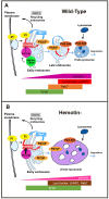
Fig 7. Model for the role of Hemotin in phagocytic processing.
Simplified models of endosomal maturation, modified from [18] depicting the role of the proteins and markers analysed in this work. A) Wild-type endosomal trafficking is regulated by different phosphorylation states of PI. Phosphorylation of PI into PI(3)P is achieved in early endosomes by the class II or class III PI3 kinases PI3k68D and Vps34, respectively. In late endosomes PI(3)P is again phosphorylated to produce PI(3,5)P2. This phosphorylation step allows late endosomes to progress into degradation allowing lysosomes to fuse to late endosomes to produce multivesicular bodies. This trafficking progression can be reversed by dephosphorylation of PI(3)P or PI(3,5)P2 by myotubulurin phosphatases. Hemotin and Stannin are functional homologues that localise to early endosomes, where they bind and repress 14-3-3ζ. Our genetic and biochemical data indicates that 14-3-3ζ binds the PI3K68D kinase and promotes its function, perhaps by directly increasing its enzymatic activity, or indirectly by promoting its correct localisation in early endosomes. Since Hemotin antagonises 14-3-3ζ, it indirectly reduces the development of early endosomes through PI3K68D. B) The absence of Hemotin produces an excess of 14-3-3ζ function, which results in an excess of PI3K68D function and leads to an increase in endocytic vesicles containing PI(3)P, as detected by expansion of the area occupied by the FYVE marker. These abnormal vesicles display an abnormal maturation during phagocytosis, with excessive co-expression of early lysosomal markers (such as FYVE) and late ones (Lysotracker and Rab7), and a slower and less intense acidification of their contents, as revealed by the pHrodo pH marker.

References

    1. Aderem A, Underhill DM (1999) Mechanisms of phagocytosis in macrophages. Annu Rev Immunol 17: 593–623. - PubMed
    1. Bazzini AA, Johnstone TG, Christiano R, Mackowiak SD, Obermayer B, et al. (2014) Identification of small ORFs in vertebrates using ribosome footprinting and evolutionary conservation. EMBO J. 2014/04/08 ed. pp. embj.201488411. - PMC - PubMed
    1. Aspden JL, Eyre-Walker YC, Philips RJ, Brocard M, Amin U, et al. (2014) Extensive translation of small ORFs revealed by polysomal ribo-Seq eLife 3: e03528 10.7554/eLife.03528 - DOI - PMC - PubMed
    1. Slavoff SA, Mitchell AJ, Schwaid AG, Cabili MN, Ma J, et al. (2013) Peptidomic discovery of short open reading frame-encoded peptides in human cells. Nat Chem Biol 9: 59–64. 10.1038/nchembio.1120 - DOI - PMC - PubMed
    1. Galindo MI, Pueyo JI, Fouix S, Bishop SA, Couso JP (2007) Peptides encoded by short ORFs control development and define a new eukaryotic gene family. PLoS Biology 5: 1052–1062. - PMC - PubMed

Publication types