. 2016 May 3;7(18):25377-90.
doi: 10.18632/oncotarget.8298.

Oxidative stress at low levels can induce clustered DNA lesions leading to NHEJ mediated mutations

Affiliations

Oxidative stress at low levels can induce clustered DNA lesions leading to NHEJ mediated mutations

Vyom Sharma et al. Oncotarget. .

Abstract

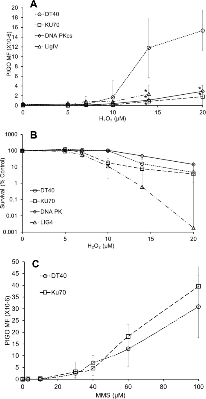
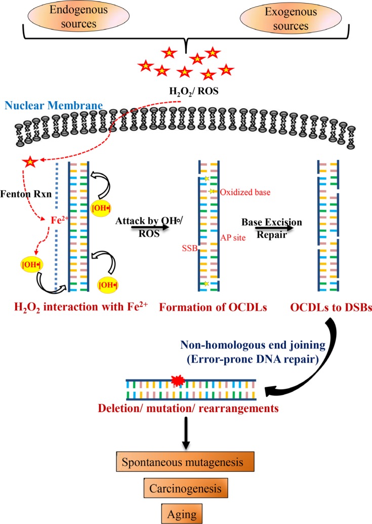
DNA damage and mutations induced by oxidative stress are associated with various different human pathologies including cancer. The facts that most human tumors are characterized by large genome rearrangements and glutathione depletion in mice results in deletions in DNA suggest that reactive oxygen species (ROS) may cause gene and chromosome mutations through DNA double strand breaks (DSBs). However, the generation of DSBs at low levels of ROS is still controversial. In the present study, we show that H2O2 at biologically-relevant levels causes a marked increase in oxidative clustered DNA lesions (OCDLs) with a significant elevation of replication-independent DSBs. Although it is frequently reported that OCDLs are fingerprint of high-energy IR, our results indicate for the first time that H2O2, even at low levels, can also cause OCDLs leading to DSBs specifically in G1 cells. Furthermore, a reverse genetic approach revealed a significant contribution of the non-homologous end joining (NHEJ) pathway in H2O2-induced DNA repair & mutagenesis. This genomic instability induced by low levels of ROS may be involved in spontaneous mutagenesis and the etiology of a wide variety of human diseases like chronic inflammation-related disorders, carcinogenesis, neuro-degeneration and aging.

Keywords: NHEJ; clustered DNA lesions; double strand breaks; mutations; oxidative stress.


Conflict of interest statement

Conflicts of Interest: None declared.

Figures

Figure 1
Figure 1. DNA damage response analysis in DT40 cells and their DNA repair mutants exposed to H2O2
Cells were exposed to different concentrations of freshly prepared H2O2 at 37°C. Survival data were log-transformed giving approximate normality. Analysis of covariance (ANCOVA) was used to test for mean intercept differences and differences in the slopes of the linear dose response curves between wild type and a series of mutant cells. Error bars indicate the 95% confidence intervals. HR- homologous recombination; NHEJ- Non-homologous end joining; BER/ NER- Base excision repair/ Nucleotide excision repair; MMR- Mismatch repair; DDCP- DNA damage checkpoint; TLS- Translesion synthesis.
Figure 2
Figure 2. DNA damage induced by low levels of H2O2
(A) 8-oxodG amounts in DT40 cells exposed to H2O2 for 30 min. The box shows 8-oxodG amounts in isogenic KU70-deficient cells under similar conditions. Data represent mean ± SD. One-way analysis of variance (ANOVA) with Dunnet's test was employed to test for statistically significant differences compared to control. In a second type of statistical analysis, a piece-wise linear model (the hockey stick model) was applied to find the breakpoint value. (B) Generation and repair of DSBs in DT40 and isogenic DSBs repair-deficient cells. To understand the repair kinetics, the H2O2 dose was increased to 50 μM for repair experiments. Cells were exposed to H2O2 for 30 min and processed further for PFGE. Bands representing DSBs were quantified using Quantiscan (Biosoft, Cambridge, UK). Relative DSB levels were obtained by comparing DSBs signals in each sample to the background signals observed for unexposed wild type DT40 cells. Data represent mean (n = 5) ± SD. *p < 0.05 when compared using Student's t test.
Figure 3
Figure 3. Induction of 53BP1 foci in G1 cells following exposure to H2O2
(A) TK6 cells were exposed to H2O2 (40 μM) for 30 min and co-immunostained with anti-53BP1 (green) and anti-cyclin A (magenta). (B) TK6 cells were pulse-labelled with BrdU for 15 min and then further incubated with H2O2 (40 μM) for 30 min. Cells were fixed and labelled with anti-53BP1 (green) antibody. BrdU-labelled cells were visualized with Alexa Fluor 555 (red) antibody. Nuclei were counterstained with DAPI (blue). The images were recorded using confocal microscopy and represents maximum intensity projections of z-stacks. 53BP1 foci are marked by white arrows. Magnification, 600×.
Figure 4
Figure 4. Formation of oxidatively induced clustered DNA lesions (OCDLs) on exposure to low doses of H2O2
(A) A representative image of OCDLs induced by 20 μM H2O2 in DT40 cells as assessed by modified PFGE. The area quantitated under each band (representing total DSBs) is shown by bracket (}). (B) Quantitative analysis of OCDL induction in DT40 and TK6 cells. Bands representing total DSBs were quantified using Quantiscan (Biosoft, Cambridge, UK). Relative DSB levels were obtained by comparing DSBs signals in each sample to the background signals observed for unexposed wild type DT40 cells. *p < 0.05; a statistical comparison of DSBs/OCDLs induction between exposed and unexposed cells under similar conditions (with/without enzyme) was conducted using a Student's t-test. Data represent mean (n = 3) ± SD.
Figure 5
Figure 5. NHEJ plays a predominant role in oxidative stress-induced mutagenesis
(A) PIG-O mutation analysis in wild type DT40 cells and NHEJ-deficient cells (KU70-, LIGIV-, and DNA PKcs-deficient) exposed to H2O2. *p < 0.05 when mutation levels were compared to wild type DT40 cells at respective doses using Student's t-test. (B) Survival of wild type DT40 cells and KU70-, LIGIV-, and DNA PKcs-deficient cells after exposure to H2O2, as assessed during the phenotype expression period in the mutation assay. (C) PIG-O mutation analysis in wild type DT40 cells and KU70- deficient cells exposed to methylmethanesulfonate. Data represent mean (n = 3) ± SD.
Figure 6
Figure 6. Schematic model for combined roles of OCDLs and error-prone NHEJ in oxidative stress-induced mutagenesis
Oxidative stress and hydroxyl radicals can be produced in cells due to a variety of endogenous and exogenous factors. H2O2 even at low concentrations can react with Fe2+ that is weakly associated with the N7 of guanine (RTGR sequence; Fenton's reaction), leading to base damages and sugar lesions. The formation of more than one hydroxyl radical by closely spaced Fe2+ ions or the redox recycling process or during replication due to loose DNA structure may damage the DNA in proximity to form OCDLs. OCDLs can be converted to DSBs with complex DNA ends during the repair process. Since OCDLs can cause complex DNA ends, they may be repaired primarily by NHEJ (Ku70-, LIGIV-, and DNA-PKcs). However, the error-prone NHEJ repair pathway, while trying to repair these DSBs, may introduce mutations in the genome. Thus, the genomic instability produced may play relevant roles in the etiology of a wide variety of human diseases including cancer, chronic inflammation-related disorders, neuro-degeneration and aging.

References

    1. Evans MD, Dizdaroglu M, Cooke MS. Oxidative DNA damage and disease: induction, repair and significance. Mutat Res. 2004;567:1–61. - PubMed
    1. Toyokuni S, Okamoto K, Yodoi J, Hiai H. Persistent oxidative stress in cancer. FEBS Lett. 1995;358:1–3. - PubMed
    1. Lieber MR, Karanjawala ZE. Ageing, repetitive genomes and DNA damage. Nat Rev Mol Cell Biol. 2004;5:69–75. - PubMed
    1. Ames BN. Endogenous DNA damage as related to cancer and aging. Mutat Res. 1989;214:41–46. - PubMed
    1. Guyton KZ, Kensler TW. Oxidative mechanisms in carcinogenesis. Br Med Bull. 1993;49:523–544. - PubMed

LinkOut - more resources