. 2016 May 20;291(21):11083-93.
doi: 10.1074/jbc.M115.711895. Epub 2016 Mar 25.

Rhein Inhibits AlkB Repair Enzymes and Sensitizes Cells to Methylated DNA Damage

Affiliations

Rhein Inhibits AlkB Repair Enzymes and Sensitizes Cells to Methylated DNA Damage

Qi Li et al. J Biol Chem. .

Abstract

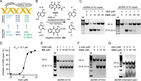
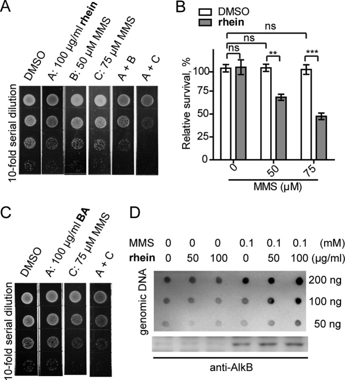
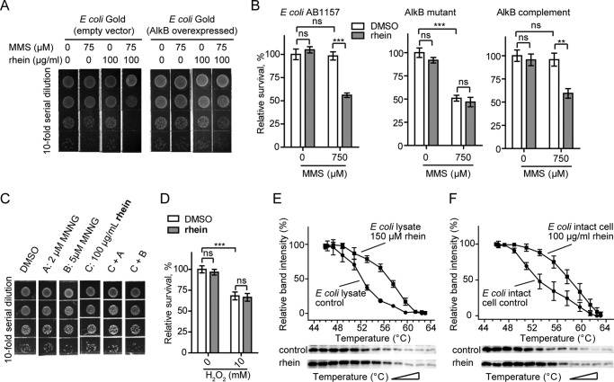
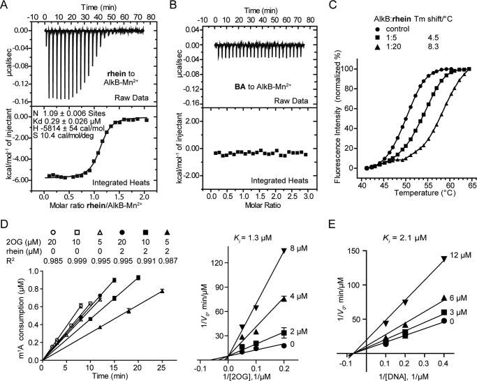
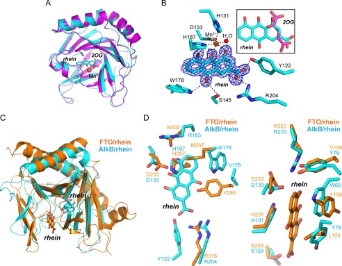
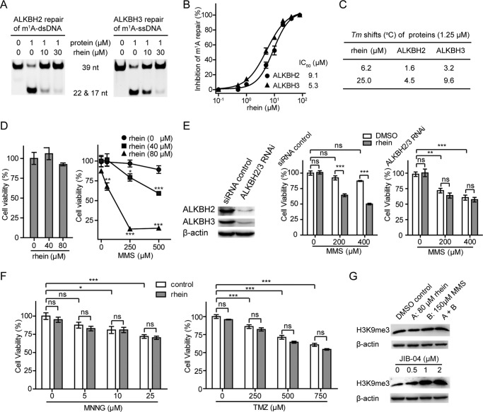
The AlkB repair enzymes, including Escherichia coli AlkB and two human homologues, ALKBH2 and ALKBH3, are iron(II)- and 2-oxoglutarate-dependent dioxygenases that efficiently repair N(1)-methyladenine and N(3)-methylcytosine methylated DNA damages. The development of small molecule inhibitors of these enzymes has seen less success. Here we have characterized a previously discovered natural product rhein and tested its ability to inhibit AlkB repair enzymes in vitro and to sensitize cells to methyl methane sulfonate that mainly produces N(1)-methyladenine and N(3)-methylcytosine lesions. Our investigation of the mechanism of rhein inhibition reveals that rhein binds to AlkB repair enzymes in vitro and promotes thermal stability in vivo In addition, we have determined a new structural complex of rhein bound to AlkB, which shows that rhein binds to a different part of the active site in AlkB than it binds to in fat mass and obesity-associated protein (FTO). With the support of these observations, we put forth the hypothesis that AlkB repair enzymes would be effective pharmacological targets for cancer treatment.

Keywords: DNA repair; DNA-protein interaction; bacteria; cancer therapy; chemical biology; enzyme inhibitor; enzyme structure.


Figures

FIGURE 1.
FIGURE 1.
Rhein selectively inhibits AlkB in vitro. A, the three major repair pathways in E. coli. DNA glycosylase is colored in cyan, DNA methyltransferase repair is in blue, and the AlkB repair is in green, respectively. B, scheme of AlkB repair methylated DNA. The structures of the inhibitor rhein and negative control BA are shown. C, rhein inhibits AlkB repair of m1A in ssDNA (left panel) and dsDNA (right panel) by using the DpnII digestion assay. The upper band is 49-nucleotide dsDNA, which contains an m1A lesion, and the lower band represents the mixture of 22- and 27-nucleotide dsDNA products after DpnII digestion. The 2OG concentration is 50 μm. D, quantitative determination of inhibitory activity using HPLC-based assay. The fitted IC50 is 12.7 μm assayed at 50 μm 2OG. The error bars are the means ± S.E. (n = 3). E, BA fails to inhibit m1A-dsDNA repair by AlkB. The 39-nucleotide m1A-containing DNA substrate was tested. F, C-Ada repair of O6mG is unimpaired in the presence of rhein. The upper band is 39-nucleotide O6mG-containg dsDNA, and the lower band is the digested fragments by PvuII. G, rhein is inactive to inhibit AlkA glycosylase. The 25-nucleotide mismatched dsDNA substrate is tested. All reactions were assayed in triplicate. nt, nucleotide.
FIGURE 2.
FIGURE 2.
Rhein sensitizes E. coli to MMS by enhancing the m3C accumulation. A, plate killing assay to show growth of E. coli Gold during exposure to 50 or 75 μm MMS with 100 μg/ml rhein, respectively. The density in the top line is A600 0.005. B, cfu count assay to show resistance of E. coli Gold to MMS in the presence of rhein. The error bars are means ± S.E. (n = 6). **, p < 0.1; ***, p < 0.001. C, growth of E. coli Gold in the presence of MMS and compound BA. D, the quantification of m3C (upper blot) and AlkB protein (lower blot) in E. coli Gold using blot assays. ns, not significant.
FIGURE 3.
FIGURE 3.
Cellular target engagement of rhein. A, plate killing assay to show the resistance of E. coli Gold that overexpresses AlkB to MMS in the presence of rhein. The starting density is A600 0.008. B, cfu count assay to show resistance of E. coli AB1157 (wild type), HK82 (AlkB mutant), and AlkB complemented HK82 to MMS by rhein. The error bars are means ± S.E. (n = 6). **, p < 0.01; ***, p < 0.001. C, rhein could not sensitize E. coli Gold to MNNG. The density of bacteria in the top line is A600 0.005. D, rhein could not sensitize E. coli growth to other DNA-damaging agents such as the oxidizing agent (H2O2). E, CETSA showing that rhein increases the thermal stability of AlkB in E. coli cell lysate. The data are presented as means ± S.E., and experiments were performed in triplicate. F, CETSA shows that rhein stabilizes AlkB in intact bacterial cells. ns, not significant.
FIGURE 4.
FIGURE 4.
Mechanistic study for inhibition of AlkB by rhein. A, isothermal titration calorimetry of rhein binding to AlkB-Mn2+ complex. Binding curves were fitted as a single binding event, and the constant is the average of two measurements. The fitted Kd is 0.29 μm. B, compound BA could not bind to AlkB-Mn2+ complex. C, differential scanning fluorimetry assay shows that rhein stabilizes AlkB by increasing Tm over 8 °C. Also shown are graphs of unfolding transition of 1.25 μm AlkB in the presence of rhein at 6.25 and 25 μm, respectively. The experiments were performed in triplicate. D, kinetics analyses of the mode of AlkB inhibition by rhein with respect to 2OG. Some of the initial rates linear fits are shown (left panel). E, kinetics analyses of AlkB inhibition by rhein with respect to m1A.
FIGURE 5.
FIGURE 5.
Structural insights into the mode of rhein binding to AlkB. A, structure alignment of the AlkB-rhein (PDB code 4RFR) and 2OG-bound AlkB (PDB code 3I3Q) performed in PyMOL with RMSD = 0.28 Å. The AlkB-rhein structure is colored cyan, AlkB/2OG is magenta, and the oxygen atom is red, respectively. Mn2+ is shown as a sphere and colored orange. Rhein and 2OG are shown as sticks. B, an m|Fo| − D|Fc| map was calculated within the PHENIX program suite after omission of rhein from the complex model and subsequent simulated annealing. The map density is contoured to 3.0 σ. The coordination of Mn2+ by ligands and hydrogen bonding are denoted by dotted dark lines. The map is shown in blue. The superimposition of rhein and 2OG is presented. C, structural superimposition of AlkB-rhein and FTO-rhein complexes performed in PyMOL. The FTO/rhein (PDB code 4IE7) is colored orange. Rhein is shown as sticks. D, zoomed-in view token from C to show the pocket for rhein binding to AlkB and FTO, respectively. Rhein could not bind to FTO similarly to AlkB because of the steric clashes by Tyr295 and Met297 (left panel). A likely binding pocket is observed in AlkB for rhein binding similarly in FTO (right panel).
FIGURE 6.
FIGURE 6.
Rhein inhibits ALKBH2 and ALKBH3 in vitro and sensitizes U87 cells to MMS. A, DpnII digestion assay to show rhein inhibits ALKBH2 and ALKBH3 repair of m1A in 39 nucleotide dsDNA and ssDNA, respectively. The 2OG concentration is 50 μm. B, quantitative determination of rhein inhibition of ALKBH repair using HPLC-based assay. The IC50 is fitted at 9.1 μm for ALKBH2 repair of dsDNA and 5.3 μm for ALKBH3 repair of ssDNA, respectively. This is assayed at 50 μm 2OG. The error bars are means ± S.E. (n = 3). C, Tm shifts of ALKBH2 and ALKBH3 by rhein. D, assessment of the growth of U87 cells in the presence of rhein (left panel) and under the combined treatment of MMS and rhein (right panel) using MTT assay. All the t tests were carried out between combination-treated groups and those adding MMS alone. E, the expression of ALKBH2 and ALKBH3 were silenced in U87 cells. Rhein sensitization of the proliferation of U87 cells to MMS is dependent on ALKBH2 and ALKBH3 enzymes. F, rhein is inactive to sensitizing U87 cells to MNNG or TMZ by MTT assay. G, Western blot analyses to monitor the amount of H3K9me3 in the presence of rhein and MMS (upper panel) and under the treatment of JIB-04 (lower panel). The error bars are means ± S.E. (n = 6). *, p < 0.05; **, p < 0.01; ***, p < 0.001. All tests were performed in triplicate. ns, not significant; nt, nucleotide.

Similar articles

Cited by

References

    1. Sedgwick B. (2004) Repairing DNA-methylation damage. Nat. Rev. Mol. Cell Biol. 5, 148–157 - PubMed
    1. Rose N. R., McDonough M. A., King O. N., Kawamura A., and Schofield C. J. (2011) Inhibition of 2-oxoglutarate dependent oxygenases. Chem. Soc. Rev. 40, 4364–4397 - PubMed
    1. Zhang Y., Chen F. X., Mehta P., and Gold B. (1993) Groove- and sequence-selective alkylation of DNA by sulfonate esters tethered to lexitropsins. Biochemistry 32, 7954–7965 - PubMed
    1. Loechler E. L., Green C. L., and Essigmann J. M. (1984) In vivo mutagenesis by O6-methylguanine built into a unique site in a viral genome. Proc. Natl. Acad. Sci. U.S.A. 81, 6271–6275 - PMC - PubMed
    1. Shah D., and Gold B. (2003) Evidence in Escherichia coli that N3-methyladenine lesions and cytotoxicity induced by a minor groove binding methyl sulfonate ester can be modulated in vivo by netropsin. Biochemistry 42, 12610–12616 - PubMed

Publication types

MeSH terms

LinkOut - more resources