Review
. 2017 Mar:156:41-49.
doi: 10.1016/j.exer.2016.03.016. Epub 2016 Mar 23.

The molecular mechanisms underlying lens fiber elongation

Affiliations
Review

The molecular mechanisms underlying lens fiber elongation

Dylan S Audette et al. Exp Eye Res. 2017 Mar.

Abstract

Lens fiber cells are highly elongated cells with complex membrane morphologies that are critical for the transparency of the ocular lens. Investigations into the molecular mechanisms underlying lens fiber cell elongation were first reported in the 1960s, however, our understanding of the process is still poor nearly 50 years later. This review summarizes what is currently hypothesized about the regulation of lens fiber cell elongation along with the available experimental evidence, and how this information relates to what is known about the regulation of cell shape/elongation in other cell types, particularly neurons.

Keywords: Actin; Cell shape; Cytoskeleton; Differentiation; Tubulin.


Figures

Figure 1
Figure 1
A) A cow lens placed over the National Eye Institute logo showing both the clarity and refractive properties of the ocular lens. B) Parts of the lens. The lens is composed of two cell types: a monolayer of epithelial cells seen on the anterior surface, which proliferate and differentiate to fiber cells which make up the majority of the lens. Light yellow– light; dark grey– lens capsule; yellow– central epithelium; orange– germinative zone; red– transition zone; purple– meridional row region/bow region; light blue– outer cortical fiber cells; light green-inner cortical fiber cells; dark green– beginning nuclear fiber cells; light gray– nuclear fiber cells including the primary fiber cells which are found at the very center.
Figure 2
Figure 2
Lens fiber cells have a hexagonal geometry when viewed in cross section. (A) An equatorial cross section of a mouse lens stained with fluorescent wheat germ and viewed with a confocal microscope. The hexagonal geometry of cells is highlighted in the center of the image showing the two broad sides of the cell labeled in red, the four short sides labeled in blue and the six vertices represented as yellow dots. Scale bar= 7.5μm. (B) A scanning electron micrograph showing lens fiber cells cut across their major axis allowing for viewing of their hexagonal geometry. The yellow vertices and white arrows show that there are membrane protrusions seen along these edges. Red– broad side; blue– short side; yellow– vertices/membrane protrusions; Scale bar= 5μm
Figure 3
Figure 3
Scanning electron micrographs showing ball and sockets on newly differentiated fiber cells and the formation of elaborate membrane protrusions in mouse lenses. (A) An electron micrograph showing straight fiber cells dotted with ball and sockets along the broad side of the cell in the youngest lens fiber cell layers. (B) As fiber cells mature, ball and sockets are seen along the broad sides of cells, and membrane protrusions (arrowheads) are first seen along the vertices of these cells. C) Deeper in the lens cortex, morphologically obvious ball and socket junctions are not seen, but the membrane protrusions become more obvious (arrowheads) D) In the deepest layers of the lens cortex, the membrane protrusions become even more elaborate (arrowheads). arrowheads- membrane protrusions; b– ball; s– socket; Scale bar for all panels= 5μm.
Figure 4
Figure 4
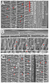
Scanning electron micrographs showing longitudinal views of C57Bl/6 mouse lens fiber cells which highlight the changes in fiber cell morphology that occur as fiber cells mature. (A) A longitudinal view of fiber cells from the lens capsule to a depth of about 150 μm within the lens showing where different structures arise. Note though, the locations of the structures is likely to differ between mouse genetic backgrounds. (B) The interface between cells developing from young cortical fiber cells with ball and sockets to more mature fiber cells which have prominent membrane protrusions. (C) A longitudinal view of the lens at a depth of about 200 to 350 μm showing that the paddles are now more prominent and distinct membrane protrusions can also be seen along these paddles. Red p= paddle; arrowheads= membrane protrusions; b= ball; s= socket; scale bar= 10 μm
Figure 5
Figure 5
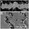
Scanning electron micrographs showing that mouse lens nuclear fiber cells have elaborate “membrane furrows” on their broad sides. (A) A single nuclear fiber cell isolated from the beginning of the lens nucleus that has a slight undulation, along with membrane furrows on its broad sides and distinct membrane protrusions at the vertices. (B) A higher magnification image of the lens nucleus showing that membrane furrows even extend out to the membrane protrusions. arrow= membrane furrows; arrowheads= membrane protrusions; scale bar= 5μm

References

    1. Al-Ghoul KJ, Lindquist TP, Kirk SS, Donohue ST. A novel terminal web-like structure in cortical lens fibers: architecture and functional assessment. Anat Rec (Hoboken) 2010;293:1805–15. doi: 10.1002/ar.21216. - DOI - PMC - PubMed
    1. Al-Ghoul KJ, Nordgren RK, Kuszak AJ, Freel CD, Costello MJ, Kuszak JR. Structural Evidence of Human Nuclear Fiber Compaction as a Function of Ageing and Cataractogenesis. Exp Eye Res. 2001;72:199–214. doi: 10.1006/exer.2000.0937. - DOI - PubMed
    1. Alizadeh A, Clark JI, Seeberger T, Hess J, Blankenship T, Spicer A, FitzGerald PG. Targeted genomic deletion of the lens-specific intermediate filament protein CP49. Invest Ophthalmol Vis Sci. 2002;43:3722–7. - PubMed
    1. Audette DS, Anand D, So T, Rubenstein TB, Lachke SA, Lovicu FJ, Duncan MK. Prox1 and fibroblast growth factor receptors form a novel regulatory loop controlling lens fiber differentiation and gene expression. Development. 2016;143:318–28. doi: 10.1242/dev.127860. - DOI - PMC - PubMed
    1. Augusteyn RC. On the growth and internal structure of the human lens. Exp Eye Res. 2010;90:643–54. doi: 10.1016/j.exer.2010.01.013. - DOI - PMC - PubMed

Publication types