. 2016 May 8;428(9 Pt B):1833-45.
doi: 10.1016/j.jmb.2016.03.016. Epub 2016 Mar 22.

Structural Dynamics and Mechanochemical Coupling in DNA Gyrase

Affiliations

Structural Dynamics and Mechanochemical Coupling in DNA Gyrase

Aakash Basu et al. J Mol Biol. .

Abstract

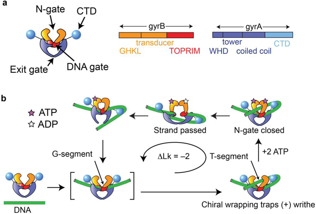
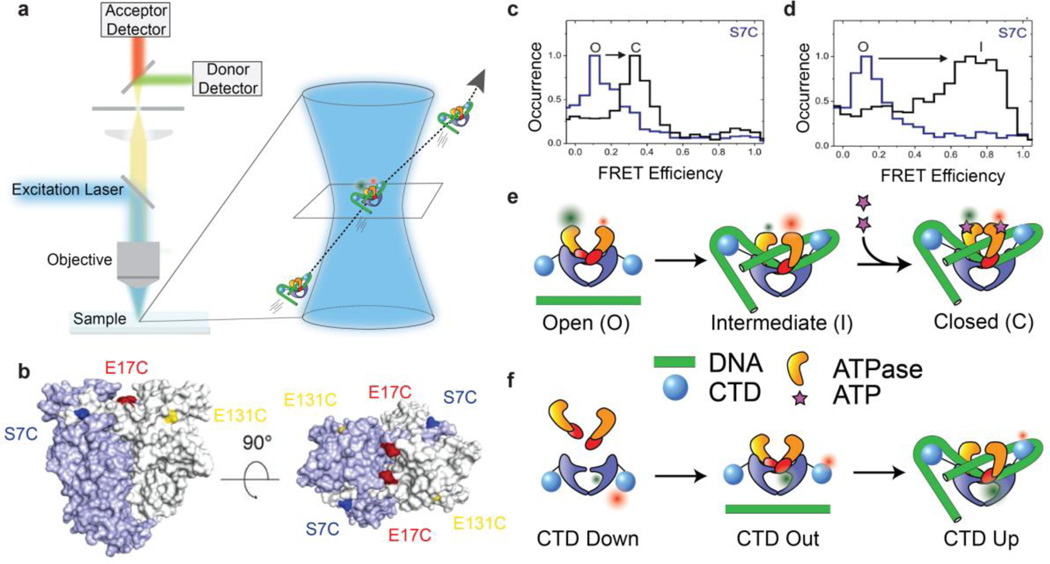
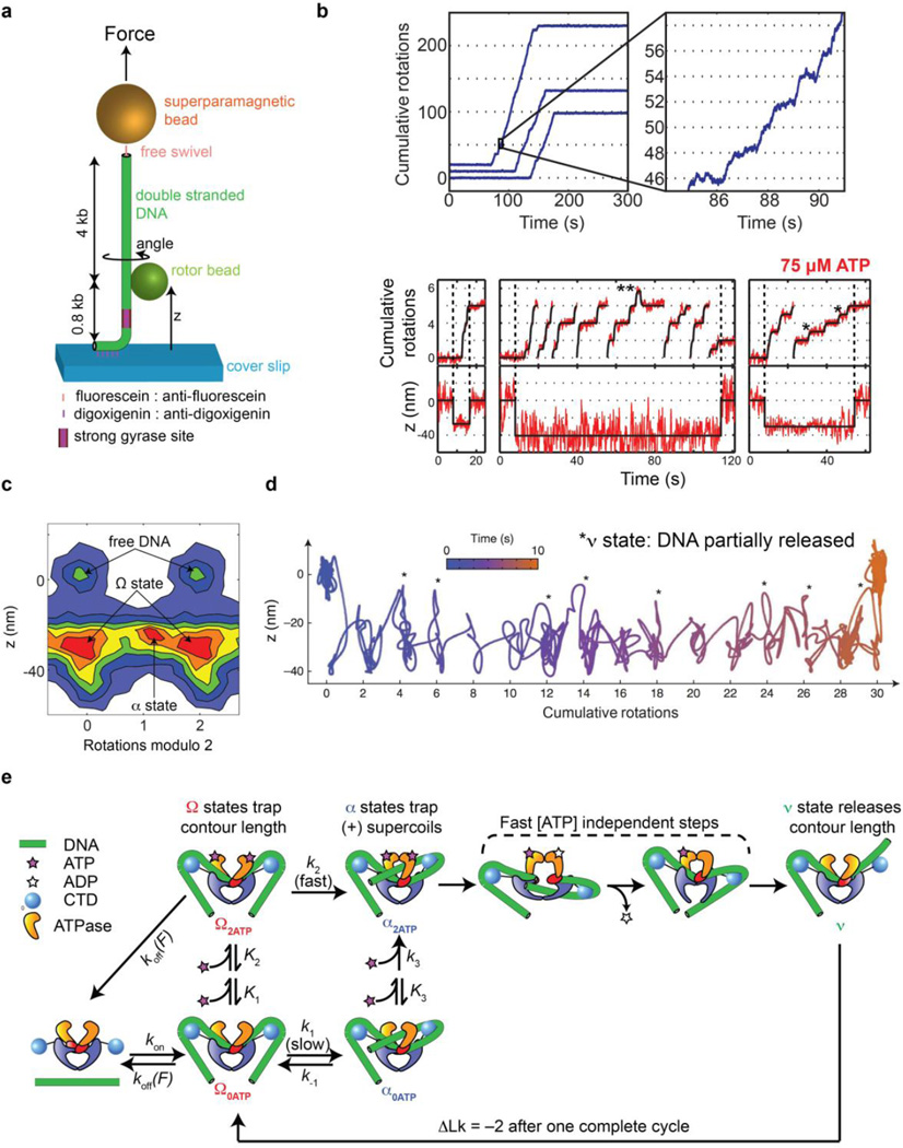
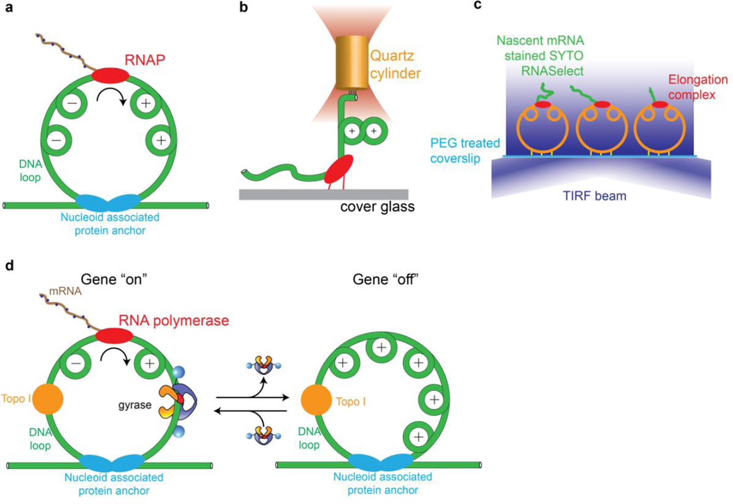
Gyrase is a molecular motor that harnesses the free energy of ATP hydrolysis to perform mechanical work on DNA. The enzyme specifically introduces negative supercoiling in a process that must coordinate fuel consumption with DNA cleavage and religation and with numerous conformational changes in both the protein and DNA components of a large nucleoprotein complex. Here we present a current understanding of mechanochemical coupling in this essential molecular machine, with a focus on recent diverse biophysical approaches that have revealed details of molecular architectures, new conformational intermediates, structural transitions modulated by ATP binding, and the influence of mechanics on motor function. Recent single-molecule assays have also illuminated the reciprocal relationships between supercoiling and transcription, an illustration of mechanical interactions between gyrase and other molecular machines at the heart of chromosomal biology.

Keywords: FRET; magnetic tweezers; molecular motor; single-molecule; topoisomerase.


Figures

Figure 1
Figure 1
Composition and basic mechanism of DNA gyrase. (a) Cartoon showing domain organization. Gyrase is an A2B2 heterotetramer. Interfaces between the subunits form three gates that can be opened and closed. (b) Outline of the enzymatic cycle. The G-segment binds to the central DNA gate. Chiral wrapping presents a proximal T-segment within the N-gate cavity. ATP binding induces N-gate closure, followed by passage of the T-segment through the transiently cleaved G-segment and expulsion through the C-gate. One round of strand passage leads to the introduction of two negative supercoils.
Figure 2
Figure 2
Recent structures illuminate the architectures of gyrase and related type II topoisomerases. (a) 23 Å CryoEM map of the T. thermophilus gyrase holoenzyme in complex with 155 bp DNA, ciprofloxacin, and AMPPNP (reproduced from [21]). The domain architecture can be seen together with density attributable to DNA wrapped around the CTDs. Crystal structures of protein components and modeled DNA duplex (green) have been fit to the density. The closed N-gate is shown in a domain-swapped configuration first observed in (b) a crystal structure (reproduced from [22]) of the related enzyme S. cerevisiae topo II in complex with G-segment DNA (green) and AMPPNP. The DNA-gate and the C-gate are also seen in closed configurations in these structures.
Figure 3
Figure 3
Single-molecule FRET reveals DNA and nucleotide dependent conformations of B. subtilis DNA gyrase. (a) Schematic of confocal smFRET microscopy (not to scale). Labeled complexes diffusing through the femtoliter confocal volume produce brief bursts of fluorescence that are collected on donor and acceptor channels to measure distributions of FRET efficiencies. (b) FRET labeling positions used for probing N-gate conformations (reproduced from [27]). (c–d) N-gate FRET histograms for gyrase using the S7C labeling position (reproduced from [27]), showing three N-gate conformations labeled O (open), C (closed), and I (intermediate/narrowed). For this labeling position, FRET is lower in the closed state than the intermediate state, explained by the N-terminal location of S7C in the intertwined dimerized ATPase domains. (c) No nucleotide (blue) vs ADPNP (black), in the absence of DNA. (d) No DNA (black) vs relaxed plasmid DNA (blue), in the absence of nucleotide. (e) Cartoons of N-gate conformations probed by FRET. (f) Cartoons of CTD positions based on smFRET measurements between the gyrA CTD and the core enzyme [30]. CTDs are positioned toward the exit gate in the gyrA dimer alone, move out slightly when gyrB is bound, and swing up when the enzyme is complexed with DNA.
Figure 4
Figure 4
Rotor bead tracking reveals new conformations and ATP-dependent dynamics of E. coli DNA gyrase. (a) The rotor bead tracking (RBT) assay. DNA is stretched using a magnetic bead, and a submicron rotor bead is attached to the side of the molecule and tracked using fluorescence [35] [36] or evanescent scattering [37] videomicroscopy to measure changes in DNA angle and extension (z) in real time. (b) RBT traces (reproduced from [36]) in the presence of DNA gyrase under in 1 mM ATP (above) or 75 µM ATP (below). Individual gyrase encounters lead to bursts of stepwise rotation, corresponding to processive negative supercoiling. [ATP]-dependent dwells are seen at the even rotation mark and also at an intermediate angle (*) corresponding to a chirally wrapped intermediate. (c) 2D histogram (reproduced from [36]) of paired (angle,z) values during gyrase activity in presence of 75 µM ATP, showing distinct conformational states visited by the enzyme. Angles are shown modulo 2 rotations. The Ω state is significantly contracted in z but lies at the ~0 rotation mark, which is explained by sequestering DNA contour without trapping writhe. The α state can also be seen at the ~1 rotation mark, corresponding to trapping (+) writhe prior to strand passage. (d) High-resolution dynamics of gyrase at 1 mM ATP using gold rotor bead tracking (reproduced from [37]). A single processive burst is shown in the (angle, z) plane. Major dwells are interrupted by brief excursions to a state (*) that releases significant contour length. (e) Branched kinetic model for structural transitions and ATP coupling in DNA gyrase [36, 37]. The kinetics of processive supercoiling are dominated by the transition from Ω to α, which can occur slowly and reversibly in the absence of ATP or quickly when 2 ATP are bound. Subsequent strand passage also requires the presence of 2 ATP. DNA is partially released after strand passage and recaptured to begin a new round of supercoiling.
Figure 5
Figure 5
Mechanical interplay of gyrase, transcription, and DNA supercoiling investigated using single-molecule methods. (a) Helical tracking of the advancing transcription complex leads to twin supercoiling domains in a constrained DNA duplex [54, 63]. (b) An optical torque wrench assay [64] showed that RNA polymerase stalls due to positive supercoils that accumulate ahead of the enzyme, with a measured stall torque of ~10 pN nm. (c) Single-molecule assay for transcription on tethered constrained circular templates [54]. Fluorescence accumulates during transcription due to an RNA-binding dye. Dynamics can be investigated in the presence of gyrase and/or topoisomerase I. (d) Model for transcriptional bursting based on single-molecule measurements [54]. Topoisomerase I constitutively relieves (−) supercoils behind the transcription complex, leading to the accumulation of (+) supercoils in a constrained chromosomal loop. When gyrase is bound, (+) supercoils are relaxed and transcription can proceed. When gyrase dissociates, accumulated (+) supercoils inhibit transcription, intermittently shutting off gene expression.

References

    1. Wang JC. Cellular roles of DNA topoisomerases: a molecular perspective. Nat Rev Mol Cell Biol. 2002;3:430–440. - PubMed
    1. Nollmann M, Crisona NJ, Arimondo PB. Thirty years of Escherichia coli DNA gyrase: from in vivo function to single-molecule mechanism. Biochimie. 2007;89:490–499. - PubMed
    1. Schoeffler AJ, Berger JM. DNA topoisomerases: harnessing and constraining energy to govern chromosome topology. Quarterly reviews of biophysics. 2008;41:41–101. - PubMed
    1. Dong KC, Berger JM. Structural basis for gate-DNA recognition and bending by type IIA topoisomerases. Nature. 2007;450:1201–1205. - PubMed
    1. Bax BD, Chan PF, Eggleston DS, Fosberry A, Gentry DR, Gorrec F, et al. Type IIA topoisomerase inhibition by a new class of antibacterial agents. Nature. 2010;466:935–940. - PubMed

Publication types

LinkOut - more resources