. 2017 Jun 15;426(2):176-187.
doi: 10.1016/j.ydbio.2016.02.032. Epub 2016 Mar 23.

Genome-wide analysis of dorsal and ventral transcriptomes of the Xenopus laevis gastrula

Affiliations

Genome-wide analysis of dorsal and ventral transcriptomes of the Xenopus laevis gastrula

Yi Ding et al. Dev Biol. .

Abstract

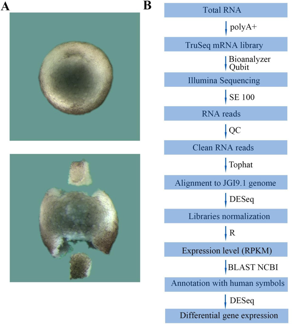
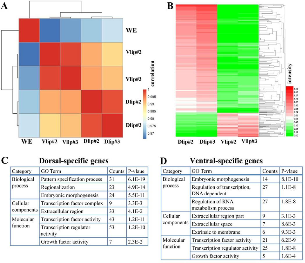
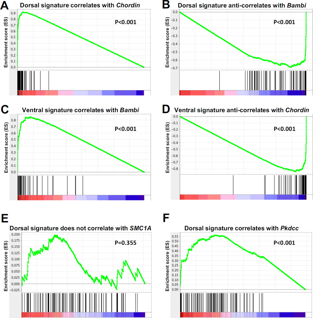
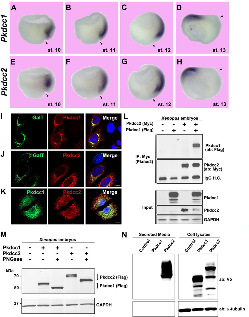
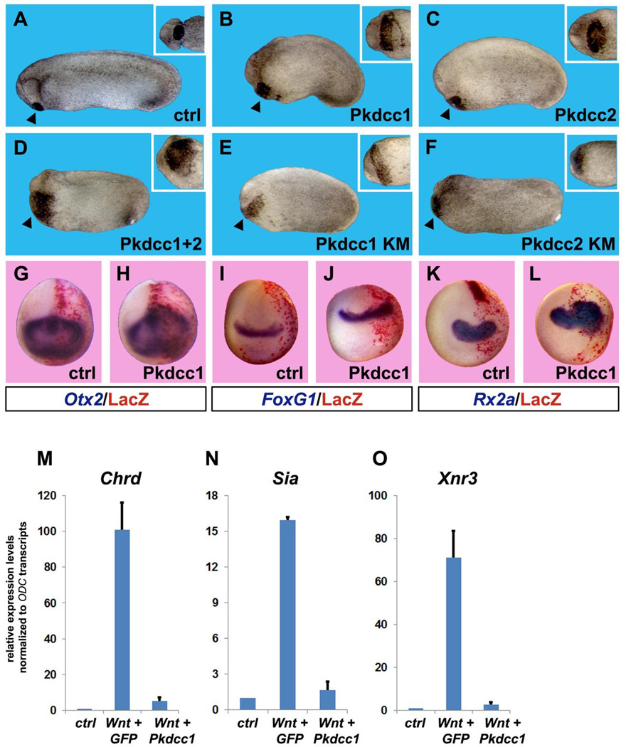
RNA sequencing has allowed high-throughput screening of differential gene expression in many tissues and organisms. Xenopus laevis is a classical embryological and cell-free extract model system, but its genomic sequence had been lacking due to difficulties arising from allotetraploidy. There is currently much excitement surrounding the release of the completed X. laevis genome (version 9.1) by the Joint Genome Institute (JGI), which provides a platform for genome-wide studies. Here we present a deep RNA-seq dataset of transcripts expressed in dorsal and ventral lips of the early Xenopus gastrula embryo using the new genomic information, which was further annotated by blast searches against the human proteome. Overall, our findings confirm previous results from differential screenings using other methods that uncovered classical dorsal genes such as Chordin, Noggin and Cerberus, as well as ventral genes such as Sizzled, Ventx, Wnt8 and Bambi. Complete transcriptome-wide tables of mRNAs suitable for data mining are presented, which include many novel dorsal- and ventral-specific genes. RNA-seq was very quantitative and reproducible, and allowed us to define dorsal and ventral signatures useful for gene set expression analyses (GSEA). As an example of a new gene, we present here data on an organizer-specific secreted protein tyrosine kinase known as Pkdcc (protein kinase domain containing, cytoplasmic) or Vlk (vertebrate lonesome kinase). Overexpression experiments indicate that Pkdcc can act as a negative regulator of Wnt/ β-catenin signaling independently of its kinase activity. We conclude that RNA-Seq in combination with the X. laevis complete genome now available provides a powerful tool for unraveling cell-cell signaling pathways during embryonic induction.

Keywords: Dorsal–Ventral patterning; RNA-Seq; Secreted Tyrosine kinase; Spemann organizer; Vlk; Xenopus laevis.


Figures

Fig. 1
Fig. 1
D-V dissections and experimental flowchart. (A) Dorsal and ventral lip dissection. The upper panel shows a wild-type stage 10.5 Xenopus embryo, while the lower panel shows dorsal (Dlip, top) and ventral lips (Vlip, bottom) dissected from a sibling stage 10.5 embryo. (B) Outline of the RNA-seq analysis pipeline described in materials and methods.
Fig. 2
Fig. 2
Distribution of transcripts sequenced from dorsal and ventral lips. (A) Curve showing normal distribution of fold change of dorsal transcripts. The curve was created using 27,594 mRNAs obtained from the first experiment after selecting for abundance of expression; the ordinate indicates the probability that a gene is located in the curve by chance (probability mass function). Genes were organized by the dorsal/ventral natural logarithm, providing dorsal genes with positive values, while assigning negative numbers to ventral genes. (B) A normal Gaussian curve was obtained by artificially removing about 11,000 transcripts expressed at similar levels in both dorsal and ventral lips. The graph indicates that the data obtained was evenly distributed throughout both dorsal and ventral lips and defined the standard deviation. The most dorsal gene was Chordin, 9 standard deviations above the mean, while the most ventrally expressed gene was Sizzled, 8 standard deviations above the mean. (C) Box and whisker plots reveal transcripts upregulated upon bisection of dorsal (Dorsal/WE) or ventral (Ventral/WE) lips compared to uncut wild type whole embryos (WE), and dorsal and ventral transcriptomes (Dorsal/Ventral and Ventral/Dorsal). Only some of the outliers above the whisker are annotated, with Short on the right and Long subgenome genes on the left. Chordin is referred to by its abbreviation chrd in this Figure and throughout the Supplementary Tables and Xnr3 in the main text is referred to as nodal 3 here. Note that cutting the embryo triggers the expression of injury response genes in both fragments.
Fig. 3
Fig. 3
The D-V transcriptomes obtained by RNA-seq are highly reproducible. (A) Correlation plot of duplicate dorsal and ventral lip RNA-seq experiments from different egg clutches shows high reproducibility of the method. The correlation scores were calculated as the Pearson Correlation Coefficient (PCC) and color-coded as shown in the scale bar on the right of the panel. Note that Vlip and Dlip samples clustered into groups based on PCC. (B) Heatmap of differentially expressed genes between dorsal and ventral lip libraries. The expression data (RPKM) of each gene across different samples was scaled and represented as z-scores. The expression levels (expressed as logarithm of RPKM) are indicated in the scale bar on the right panel. (C) Table showing the top gene ontology (GO) terms associated with dorsal-specific genes. (D) Table of GO terms for ventral-specific transcripts.
Fig. 4
Fig. 4
Gene Set Enrichment Analysis (GSEA) of dorsal and ventral gene signatures. (A–B) The dorsal gene signature (comprised of 107 genes) positively correlated with Chordin (P<0.001), a Spemann organizer marker, and negatively correlated (or anti-correlated) with BAMBI (P<0.001), a marker of ventral tissues. (C–D) The ventral signature (which contains 70 genes) positively correlated with BAMBI (P<0.001), while it negatively correlated with Chordin (P<0.001). (E) SMC1A, used here as a negative control, did not correlate with the dorsal (P=0.355) nor the ventral (not shown) signature. (F) Pkdcc, a novel dorsal gene described here, significantly correlated with the dorsal gene signature (P<0.001).
Fig. 5
Fig. 5
Pkdcc1 and 2 are expressed in the Spemann Organizer and localize to the Golgi apparatus. (A–H) Whole-mount in situ hybridization on hemisectioned Xenopus gastrula embryos showing Pkdcc1 and Pkdcc2 expression pattern from early to late gastrula (stage 10–13). Note the more anterior localization (in anterior endomesoderm) of Pkdcc2 compared to Pkdcc1 at stage 10. By the end of gastrulation Pkdcc1/2 expression was restricted to the prechordal plate. Arrowheads indicate dorsal lip position. (I–K) Immunofluorescence on HeLa cells overexpressing Pkdcc1-Flag and Pkdcc2-myc shows that the two kinases co-localize with the Golgi marker Galactosyl-Transferase (GalT). The two kinases also showed an overlapping subcellular localization. Nuclei were stained with DAPI (merge). Scale bars represent 10 µm. (L) Immunoprecipitation experiment showing physical interaction between Pkdcc1 and Pkdcc2 in Xenopus embryos. Pkdcc1-flag and/or Pkdcc2-myc mRNAs were injected at 4-cell stage and embryos were lysed at stage 12. One third of the total protein extract was saved and used as input. (M) Pkdcc1 and 2 are glycosylated, as shown by the electrophoretic mobility shift caused by PNGase F treatment. (N) Analysis of V5-tagged Pkdcc1 and 2 secretion in HEK293T cells showing that Pkdcc2 was found in the culture medium, indicating extensive secretion.
Fig. 6
Fig. 6
Overexpression of Pkdcc enlarges anterior tissues and inhibits canonical Wnt signaling. (A–C) Overexpression of 1 ng of Pkdcc1 or Pkdcc2 mRNA caused expansion of anterior structures such as the cement gland (see insets). Percent of embryos showing enlarged cement gland and head were 0% n=60 for controls; 81% n=54 for Pkdcc1; 84% n=38 for Pkdcc2. (D) This effect is synergistic, since injection of 500 pg each of Pkddc1 and 2 caused an even greater enlargement of the cement gland (98% of the embryos showed this phenotype, n=50). (E–F) Kinase mutant (KM) forms of Pkdcc1 and 2 also caused an increase of cement gland tissues, suggesting that the kinase activity is dispensable for Pkdcc-mediated anteriorizing effects. Percent of embryos showing enlarged cement glands were: 82% n=27 for Pkdcc1-KM; 83% n=24 for Pkdcc2-KM. All embryos are shown from lateral views, insets show ventral view of the cement gland from the same embryos. Arrowheads point to cement gland. (G–L) Whole-mount in situ hybridizations for anterior neural markers show that Pkdcc1 mRNA overexpression expands Otx2, FoxG1 (also known as BF1) and Rx2a. The injected side was identified through Red Gal staining of the lineage tracer nuclear Lacz (nLacz) coinjected with Pkdcc1. Control embryos were injected with nLacz only. Percentage of Pkdcc1-injected embryos showing expanded neural markers: Otx2 66% n=27; FoxG1 76% n=47; Rx2a 92% n=38. (M–O) Pkdcc1 abolishes canonical Wnt signaling. Quantitative RT-PCR analysis performed on animal cap explants co-injected with xWnt8 and Pkdcc1 mRNAs shows that Pkdcc1 greatly reduces induction of canonical Wnt target genes Chordin (Chrd), Siamois (Sia) and Nodal related 3 (Xnr3). GFP mRNA was used as a negative control. Transcript levels of the housekeeping gene ODC were used for normalization. Error bars indicate standard deviations derived from three independent experiments.

References

    1. Anders S, Huber W. Differential expression analysis for sequence count data. Genome Biol. 2010;11:R106. - PMC - PubMed
    1. Andreazzoli M, Gestri G, Angeloni D, Menna E, Barsacchi G. Role of Xrx1 in Xenopus eye and anterior brain development. Development. 1999;126:2451–2460. - PubMed
    1. Bachiller D, Klingensmith J, Kemp C, Belo JA, Anderson RM, May SR, McMahon JA, McMahon AP, Harland RM, Rossant J, De Robertis EM. The organizer factors Chordin and Noggin are required for mouse forebrain development. Nature. 2000;403:658–661. - PubMed
    1. Bier E, De Robertis EM. BMP gradients: A paradigm for morphogen-mediated developmental patterning. Science. 2015;348:aaa5838. - PubMed
    1. Blum M, De Robertis EM, Wallingford JB, Niehrs C. Morpholinos: antisense and sensibility. Dev. Cell. 2015;35:145–149. - PubMed

MeSH terms