. 2016 Apr 26;7(17):24483-94.
doi: 10.18632/oncotarget.8231.

Phosphorylation of TOPK at Y74, Y272 by Src increases the stability of TOPK and promotes tumorigenesis of colon

Affiliations

Phosphorylation of TOPK at Y74, Y272 by Src increases the stability of TOPK and promotes tumorigenesis of colon

Juanjuan Xiao et al. Oncotarget. .

Abstract

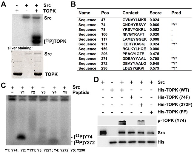
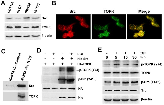
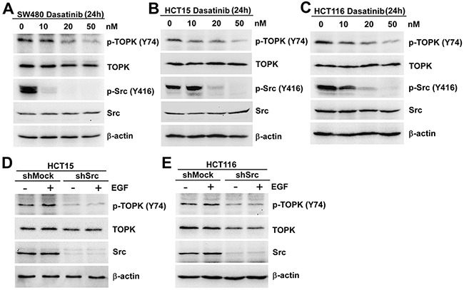
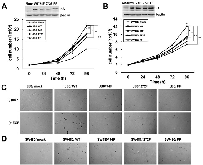
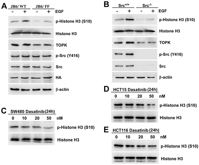
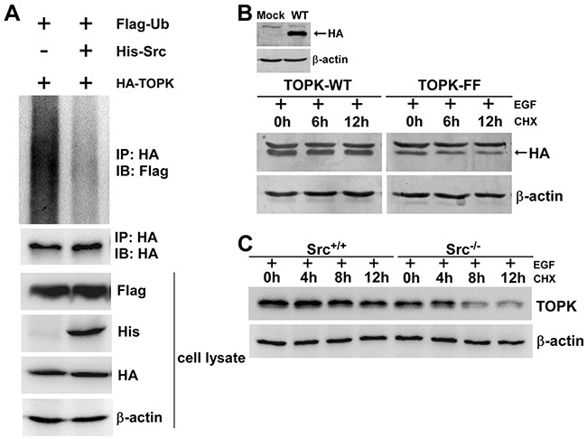
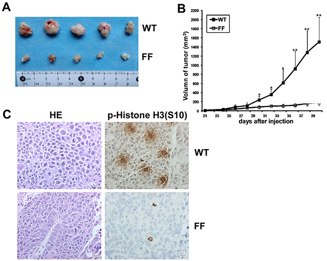
T-LAK cell-originated protein kinase (TOPK), a serine/threonine protein kinase, is highly expressed in a variety of tumors and associated with a poor prognosis of human malignancies. However, the activation mechanism of TOPK is still unrevealed. Herein, first we found that Src directly bound with and phosphorylated TOPK at Y74 and Y272 in vitro. Anti-phospho-TOPK at Y74 was prepared, the endogenous phosphorylation of TOPK at Y74 was detected in colon cancer cells, and the phosphorylation was inhibited in cells expressing low levels of Src. Subsequently, we stably transfected Y74 and Y272 double mutated TOPK (TOPK-FF) into JB6 or SW480 cells, and observed that both the anchorage-independent growth ability and tumorigenesis of TOPK-FF cells were suppressed compared with those of wild type TOPK (TOPK-WT) ex vivo and in vivo. The phosphorylation level of TOPK substrate, Histone H3 at Ser10 also decreased dramatically ex vivo or in vivo. Moreover, we showed that Src could inhibit the ubiquitination of TOPK. Transiently expressed TOPK-WT was more stable than TOPK-FF in pause and chase experiment. Endogenous TOPK was more stable in Src wild type (Src+/+) MEFs than in Src knockout (Src-/-). Taken together, our results indicate that Src is a novel upstream kinase of TOPK. The phosphorylation of TOPK at Y74 and Y272 by Src increases the stability and activity of TOPK, and promotes the tumorigenesis of colon cancer. It may provide opportunities for TOPK based prognosis and targeted therapy for colon cancer patients.

Keywords: Src; TOPK; colon cancer; stability; tumorigenesis.


Conflict of interest statement

The authors declare no conflict of interest.

Figures

Figure 1
Figure 1. Src phosphorylates TOPK at Y74 and Y272 in vitro
A. Active Src phosphorylated inactive TOPK in vitro in the presence of [γ-32P] ATP as visualized by autoradiograph. B. Potential phosphorylated tyrosine sites of TOPK were predicted by NetPhos 2.0 software program. C. Src phosphorylated TOPK at Y74 or Y272 in peptide mapping. Five synthesized peptides containing potential tyrosine sites were used as substrates in an in vitro kinase assay with active Src in the presence of [γ-32P] ATP and the results were visualized by autoradiography. D. Validation of anti phospho-TOPK (Y74) (p-TOPK (Y74)) in an in vitro kinase assay. Wild type His-TOPK (WT), single mutant His-TOPK (74F), single mutant His-TOPK (272F) or double mutant His-TOPK (FF) as shown was used as substrate for active Src. Reactive products were resolved by SDS-PAGE and visualized by Western blot with p-TOPK (Y74).
Figure 2
Figure 2. Src binds with TOPK and phosphorylates TOPK at Y74 ex vivo
A. The levels of Src and TOPK in four colon cancer cell lines were shown. B. Colocalization of Src and TOPK was visualized by confocal microscope in SW480 cells. Cytoplasmic and nuclear staining of Src and TOPK was mostly merged together. C. Ni-NTA-His-TOPK bound with endogenous Src of SW480 cells. D. Src promoted phosphorylation of TOPK in 293T cells induced by EGF after co-transfected with pcDNA4-His-Src and pcDNA3-HA-TOPK (EGF 80 ng/ml; 30 min). E. EGF induced a time-dependent phosphorylation of TOPK at Y74 in SW480 cells (EGF 80 ng/ml; 0 min, 5 min, 15 min, 30 min).
Figure 3
Figure 3. The phosphorylation of TOPK at Y74 was inhibited in colon cancer cells expressing low levels of Src
SW480 A. HCT15 B. and HCT116 C. cells were treated with Dasatinib for 24 h in a dose-dependent manner and the samples were resolved by SDS-PAGE and analyzed by Western blot respectively. The stable shMock and shSrc in HCT15 D. or HCT116 E. cells were treated with EGF (80 ng/ml; 30 min), and analyzed by Western blot, respectively. Data are representatives of results from triplicate experiments.
Figure 4
Figure 4. The phosphorylation of TOPK at Y74 and Y272 by Src promotes carcinogenesis ex vivo
A. Growth curves of JB6 cells stably expressing pcDNA3-Mock (JB6/ Mock), pcDNA3-TOPK-WT (JB6/ WT), pcDNA3-TOPK-74F (JB6/ 74F), pcDNA3-TOPK-272F (JB6/ 272F), or double-mutant pcDNA3-TOPK-FF (JB6/ FF). Inset (top) showed verification of the cell lines identified by Western blot. Data are represented as means±SD of triplicate experiments. *, significantly (P < 0.05) decrease in cell number in JB6/ 74F, JB6/ 272F or JB6/ FF cells compared with JB6/ WT cells respectively. **, significantly (P < 0.01) increase in cell number in JB6/ WT cells compared with JB6/ Mock cells. B. Growth curves of SW480 cells stably expressing pcDNA3-mock (SW480/ Mock), pcDNA3-TOPK-WT (SW480/ WT), pcDNA3-TOPK-74F (SW480/ 74F), pcDNA3-TOPK-272F (SW480/ 272F), or double-mutant pcDNA3-TOPK-FF (SW480/ FF). Inset (top) showed verification of the cell lines identified by Western blot. Data are represented as means±SD of triplicate experiments. *, respective (P < 0.05) decrease in cell number in SW480/ 74F or 272F compared with SW480/ WT cells. **, significantly (P < 0.01) decrease in cell number in SW480/ FF cells compared with SW480/ WT cells and significantly (P < 0.01) increase in cell number in SW480/ WT cells compared with SW480/ Mock cells. C. Transfectants of JB6/ Mock, WT, 74F, 272F, or FF were compared for EGF-induced colony formation in soft agar. D. Transfectants of SW480/ Mock, WT, 74F, 272F, or FF were compared for colony formation in soft agar.
Figure 5
Figure 5. The phosphorylation of TOPK at Y74, Y272 by Src enhances the activity of TOPK
A. JB6/ WT and JB6/ FF cells were treated with EGF (20 ng/ml; 15 min), and the samples were analyzed by Western blot. B. Src+/+ and Src−/− MEFs were treated with EGF (20 ng/ml; 15 min), and analyzed by Western blot. SW480 C. HCT15 D. and HCT116 E. cells were treated with Dasatinib for 24 h in a dose-dependent manner. Then Histones were isolated, resolved by SDS-PAGE and analyzed by Western blot, respectively. Data are representatives of results from triplicate experiments.
Figure 6
Figure 6. The phosphorylation of TOPK by Src enhances the stability of TOPK
A. Phosphorylation of TOPK by Src prevented TOPK ubiquitination. HEK293T cells were transfected with pCMV-Flag-ubiquitin (Flag-Ub), pcDNA3-HA-TOPK (HA-TOPK), and pcDNA4-His-Src (His-Src) as indicated. The cells were harvested 48 h after transfection. Then the samples were immunoprecipitated (IP) with anti-HA and detected with anti-Flag by Western blot. The transfection efficiency and equal protein loading were verified by Western blot using the whole cell lysate. B. HEK293T cells were transfected with pcDNA3-HA-TOPK wild type (TOPK-WT) and pcDNA3-HA-TOPK double mutant (TOPK-FF), and then were treated with EGF (80 ng/ml; 30 min) followed by addition of CHX (100 μg/ml) to prevent new protein synthesis. The time-dependent stability of TOPK was detected with anti-HA by Western blot. C. Src+/+ and Src−/− MEFs were treated with EGF (20 ng/ml; 15 min) followed by addition of CHX (100 μg/ml) to prevent new protein synthesis. Lysates were collected at the indicated time points. The protein level of TOPK was detected by Western blot.
Figure 7
Figure 7. Src phosphorylates TOPK to promote the tumorigenesis of colon cancer in vivo
A. Tumors dissected from SW480/ WT or SW480/ FF group were shown. B. Tumor growth curve from mice injected with SW480/ WT or SW480/ FF cells. Data are expressed as means±SE of 5 mice in each group. The asterisk indicated a significant increase in tumor size in SW480/ WT injected mice compared with SW480/ FF injected mice. *P < 0.05, **P < 0.01. C. Left, H&E stained tumor sections from mice injected with either SW480/ WT or SW480/ FF cells. Poorly differentiated adenocarcinoma cell clusters were found in the two groups. Right, IHC analysis of p-Histone H3 (S10) expression in tumor sections from mice injected with either SW480/ WT or SW480/ FF cells. Nuclear expression of p-Histone H3 (S10) was detected in the tumor sections from SW480/ WT cells, while very weak staining of p-Histone H3 (S10) was detected from SW480/ FF cells. Magnification, 400×.

References

    1. Abe Y, Matsumoto S, Kito K, Ueda N. Cloning and expression of a novel MAPKK-like protein kinase, lymphokine-activated killer T-cell-originated protein kinase, specifically expressed in the testis and activated lymphoid cells. J Biol Chem. 2000;275:21525–21531. - PubMed
    1. Gaudet S, Branton D, Lue RA. Characterization of PDZ-binding kinase, a mitotic kinase. Proc Natl Acad Sci. 2000;97:5167–5172. - PMC - PubMed
    1. Matsumoto S, Abe Y, Fujibuchi T, Takeuchi T, Kito K, Ueda N, Shigemoto K, Gyo K. Characterization of a MAPKK-like protein kinase PBK/TOPK. Biochem Biophys Res Commun. 2004;325:997–1004. - PubMed
    1. Park JH, Nishidate T, Nakamura Y, Katagiri T. Critical roles of T-LAK cell-originated protein kinase in cytokinesis. Cancer Sci. 2010;101:403–411. - PMC - PubMed
    1. Fukukawa C, Ueda K, Nishidate T, Katagiri T, Nakamura Y. Critical roles of LGN/GPSM2 phosphorylation by PBK/TOPK in cell division of breast cancer cells. Genes Chromosomes Cancer. 2010;49:861–872. - PubMed

MeSH terms