

The non-coding RNA composition of the mitotic chromosome by 5'-tag sequencing
- PMID: 27016738
- PMCID: PMC4889943
- DOI: 10.1093/nar/gkw195
The non-coding RNA composition of the mitotic chromosome by 5'-tag sequencing
Abstract
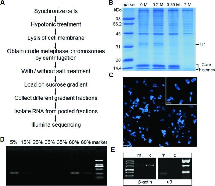
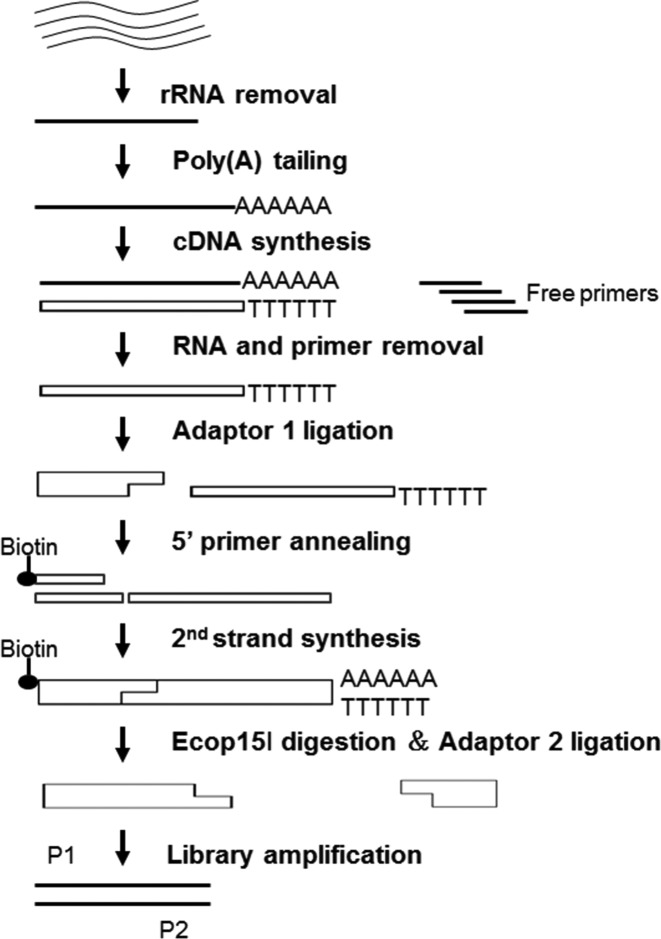
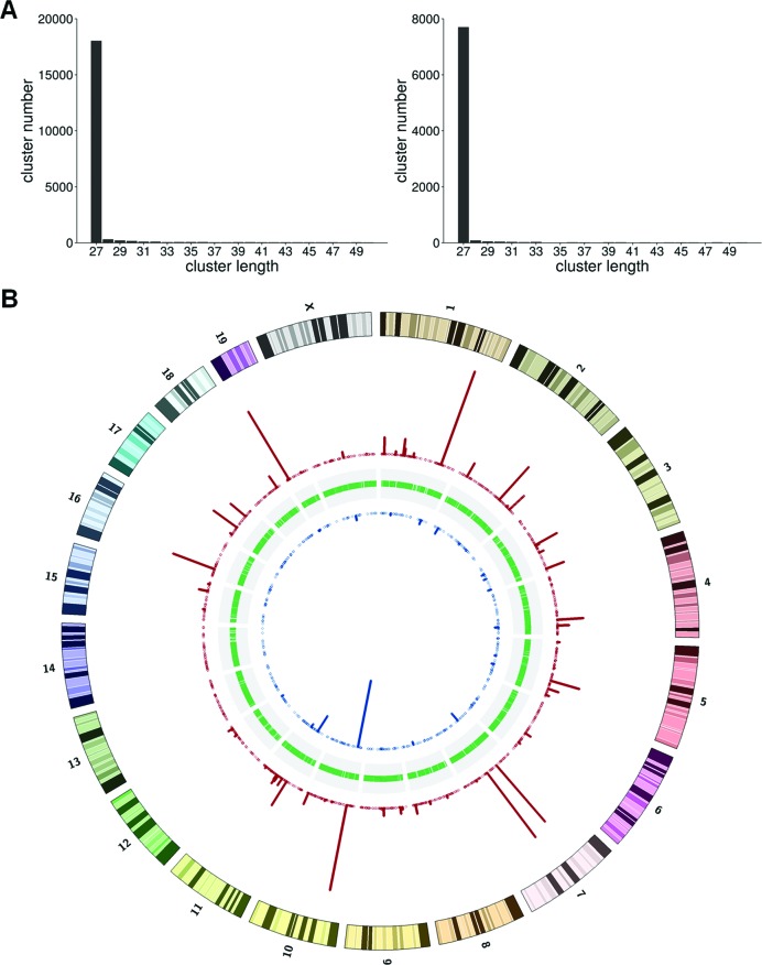
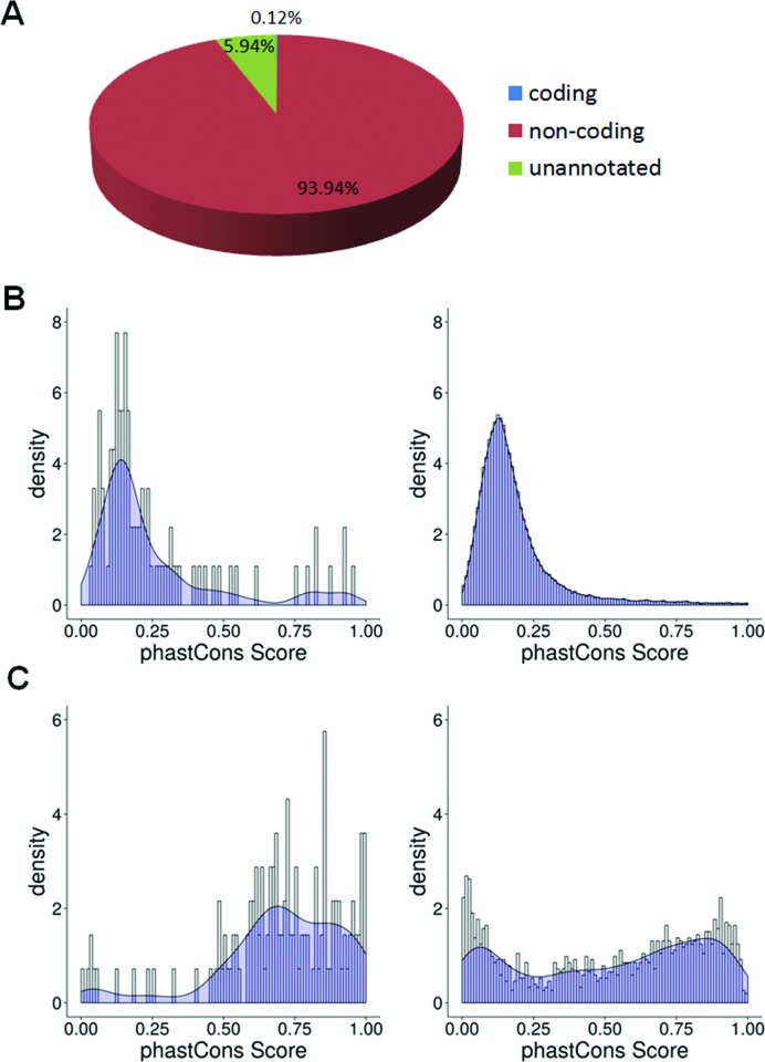
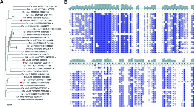
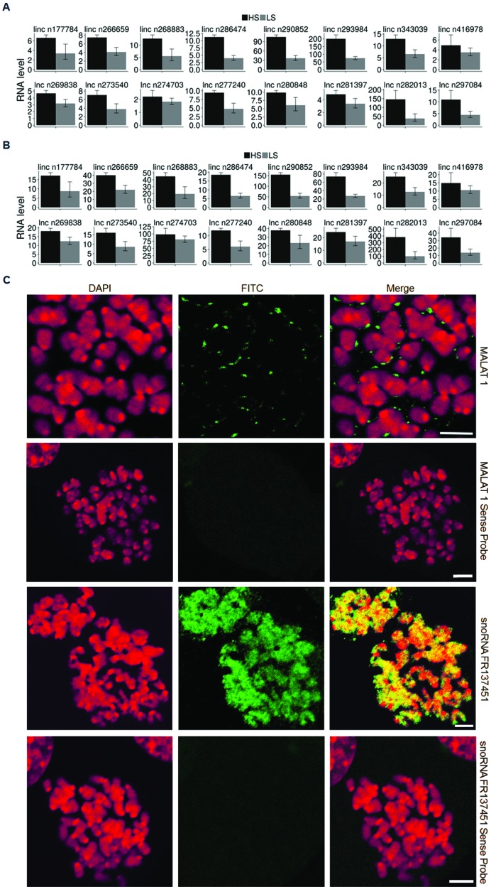
Mitotic chromosomes are one of the most commonly recognized sub-cellular structures in eukaryotic cells. Yet basic information necessary to understand their structure and assembly, such as their composition, is still lacking. Recent proteomic studies have begun to fill this void, identifying hundreds of RNA-binding proteins bound to mitotic chromosomes. However, by contrast, there are only two RNA species (U3 snRNA and rRNA) that are known to be associated with the mitotic chromosome, suggesting that there are many mitotic chromosome-associated RNAs (mCARs) not yet identified. Here, using a targeted protocol based on 5'-tag sequencing to profile the mammalian mCAR population, we report the identification of 1279 mCARs, the majority of which are ncRNAs, including lncRNAs that exhibit greater conservation across 60 vertebrate species than the entire population of lncRNAs. There is also a significant enrichment of snoRNAs and specific SINE RNAs. Finally, ∼40% of the mCARs are presently unannotated, many of which are as abundant as the annotated mCARs, suggesting that there are also many novel ncRNAs in the mCARs. Overall, the mCARs identified here, together with the previous proteomic and genomic data, constitute the first comprehensive catalogue of the molecular composition of the eukaryotic mitotic chromosomes.
© The Author(s) 2016. Published by Oxford University Press on behalf of Nucleic Acids Research.
Figures
Similar articles
-
Quantitative catalogue of mammalian mitotic chromosome-associated RNAs.Sci Data. 2024 Jan 6;11(1):43. doi: 10.1038/s41597-023-02884-8. Sci Data. 2024. PMID: 38184632 Free PMC article.
-
The actin-related protein hArp8 accumulates on the mitotic chromosomes and functions in chromosome alignment.Exp Cell Res. 2008 Feb 15;314(4):859-68. doi: 10.1016/j.yexcr.2007.11.020. Epub 2007 Dec 4. Exp Cell Res. 2008. PMID: 18163988
-
Interphase chromatin LINEd with RNA.Cell. 2014 Feb 27;156(5):864-5. doi: 10.1016/j.cell.2014.02.005. Cell. 2014. PMID: 24581484
-
The decalog of long non-coding RNA involvement in cancer diagnosis and monitoring.Crit Rev Clin Lab Sci. 2014 Dec;51(6):344-57. doi: 10.3109/10408363.2014.944299. Epub 2014 Aug 15. Crit Rev Clin Lab Sci. 2014. PMID: 25123609 Review.
-
The Host RNAs in Retroviral Particles.Viruses. 2016 Aug 19;8(8):235. doi: 10.3390/v8080235. Viruses. 2016. PMID: 27548206 Free PMC article. Review.
Cited by
-
The regulatory roles of small nucleolar RNAs within their host locus.RNA Biol. 2024 Jan;21(1):1-11. doi: 10.1080/15476286.2024.2342685. Epub 2024 Apr 16. RNA Biol. 2024. PMID: 38626213 Free PMC article. Review.
-
Genome folding principles revealed in condensin-depleted mitotic chromosomes.bioRxiv [Preprint]. 2023 Nov 13:2023.11.09.566494. doi: 10.1101/2023.11.09.566494. bioRxiv. 2023. Update in: Nat Genet. 2024 Jun;56(6):1213-1224. doi: 10.1038/s41588-024-01759-x. PMID: 38014261 Free PMC article. Updated. Preprint.
-
Condensation patterns of prophase/prometaphase chromosome are correlated with H4K5 histone acetylation and genomic DNA contents in plants.PLoS One. 2017 Aug 30;12(8):e0183341. doi: 10.1371/journal.pone.0183341. eCollection 2017. PLoS One. 2017. PMID: 28854212 Free PMC article.
-
Ribosomal RNA regulates chromosome clustering during mitosis.Cell Discov. 2022 May 31;8(1):51. doi: 10.1038/s41421-022-00400-7. Cell Discov. 2022. PMID: 35637200 Free PMC article.
-
Three-dimensional genome organization via triplex-forming RNAs.Nat Struct Mol Biol. 2021 Nov;28(11):945-954. doi: 10.1038/s41594-021-00678-3. Epub 2021 Nov 10. Nat Struct Mol Biol. 2021. PMID: 34759378
References
-
- Flemming W. Zellsubstanz, Kern und Zelltheilung. Leipzig: Verlag Von F.C.W. Vogel; 1882.
-
- Vagnarelli P. Chromatin reorganization through mitosis. Adv. Protein Chem. Struct. Biol. 2013;90:179–224. - PubMed
-
- Mora-Bermúdez F., Gerlich D., Ellenberg J. Maximal chromosome compaction occurs by axial shortening in anaphase and depends on Aurora kinase. Nat. Cell Biol. 2007;9:822–831. - PubMed
Publication types
MeSH terms
Substances
LinkOut - more resources
Full Text Sources
Other Literature Sources
Molecular Biology Databases
