Review
. 2016 Mar;35(1):75-91.
doi: 10.1007/s10555-016-9618-0.

Progression and metastasis of lung cancer

Affiliations
Review

Progression and metastasis of lung cancer

Helmut H Popper. Cancer Metastasis Rev. 2016 Mar.

Abstract

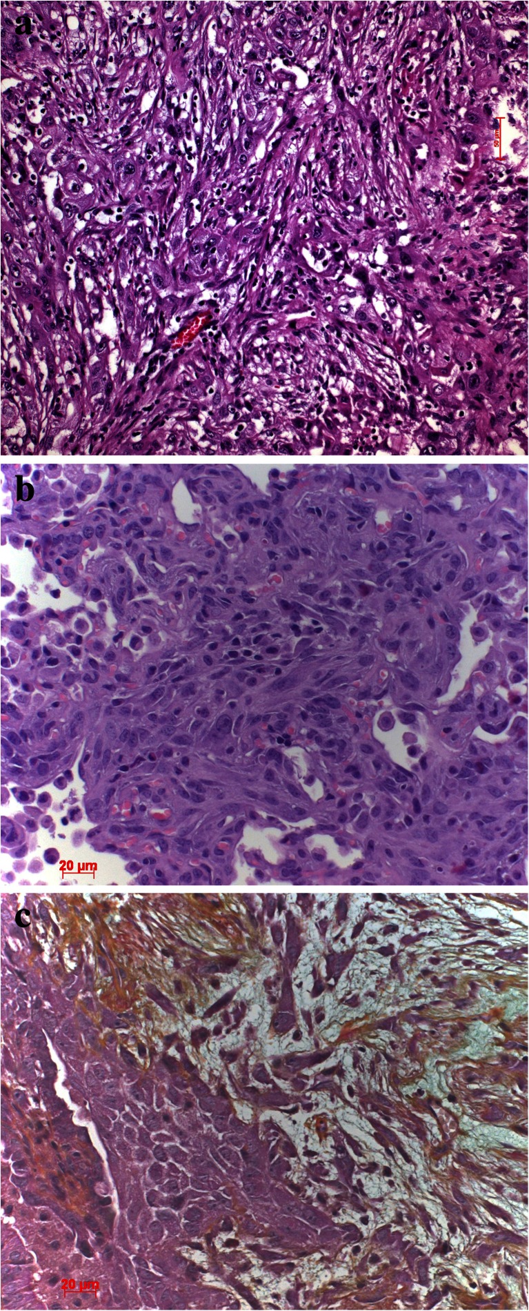
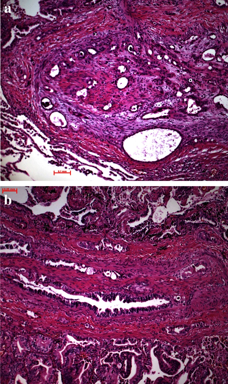
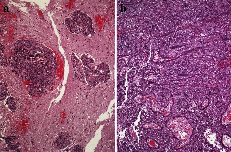
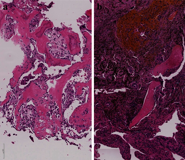
Metastasis in lung cancer is a multifaceted process. In this review, we will dissect the process in several isolated steps such as angiogenesis, hypoxia, circulation, and establishment of a metastatic focus. In reality, several of these processes overlap and occur even simultaneously, but such a presentation would be unreadable. Metastasis requires cell migration toward higher oxygen tension, which is based on changing the structure of the cell (epithelial-mesenchymal transition), orientation within the stroma and stroma interaction, and communication with the immune system to avoid attack. Once in the blood stream, cells have to survive trapping by the coagulation system, to survive shear stress in small blood vessels, and to find the right location for extravasation. Once outside in the metastatic locus, tumor cells have to learn the communication with the "foreign" stroma cells to establish vascular supply and again express molecules, which induce immune tolerance.

Keywords: Angiogenesis; Bone; Brain; Circulation; Epithelial-mesenchymal transition; Hypoxia; Lung cancer; Metastasis; Migration.


Figures

Fig. 1
Fig. 1
Angiogenesis in preneoplastic lesions, a atypical adenomatous hyperplasia has no new vessels but instead relies on the normal vascular architecture of preexisting alveolar septa; in the vascular variant of squamous cell dysplasia, b the preneoplastic cells induce angiogenesis using vascular growth factors produced by the dysplastic cells
Fig. 2
Fig. 2
Desmoplastic stroma reaction is almost absent in this well-differentiated lepidic predominant adenocarcinoma (a) whereas prominent in this squamous cell carcinoma (b)
Fig. 3
Fig. 3
Experimental adenocarcinoma in a mouse. Carcinoma is induced by mutant KRAS. At a certain size of the in situ adenocarcinoma, central hypoxic necrosis develops (a), which is the prerequisite for invasion (b)
Fig. 4
Fig. 4
Desmoplastic stroma supports invasion and guides the carcinoma cells in this squamous cell carcinoma (a), whereas scar tissue inhibits invasion as in this adenocarcinoma example (b). The only way for the carcinoma cells is invasion into lymphatics, which happened in the center
Fig. 5
Fig. 5
Epithelial to mesenchymal transition (EMT) is common in pleomorphic carcinomas of the lung (a); this can also be demonstrated by cytokeratin immunohistochemistry, showing epithelial tumor cells positively stained on the left side, whereas tumor cells at the right side have lost cytokeratin and acquired vimentin (b)
Fig. 6
Fig. 6
Tumor cell migration, a this small-cell neuroendocrine carcinoma moves in small-cell groups, whereas the adenocarcinoma (b) moves almost as single cell
Fig. 7
Fig. 7
Tumor cell migration, a this mixed small- and large-cell neuroendocrine carcinoma migrates as single- or small-cell clusters, whereas the small-cell neuroendocrine carcinoma in b migrates in small complexes in this very early stage; experimental mouse model (slides provided by A. Gazdar)
Fig. 8
Fig. 8
Tumor cell migration, a a mucinous adenocarcinoma moves in larger-cell complexes along the alveolar walls, still using the supply by the alveolar septa and b an unusual 3D complex of squamous cells moving as spheroids
Fig. 9
Fig. 9
a EMT in a pleomorphic carcinoma with spindle cells and b, c EMT in mouse model of KRAS-induced adenocarcinomas with additional expression of mutant TP53. c Movat stain, which better demonstrates the invasion of the spindle tumor cells into the desmoplastic stroma
Fig. 10
Fig. 10
Vascular invasion, a tumor cells are scattered in acinar complexes within the intima of this pulmonary artery and b large acinar and papillary adenocarcinoma complexes can be seen within these blood vessels, demonstrating the other example of invasion as large tumor cell complexes
Fig. 11
Fig. 11
Brain metastasis, a cells of an adenocarcinoma interacting with astrocytes and microglial cells and b large adenocarcinoma complexes have acquired huge areas of the brain but in addition, imitate ependymal structures
Fig. 12
Fig. 12
Bone metastasis, a adenocarcinoma cell complexes have induced an impressive activity of osteoclasts, resulting in lytic bone lesions; b adenocarcinoma metastases have induced bleeding and a massive inflammatory reaction, which also results in lytic bone lesions

References

    1. Gabor S, Renner H, Popper H, Anegg U, Sankin O, Matzi V, Lindenmann J, Smolle Juttner FM. Invasion of blood vessels as significant prognostic factor in radically resected T1-3N0M0 non-small-cell lung cancer. European Journal of Cardio-Thoracic Surgery. 2004;25:439–442. doi: 10.1016/j.ejcts.2003.11.033. - DOI - PubMed
    1. Shin DY, Na II, Kim CH, Park S, Baek H, Yang SH. EGFR mutation and brain metastasis in pulmonary adenocarcinomas. Journal of Thoracic Oncology. 2014;9:195–199. doi: 10.1097/JTO.0000000000000069. - DOI - PubMed
    1. Tamura T, Kurishima K, Nakazawa K, Kagohashi K, Ishikawa H, Satoh H, Hizawa N. Specific organ metastases and survival in metastatic non-small-cell lung cancer. Molecular and Clinical Oncology. 2015;3:217–221. - PMC - PubMed
    1. Hendriks LE, Smit EF, Vosse BA, Mellema WW, Heideman DA, Bootsma GP, Westenend M, Pitz C, de Vries GJ, Houben R, Grunberg K, Bendek M, Speel EJ, Dingemans AM. EGFR mutated non-small cell lung cancer patients: more prone to development of bone and brain metastases? Lung Cancer. 2014;84:86–91. doi: 10.1016/j.lungcan.2014.01.006. - DOI - PubMed
    1. Wilbertz T, Wagner P, Petersen K, Stiedl AC, Scheble VJ, Maier S, Reischl M, Mikut R, Altorki NK, Moch H, Fend F, Staebler A, Bass AJ, Meyerson M, Rubin MA, Soltermann A, Lengerke C, Perner S. SOX2 gene amplification and protein overexpression are associated with better outcome in squamous cell lung cancer. Modern Pathology. 2011;24:944–953. doi: 10.1038/modpathol.2011.49. - DOI - PubMed

Publication types