. 2016 Mar 28;11(3):e0152611.
doi: 10.1371/journal.pone.0152611. eCollection 2016.

Crystal Structure of Human Myotubularin-Related Protein 1 Provides Insight into the Structural Basis of Substrate Specificity

Affiliations

Crystal Structure of Human Myotubularin-Related Protein 1 Provides Insight into the Structural Basis of Substrate Specificity

Seoung Min Bong et al. PLoS One. .

Erratum in

Abstract

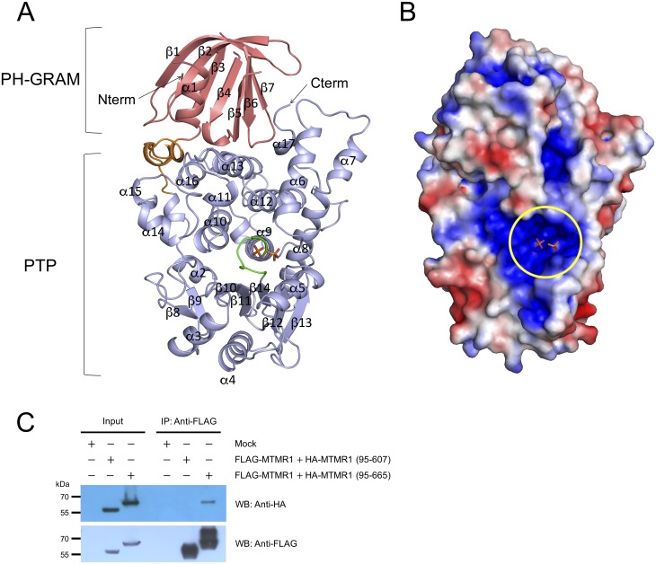
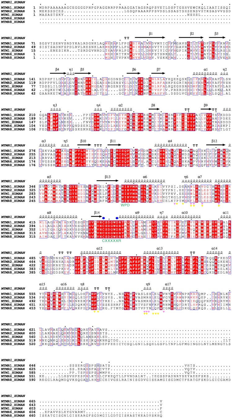
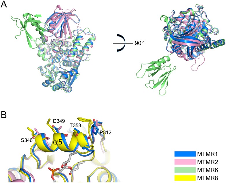
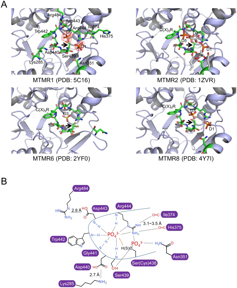
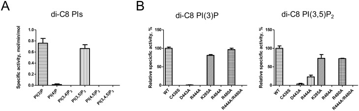
Myotubularin-related protein 1 (MTMR1) is a phosphatase that belongs to the tyrosine/dual-specificity phosphatase superfamily. MTMR1 has been shown to use phosphatidylinositol 3-monophosphate (PI(3)P) and/or phosphatidylinositol 3,5-bisphosphate (PI(3,5)P2) as substrates. Here, we determined the crystal structure of human MTMR1. The refined model consists of the Pleckstrin homology (PH)-GRAM and phosphatase (PTP) domains. The overall structure was highly similar to the previously reported MTMR2 structure. Interestingly, two phosphate molecules were coordinated by strictly conserved residues located in the C(X)5R motif of the active site. Additionally, our biochemical studies confirmed the substrate specificity of MTMR1 for PI(3)P and PI(3,5)P2 over other phosphatidylinositol phosphates. Our structural and enzymatic analyses provide insight into the catalytic mechanism and biochemical properties of MTMR1.


Conflict of interest statement

Competing Interests: The authors have declared that no competing interests exist.

Figures

Fig 1
Fig 1. Overall structure of MTMR1.
(A) A ribbon diagram of MTMR1. The PH-GRAM domain is depicted in pink, the PTP domain is in light blue, the linker is in orange, and the C(X)5R motif is in green. Two phosphate molecules are drawn. (B) The surface potential representation of MTMR1 with charge levels from -3kT/e (red) to +3kT/e (blue). The active site pocket containing the two phosphate molecules is indicated as a yellow circle. The surface potential representation was generated using PDB2PQR and APBS [48, 49]. The figures were drawn using PyMol (Schrödinger, LLC). (C) Immunoprecipitation and western blot analysis with FLAG- and HA-tagged MTMR1. A strong interaction was observed between FLAG-MTMR1 and HA-MTMR1 (95–665), suggesting the formation of dimers, but no interaction was found with CC domain-truncated MTMR1 (95–607) constructs.
Fig 2
Fig 2. Alignments of the amino acid sequences of the MTMRs.
The C(X)5R motif and residues corresponding to the WPD motif of the PTPs are indicated in green letters. The catalytically important residues are marked in blue circles. Residues involved in the dimeric interfaces for MTMR2 and MTMR8 are marked with pink (MTMR2) and yellow (MTMR8) triangles, respectively. This figure was drawn using Espript (http://espript.ibcp.fr) [50].
Fig 3
Fig 3. Comparison of the MTMR1, MTMR2, MTMR6, and MTMR8 structures.
(A) Overall comparison of the MTMR1, MTMR2, and MTMR6 structures that contain the PH-GRAM and PTP domains. The sequence identities to MTMR1 are 67.6% for 678 MTMR2 residues, 36.9% for 694 MTMR6 residues, and 39.7% for 638 MTMR8 residues. (B) Comparison of the structures of the α5 helix, which is thought to be important for membrane association. Each MTMR is colored aqua, pink, lime, or yellow. The chain A molecule of each MTMR was used for the structural comparisons.
Fig 4
Fig 4. Active sites of the known MTMR structures.
(A) Active sites of individual MTMRs. The residues on the C(X)5R loop and other important residues are shown using green stick models. The phosphates (in MTMR1 and MTMR8), sulfate (in MTMR6), or PI(3,5)P2 (in MTMR2) molecules are also depicted as stick models. The electron density map for the phosphate molecules in MTMR1 is shown with a red mesh. Black arrows point to the cysteine residue in the C(X)5R motif of the MTMRs that is mutated to serine in the MTMR structures, except for MTMR6, for crystallization purposes. (B) Illustrated representation of the phosphate-bound active site in the MTMR1 structure. The hydrogen bond distances between residues in the C(X)5R motif and the phosphate ions were approximately 2.5–3.0 Å.
Fig 5
Fig 5. Enzymatic activity of MTMR1.
(A) Specific activity of MTMR1 against di-C8 PIs. (B) Comparison of the specific activities of wild-type and mutant (C438S, D443A, R444A, K285A, R484A, R480A, and R444A/R480A) MTMR1 against di-C8 PI(3)P and di-C8 PI(3,5)P2. The reactions were performed in triplicate, and the specific activities are presented as moles of phosphate released per minute per mole of enzyme ± SD.

References

    1. Robinson FL, Dixon JE. Myotubularin phosphatases: policing 3-phosphoinositides. Trends Cell Biol. 2006;16(8):403–12. 10.1016/j.tcb.2006.06.001 . - DOI - PubMed
    1. Majerus PW, York JD. Phosphoinositide phosphatases and disease. J Lipid Res. 2009;50 Suppl:S249–54. 10.1194/jlr.R800072-JLR200 - DOI - PMC - PubMed
    1. McCrea HJ, De Camilli P. Mutations in phosphoinositide metabolizing enzymes and human disease. Physiology (Bethesda). 2009;24:8–16. 10.1152/physiol.00035.2008 - DOI - PMC - PubMed
    1. Attree O, Olivos IM, Okabe I, Bailey LC, Nelson DL, Lewis RA, et al. The Lowe's oculocerebrorenal syndrome gene encodes a protein highly homologous to inositol polyphosphate-5-phosphatase. Nature. 1992;358(6383):239–42. 10.1038/358239a0 . - DOI - PubMed
    1. Hoopes RR Jr., Shrimpton AE, Knohl SJ, Hueber P, Hoppe B, Matyus J, et al. Dent Disease with mutations in OCRL1. Am J Hum Genet. 2005;76(2):260–7. 10.1086/427887 - DOI - PMC - PubMed

Publication types

MeSH terms

Substances