. 2016 May;22(5):464-72.
doi: 10.1089/ten.TEC.2015.0581. Epub 2016 Apr 20.

Surface Chemistry and Microtopography of Parylene C Films Control the Morphology and Microtubule Density of Cardiac Myocytes

Affiliations

Surface Chemistry and Microtopography of Parylene C Films Control the Morphology and Microtubule Density of Cardiac Myocytes

Tatiana Trantidou et al. Tissue Eng Part C Methods. 2016 May.

Abstract

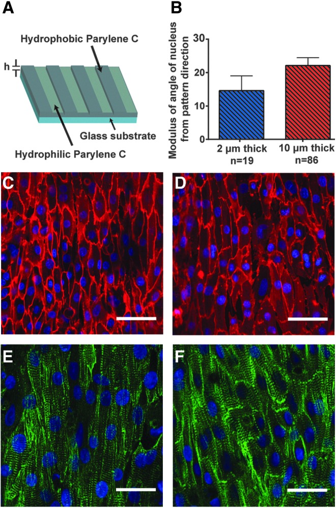
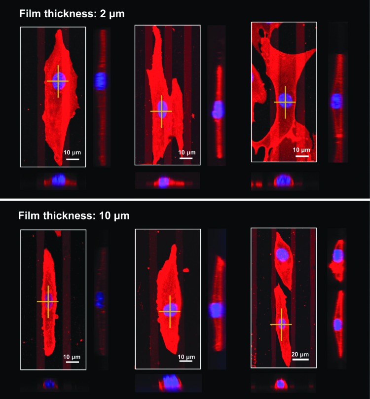
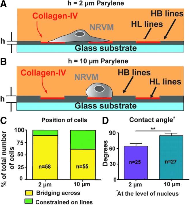
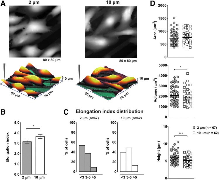
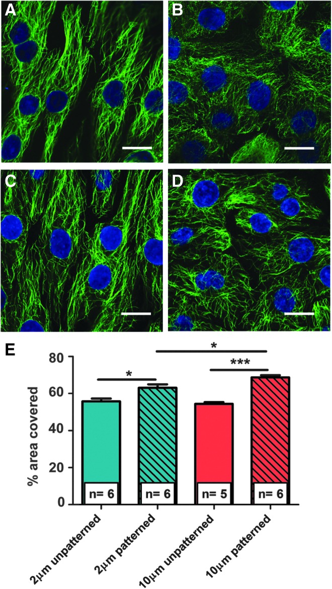
Cell micropatterning has certainly proved to improve the morphological and physiological properties of cardiomyocytes in vitro; however, there is little knowledge on the single cell-scaffold interactions that influence the cells' development and differentiation in culture. In this study, we employ hydrophobic/hydrophilic micropatterned Parylene C thin films (2-10 μm) as cell microscaffolds that can control the morphology and microtubule density of neonatal rat ventricular myocytes (NRVM) by regulating their adhesion area on Parylene through a thickness-dependent hydrophobicity. Structured NRVM on thin films tend to bridge across the hydrophobic areas, demonstrating a more spread-out shape and sparser microtubule organization, while cells on thicker films adopt a cylindrical (in vivo-like) shape (contact angles at the level of the nucleus are 64.51° and 84.73°, respectively) and a significantly (p < 0.05) denser microtubule structure. Ion scanning microscopy on NRVM revealed that cells on thicker membranes were significantly (p < 0.05) smaller in volume, but more elongated. The cylindrical shape and a significantly denser microtubule structure indicate the ability to influence cardiomyocyte phenotype using patterning and manipulation of hydrophilicity. These combined bioengineering strategies are promising tools in the generation of more representative cardiomyocytes in culture.


Figures

<b>FIG. 1.</b>
FIG. 1.
Neonatal rat ventricular myocytes (NRVM) on the microengineered Parylene C constructs. (A) Schematic representation of the fabrication process of the microengineered constructs. h represents the Parylene C film thickness. (B) Quantification of NRVM alignment on 2 μm-thick (blue) and 10 μm-thick (red) micropatterned Parylene C surfaces. n represents the number of cells. Data derived from two isolations. Error bars indicate standard error of mean (SEM). Representative immunofluorescence of NRVM cultured on (C, E) 2 μm-thick and (D, F) 10 μm-thick Parylene C constructs. Red, membrane stain cell mask orange. Green- alpha sarcomeric actinin antibody. Blue, nuclear stain Hoechst 33258 in (C, D) and DAPI in (E, F). Bars-50 μm in (C, D) and 30 μm in (E, F). Color images available online at www.liebertpub.com/tec
<b>FIG. 2.</b>
FIG. 2.
Representative immunofluorescence images and cross-sectional views (taken from the corresponding crosses) of NRVM. Cells are cultured on 2 μm-thick (top) and 10 μm-thick (bottom) Parylene C micropatterned constructs. Red, membrane stain cell mask orange. Blue, nuclear stain Hoechst 33258. Color images available online at www.liebertpub.com/tec
<b>FIG. 3.</b>
FIG. 3.
Morphology and position analysis of cultured NRVM. Schematics showing the position of NRVM on (A) the h = 2 μm and (B) h = 10 μm thick micropatterned Parylene C substrates. (C) Statistics of the cells position on the micropatterned constructs. (D) Corresponding contact angle data. Error bars indicate SEM. **p < 0.01. n represents the number of cells (Data derived from three isolations). Color images available online at www.liebertpub.com/tec
<b>FIG. 4.</b>
FIG. 4.
Topographical images obtained with hopping mode ion conductance microscopy of cultured NRVM. (A) Cells are cultured on 2 μm-thick and 10 μm-thick Parylene C constructs. The scanning pipette is depicted in the 3-dimensional reconstruction of the scans. (B, C) Elongation index, (D) surface, volume, and height of NRVM in relation to film thickness. n represents the number of cells. *p < 0.05, ***p < 0.001 (Data derived from two isolations. Error bars indicate SEM). Color images available online at www.liebertpub.com/tec
<b>FIG. 5.</b>
FIG. 5.
Representative immunofluorescence images of NRVM stained for microtubules. (A) 2 μm-thick patterned substrates, (B) 2 μm-thick unpatterned substrates, (C) 10 μm-thick patterned substrates, and (D) 10 μm-thick unpatterned Parylene C substrates. Green, mouse anti α-tubulin antibody. Blue, nuclear stain Hoechst 33258. Bars-10 μm in (A–D). (E) Quantification of microtubules. n represents the number of constructs. *p < 0.05, ***p < 0.001 (Data derived from six isolations. Error bars indicate SEM). Color images available online at www.liebertpub.com/tec

References

    1. Rao C., et al. . The effect of microgrooved culture substrates on calcium cycling of cardiac myocytes derived from human induced pluripotent stem cells. Biomaterials 34, 2399, 2013 - PMC - PubMed
    1. Trantidou T., et al. . Selective hydrophilic modification of Parylene C films: a new approach to cell micro-patterning for synthetic biology applications. Biofabrication 6, 1, 2014 - PubMed
    1. Walsh K.B., and Parks G.E. Changes in cardiac myocyte morphology alter the properties of voltage-gated ion channels. Cardiovasc Res 55, 64, 2002 - PubMed
    1. Bien H., Yin L., and Entcheva E. Cardiac cell networks on elastic microgrooved scaffolds. IEEE Eng Med Biol Mag 22, 108, 2003 - PubMed
    1. Bursac N., Parker K., Iravanian S., and Tung L. Cardiomyocyte cultures with controlled macroscopic anisotropy a model for functional electrophysiological studies of cardiac muscle. Circ Res 91, e45, 2002 - PubMed

Publication types