. 2016 May;23(5):409-15.
doi: 10.1038/nsmb.3194. Epub 2016 Mar 28.

Solid-state NMR structure of a pathogenic fibril of full-length human α-synuclein

Affiliations

Solid-state NMR structure of a pathogenic fibril of full-length human α-synuclein

Marcus D Tuttle et al. Nat Struct Mol Biol. 2016 May.

Abstract

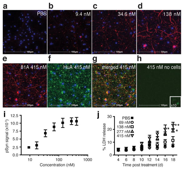
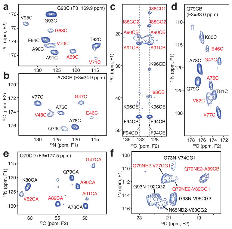
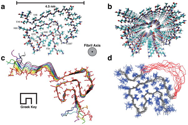
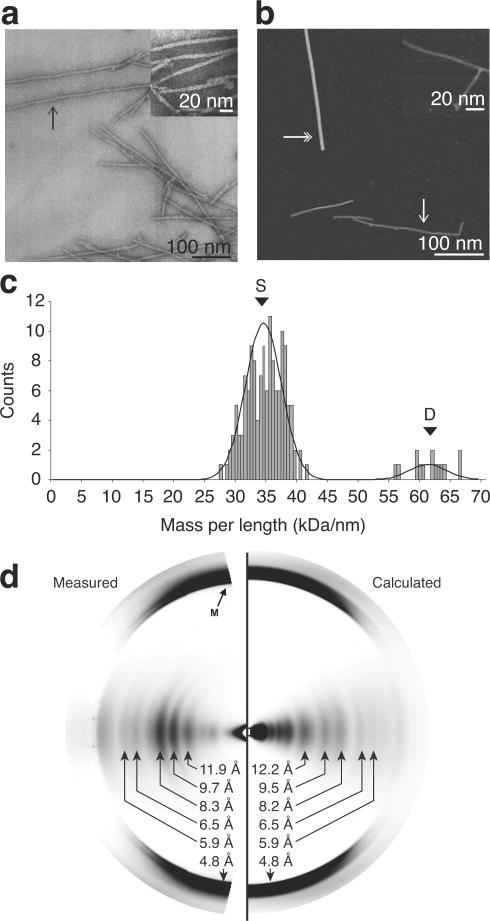
Misfolded α-synuclein amyloid fibrils are the principal components of Lewy bodies and neurites, hallmarks of Parkinson's disease (PD). We present a high-resolution structure of an α-synuclein fibril, in a form that induces robust pathology in primary neuronal culture, determined by solid-state NMR spectroscopy and validated by EM and X-ray fiber diffraction. Over 200 unique long-range distance restraints define a consensus structure with common amyloid features including parallel, in-register β-sheets and hydrophobic-core residues, and with substantial complexity arising from diverse structural features including an intermolecular salt bridge, a glutamine ladder, close backbone interactions involving small residues, and several steric zippers stabilizing a new orthogonal Greek-key topology. These characteristics contribute to the robust propagation of this fibril form, as supported by the structural similarity of early-onset-PD mutants. The structure provides a framework for understanding the interactions of α-synuclein with other proteins and small molecules, to aid in PD diagnosis and treatment.


Figures

Figure 1
Figure 1. Effects of pre-formed α-synuclein (α-syn) fibril samples on neurons
(a–e) Immunocytochemistry of fixed and extracted non-transgenic mouse primary hippocampal neurons labeled for phosphorylated α-syn (pSyn, red) with phosphorylated Ser129 specific (81A) antibody and the deoxyribonucleic acid binding dye 2-(4-amidinophenyl)-1H-indole-6-carboxamidine (DAPI, blue) after treatment with (a) phosphate buffered saline (PBS), (b–e) α-syn fibrils in PBS with concentrations (b) 9.4 nM, (c) 34.6 nM, (d) 138 nM, and (e) 415 nM. (f) Immunocytochemical staining of the neurons in condition (e) for total α-syn with the polyclonal antibody, HuA (green), and (g) the co-localization of the total α-syn signal with pSyn (merged 81A and HuA, yellow). (h) Immunostaining of total α-syn (HuA, green) and pSyn (81A, red) staining of the fibrils on a coverslip in the absence of neurons. Inset is a 10X magnification. (i) Quantitation of insoluble, pSyn signal from coverslips treated with increasing doses of α-syn fibrils. Error bars, s.e.m (n = 3 coverslips). (j) Plot of lactate dehydrogenase (LDH) detected in the media of primary neuronal cultures over time for five doses of α-syn fibrils. Error bars, s.e.m. (n = 3 wells) *P=0.05; **P=0.01 by a two-way analysis of variance with Bonferroni correction.
Figure 2
Figure 2. Long-range solid-state NMR structural restraints for an α-synuclein fibril
(a–b) Two-dimensional (2D) 15N-13C planes from three-dimensional (3D) 15N-13CO-13CX spectrum with 500 ms dipolar-assisted rotational resonance (DARR) mixing, collected with sample B (500 MHz 1H frequency, 11.111 kHz magic-angle spinning (MAS) rate, signal averaged 181.3 hours). (c) Aromatic-to-aliphatic region of 2D 13C-13C spectrum with 300 ms DARR mixing, collected with sample B (750 MHz 1H frequency, 12.5 kHz MAS rate, signal averaged 12.7 hours). (d) 2D plane from 15N-13CO-13CX spectrum with 500 ms DARR mixing collected with sample B (500 MHz 1H frequency, 11.111 kHz MAS rate, signal averaged 181.3 hours). (e) 2D plane from 15N-13CA-13CX spectrum with 500 ms DARR mixing collected with sample C (500 MHz 1H frequency, 11.111 kHz MAS rate, signal averaged 152.1 hours). (f) Region of 15N-13C spectrum for sample B with 6.4 ms transferred-echo double resonance (TEDOR) mixing (600 MHz 1H frequency, 10 kHz MAS rate, signal averaged 8.7 hours). Red labels indicate long-range correlations. distance restraints; black labels represent intraresidue and sequential correlations.
Figure 3
Figure 3. Three-dimensional structure of an α-synuclein fibril
(a) View of a central monomer from residues 44 to 96 looking down the fibril axis showing the Greek-key motif of the fibril core. (b) A view of the stacked monomers showing the sidechain alignment between each monomer down the fibril axis (c) Residues 25 to 105 of 8 monomers showing the β-sheet alignment of each monomer in the fibril and the Greek-key topology of the core. (d) Overlay of the 10 lowest energy structures showing agreement of sidechain positions within the core corresponding to an RMSD of 2.0 Å for all heavy atoms for residues 46 to 54 and 63 to 96. Residues 51–57 are shown in red with side chains removed for clarity.
Figure 4
Figure 4. Validation of α-synuclein (α-syn) fibril structure by microscopy and fiber diffraction
(a) Bright-field, negatively stained transmission electron microscopy. (b) Dark-field unstained scanning transmission electron microscopy (STEM). Single-headed arrows indicate examples of individual fibrils. Double-headed arrows indicate tobacco mosaic virus (TMV) rods, an internal control for mass-per-length (MPL) ratio determination. Insets are of higher magnification of the fibril samples. (c) Histogram of the distributions of the STEM MPL measurement of unstained, freeze-dried, α-syn fibril. The peak labeled S indicates the mass of a single fibril and the peak labeled D indicates the mass of a double fibril. (d) Experimental and calculated fiber diffraction pattern from α-syn fibrils. Black arrow labeled M indicates the cross-β meridonal diffraction near 4.8 Å resolution.

References

    1. Spillantini MG, et al. α-Synuclein in Lewy bodies. Nature. 1997;388:839–840. - PubMed
    1. Luk KC, et al. Exogenous α-synuclein fibrils seed the formation of Lewy body-like intracellular inclusions in cultured cells. Proc Natl Acad Sci USA. 2009;106:20051–20056. - PMC - PubMed
    1. Volpicelli-Daley LA, et al. Exogenous α-synuclein fibrils induce Lewy body pathology leading to synaptic dysfunction and neuron death. Neuron. 2011;72:57–71. - PMC - PubMed
    1. Desplats P, et al. Inclusion formation and neuronal cell death through neuron-to-neuron transmission of α-synuclein. Proc Natl Acad Sci USA. 2009;106:13010–13015. - PMC - PubMed
    1. Luk KC, et al. Pathological α-synuclein transmission initiates Parkinson-like neurodegeneration in nontransgenic mice. Science. 2012;338:949–953. - PMC - PubMed

Publication types