. 2016 Feb 12;12(4):454-65.
doi: 10.7150/ijbs.13379. eCollection 2016.

Alternative Roles of STAT3 and MAPK Signaling Pathways in the MMPs Activation and Progression of Lung Injury Induced by Cigarette Smoke Exposure in ACE2 Knockout Mice

Affiliations

Alternative Roles of STAT3 and MAPK Signaling Pathways in the MMPs Activation and Progression of Lung Injury Induced by Cigarette Smoke Exposure in ACE2 Knockout Mice

Yi-Han Hung et al. Int J Biol Sci. .

Abstract

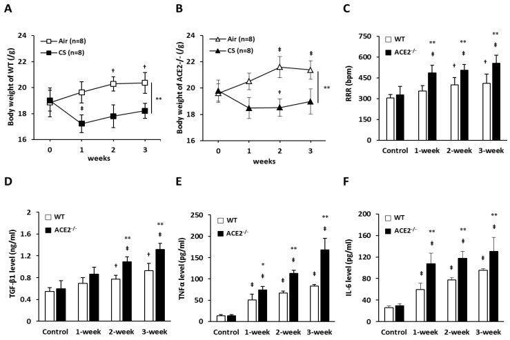
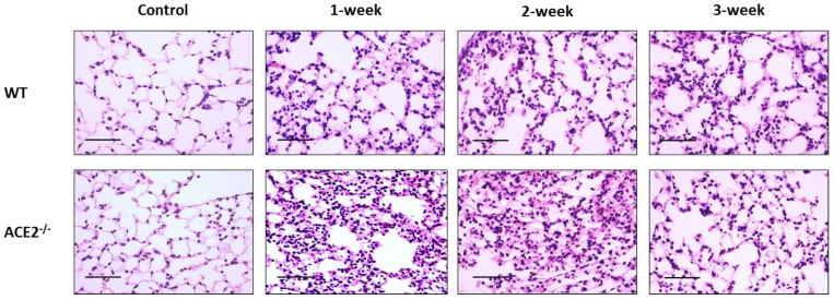
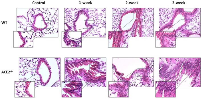
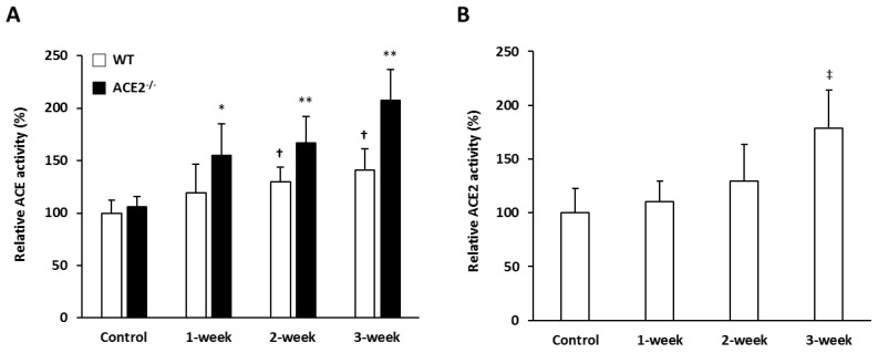
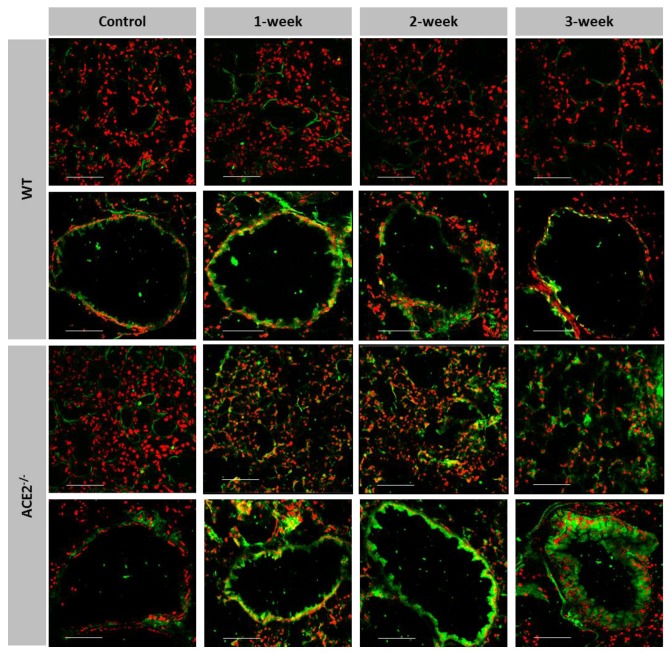
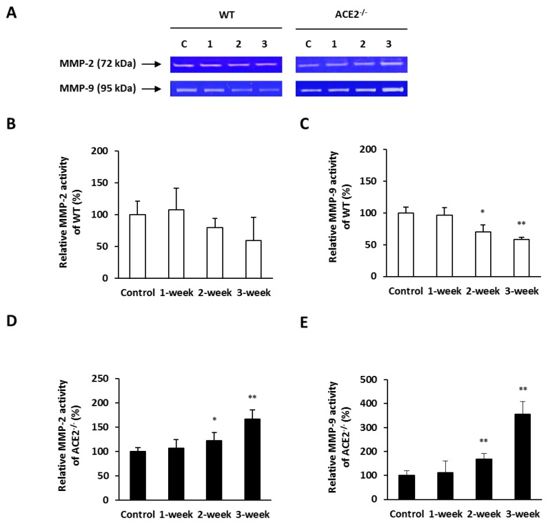
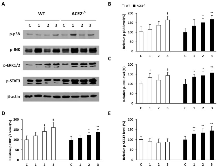
Inflammation-mediated abnormalities in the renin-angiotensin system (RAS) and expression of matrix metalloproteinases (MMPs) are implicated in the pathogenesis of lung injury. Angiotensin converting enzyme II (ACE2), an angiotensin converting enzyme (ACE) homologue that displays antagonist effects on ACE/angiotensin II (Ang II) axis, could also play a protective role against lung diseases. However, the relationship between ACE2 and MMPs activation in lung injury is still largely unclear. The purpose of this study is to investigate whether MMPs activity could be affected by ACE2 and which ACE2 derived signaling pathways could be also involved via using a mouse model with lung injury induced by cigarette smoke (CS) exposure for 1 to 3 weeks. Wild-type (WT; C57BL/6) and ACE2 KO mice (ACE2(-/-)) were utilized to study CS-induced lung injury. Increases in the resting respiratory rate (RRR), pulmonary immunokines, leukocyte infiltration and bronchial hyperplasia were observed in the CS-exposed mice. Compared to WT mice, more serious physiopathological changes were found in ACE2(-/-) mice in the first week of CS exposure. CS exposure increased pulmonary ACE and ACE2 activities in WT mice, and significantly increased ACE in ACE2(-/-) mice. Furthermore, the activity of pulmonary MMPs was decreased in CS-exposed WT mice, whereas this activity was increased in ACE2(-/-) mice. CS exposure increased the pulmonary p-p38, p-JNK and p-ERK1/2 level in all mice. In ACE2(-/-) mice, a significant increase p-STAT3 signaling was detected; however, no effect was observed on the p-STAT3 level in WT mice. Our results support the hypothesis that ACE2 deficiency influences MMPs activation and STAT3 phosphorylation signaling to promote more pulmonary inflammation in the development of lung injury.

Keywords: angiotensin converting enzyme II; cigarette smoke; lung injury; matrix metalloproteinases; renin-angiotensin system.


Conflict of interest statement

Competing Interests: The authors have declared that no competing interest exists.

Figures

Figure 1
Figure 1
Hallmark features of lung injury induced by CS exposure in the mouse model. WT (C57BL/6, n=8 for each group) mice and ACE2 KO mice (homozygous ACE2-/-, n=8 for each group) were exposed to CS or air for 3 weeks. (A) and (B) The changes of body weight of the WT and ACE2-/- mice exposed with CS or air, respectively. † p<0.05 and ‡ p<0.01 compared with the 0 week; ** p<0.01 compared with the mice after 3 weeks of CS exposure. (C) The measurements and comparisons of the resting respiration rate (RRR) of the mice exposed to CS for 3 weeks. The animals were sacrificed to isolate the lung tissues for (D) TGF-β1, (E) TNF-α and (F) IL-6 determination using an ELISA assay. All of the values are expressed as the mean±SD from each group; † p<0.05 and ‡ p<0.01 compared with the control (i.e., 0 week); * p<0.05 and ** p<0.01 compared with the WT mice at the same CS-exposed week.
Figure 2
Figure 2
Alveolar infiltration of white blood cells yielded in the experimental mice with CS-induced lung injury. The lung alveolar sections of CS-exposed WT and ACE2-/- mice were stained with haematoxylin-eosin (H&E). The non-CS treated mice were defined as the control group. Relative to the control mice, there were increases in the infiltration of white blood cells around the alveolus in the CS-exposed WT and ACE2-/- mice. (Scale=50 μm)
Figure 3
Figure 3
Changes of airway epithelial tissue yielded in the experimental mice with CS-induced lung injury. The lung bronchiole sections of CS-exposed WT and ACE2-/- mice were stained with haematoxylin-eosin (H&E). The non-CS treated mice were defined as the control group. Relative to the control mice, there were increases in the infiltration of white blood cells around the airway and airway epithelial thickening in the CS-exposed WT and ACE2-/- mice.
Figure 4
Figure 4
The pulmonary ACE and ACE2 activities were induced by CS exposure. WT mice and ACE2-/- mice were exposed to CS for 0 (Control), 1, 2 and 3 weeks, and then the animals were sacrificed to isolate lung tissues for ACE and ACE2 activity assays. The values of ACE and ACE2 activity in the control group were calculated as 100%. (A) The pulmonary ACE activities in WT and ACE2-/- mice were progressively increased with CS exposure. (B) The relative ACE2 activity in WT mice was increased upon CS exposure. All of the values are expressed as the mean±SD from each group (n=8 for each group); † p<0.05 and ‡ p<0.01 compared with the control group in WT mice; * p<0.05 and ** p<0.01 compared with the control group in ACE2-/- mice.
Figure 5
Figure 5
More gelatinase activity expressed in the pulmonary alveolus and bronchioles of CS-exposed ACE2 KO mice. The location and gelatinase activity in the lungs of WT and ACE2-/- mice exposed to CS for 1 to 3 weeks were detected using in situ zymography. The FITC signal (green) is released upon cleavage of the gelatin substrate and the nuclear signal (red) is stained. Gelatinase activity around the alveolus was significantly increased in the CS exposed ACE2-/- mice, but not in the CS-exposed WT mice. Bronchiolar gelatinase activity was significantly increased in the CS-exposed mice, especially in the ACE2-/- mice. Images were captured at low magnification (Scale=100 μm).
Figure 6
Figure 6
Pulmonary MMP-2 and MMP-9 activities elevated in CS-exposed ACE2 KO mice. The WT and ACE2-/- mice were exposed to CS for 1 to 3 weeks and sacrificed for the assays. (A) The activities of MMP-2 and MMP-9 in the lungs of WT and ACE2-/- mice were determined using a gelatin zymography assay. The value of MMP-2 and MMP-9 activities in the control mice was calculated as 100%. (B) CS exposure did not lead to a change in pulmonary MMP-2 activity, but (C) decreased pulmonary MMP-9 activity in the WT mice. (D) MMP-2 and (E) MMP-9 activities in the lungs of ACE2-/- mice were significantly upregulated upon CS exposure. All of the values are expressed as the mean±SD from each group (n=8); * p<0.05 and ** p<0.01 compared with the control group.
Figure 7
Figure 7
Pulmonary p-p38, p-JNK, p-ERK1/2 and p-STAT3 elevated in CS-exposed ACE2 KO mice. The WT and ACE2-/- mice were exposed to CS for 1 to 3 weeks and sacrificed for the assays. (A) The level of p-p38, p-JNK, p-ERK1/2 and p-STAT3 in the lungs was determined by immunoblotting. β-actin was used as the internal controls for p-p38, p-JNK, p-ERK1/2 and p-STAT3, respectively. The levels of p-p38, p-JNK, p-ERK1/2 and p-STAT3 expression in the non-CS exposed mice were calculated as 100% and defined as the control. The relative pulmonary (B) p-p38, (C) p-JNK, (D) p-ERK1/2 expression of the WT mice was increased upon CS exposure. (E) The relative p-STAT3 expression of WT mice showed no significant difference after CS exposure, but p-STAT3 expression was induced significantly by CS exposure in the lungs of ACE2-/- mice. All of the values are expressed as the mean±SD from each group; † p<0.05 and ‡ p<0.01 compared with the control group in WT mice; * p<0.05 and ** p<0.01 compared with the control group in ACE2-/- mice.

References

    1. Rubenfeld GD, Caldwell E, Peabody E. et al. Incidence and outcomes of acute lung injury. N Engl J Med. 2005;353:1685–93. - PubMed
    1. Wright JL, Cosio M, Churg A. Animal models of chronic obstructive pulmonary disease. Am J Physiol Lung Cell Mol Physiol. 2008;295:L1–15. - PMC - PubMed
    1. Wright JL, Zhou S, Churg A. Pulmonary hypertension and vascular oxidative damage in cigarette smoke exposed eNOS(-/-) mice and human smokers. Inhal Toxicol. 2012;24:732–40. - PubMed
    1. Calfee CS, Matthay MA, Eisner MD. et al. Active and passive cigarette smoking and acute lung injury after severe blunt trauma. Am J Respir Crit Care Med. 2011;183:1660–5. - PMC - PubMed
    1. Rincon M, Irvin CG. Role of IL-6 in asthma and other inflammatory pulmonary diseases. Int J Biol Sci. 2012;8:1281–90. - PMC - PubMed

Publication types

Substances