Review
. 2016 Mar;43(2):134-44.
doi: 10.5999/aps.2016.43.2.134. Epub 2016 Mar 18.

Review of Acute Traumatic Closed Mallet Finger Injuries in Adults

Affiliations
Review

Review of Acute Traumatic Closed Mallet Finger Injuries in Adults

Santiago Salazar Botero et al. Arch Plast Surg. 2016 Mar.

Abstract

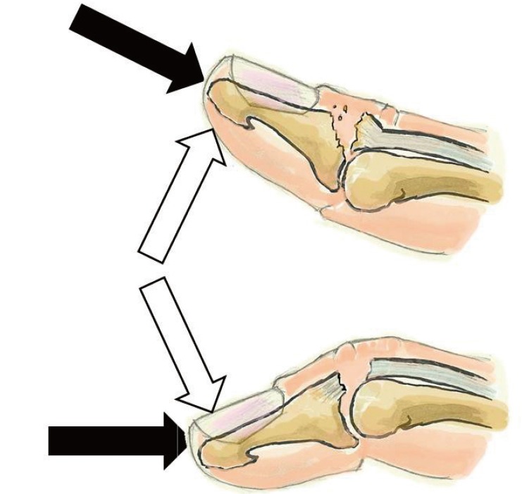
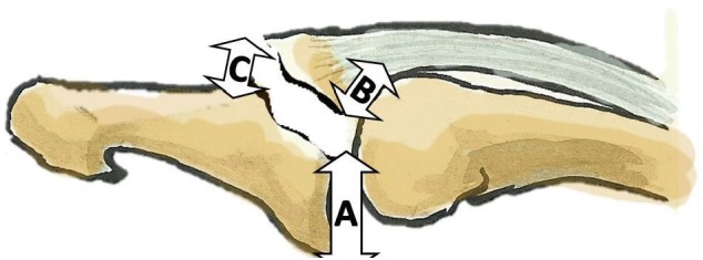
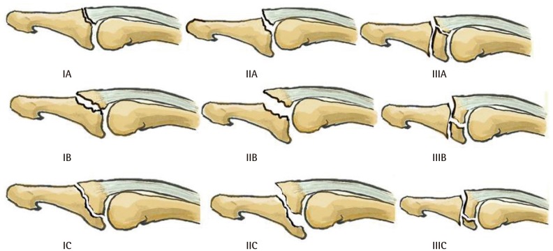
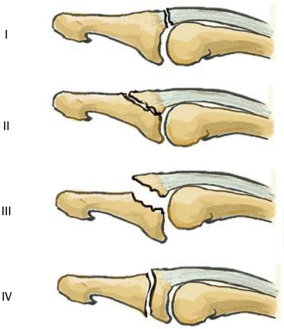
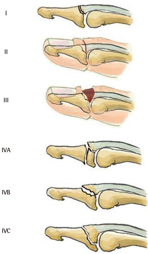
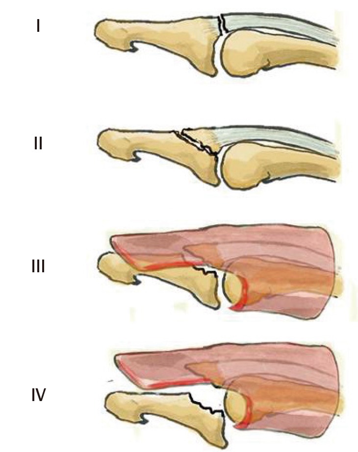
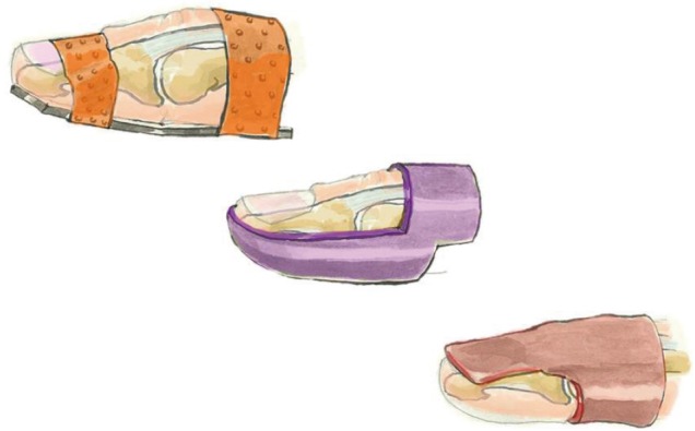
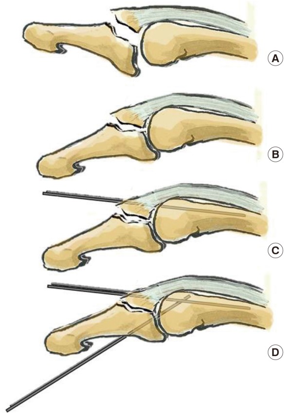
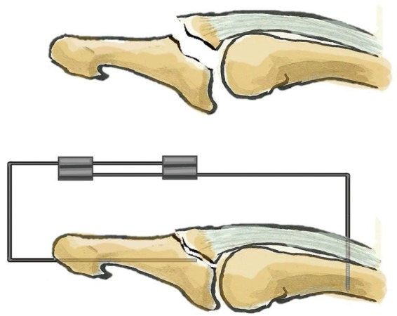
In adults, mallet finger is a traumatic zone I lesion of the extensor tendon with either tendon rupture or bony avulsion at the base of the distal phalanx. High-energy mechanisms of injury generally occur in young men, whereas lower energy mechanisms are observed in elderly women. The mechanism of injury is an axial load applied to a straight digit tip, which is then followed by passive extreme distal interphalangeal joint (DIPJ) hyperextension or hyperflexion. Mallet finger is diagnosed clinically, but an X-ray should always be performed. Tubiana's classification takes into account the size of the bony articular fragment and DIPJ subluxation. We propose to stage subluxated fractures as stage III if the subluxation is reducible with a splint and as stage IV if not. Left untreated, mallet finger becomes chronic and leads to a swan-neck deformity and DIPJ osteoarthritis. The goal of treatment is to restore active DIPJ extension. The results of a six- to eight-week conservative course of treatment with a DIPJ splint in slight hyperextension for tendon lesions or straight for bony avulsions depends on patient compliance. Surgical treatments vary in terms of the approach, the reduction technique, and the means of fixation. The risks involved are stiffness, septic arthritis, and osteoarthritis. Given the lack of consensus regarding indications for treatment, we propose to treat all cases of mallet finger with a dorsal glued splint except for stage IV mallet finger, which we treat with extra-articular pinning.

Keywords: Bony mallet; Mallet finger; Mallet fracture.


Conflict of interest statement

Philippe Liverneaux has conflict of interest with Newclip Technics and Argomedical. None of the other authors has conflit of interest.

Figures

Fig. 1
Fig. 1. Main theories explaining the mechanism of injury
In all analyses, an axial force is applied to the tip of a straight digit (black arrows), followed by extreme passive distal interphalangeal joint (DIPJ) hyperextension (white upper arrow), which would account for bony lesions, or extreme passive DIPJ hyperflexion (lower white arrow), which would account for tendinous lesions.
Fig. 2
Fig. 2. Size and displacement calculation on lateral view
Ratio of the fractured articular surface over the total articular surface of the distal phalanx base: T=B/A+B=size of fragment in %. The ratio of the gap between the distal phalanx and the bony fragment over the total articular surface of the distal phalanx base: D=C/A+B=fragment displacement in %.
Fig. 3
Fig. 3. Wehbé and Schneider classification
Type I, no volar subluxation; type II, volar subluxation; type III, growth plate fracture; subtype A, fragment size <1/3 of the articular surface; subtype B, fragment size between 1/3 and 2/3 of the articular surface; sub-type C, fragment size >2/3 of the articular surface.
Fig. 4
Fig. 4. Tubiana classification
Type I, subcutaneous tendon rupture; type II, bony avulsion at the base of the distal phalanx; type III, fracture >1/3 of the articular surface with volar subluxation; type IV, growth plate fracture.
Fig. 5
Fig. 5. Doyle classification
Type I, closed mallet finger; type II, open mallet finger; type III, open mallet finger with loss of substance; type IV, bony mallet finger; subtype A, involves the growth plate; subtype B, fragment size between 20% and 50% of the articular surface; subtype C, fragment size greater than 50% of the articular surface.
Fig. 6
Fig. 6. Modified Tubiana classification
Type I, subcutaneous tendon rupture; type II, bony avulsion at the base of the distal phalanx; type III, fracture >1/3 of the articular surface with volar subluxation reducible with a dorsal splint; type IV, fracture >1/3 of the articular surface with volar subluxation irreducible with a splint.
Fig. 7
Fig. 7. Different splint types
From left to right: volar splint, stack splint, and dorsal glued splint.
Fig. 8
Fig. 8. Ishiguro technique
We use it in type IV lesions according to the modified Tubiana classification (A). Under fluoroscopy the distal interphalangeal joint (DIPJ) is flexed (B), and a first K-wire is pinned through the extensor tendon into the middle phalanx. The wire is used as a lever that pushes on the bony fragment once the DIPJ is extended (C). A second K-wire is put across the DIPJ to avoid flexion (D).
Fig. 9
Fig. 9. Extra-articular fixed pinning
An intramedullary 1.2-mm K-wire is put through the distal phalanx percutaneously. A second threaded wire is introduced perpendicularly to the long axis of the middle phalanx from a dorsal approach and abuts the volar cortex to respect the flexor tendon. Both wires are bent and fixed together with two connectors.

References

    1. Badia A, Riano F. A simple fixation method for unstable bony mallet finger. J Hand Surg Am. 2004;29:1051–1055. - PubMed
    1. Bendre AA, Hartigan BJ, Kalainov DM. Mallet finger. J Am Acad Orthop Surg. 2005;13:336–344. - PubMed
    1. Kronlage SC, Faust D. Open reduction and screw fixation of mallet fractures. J Hand Surg Br. 2004;29:135–138. - PubMed
    1. Abouna JM, Brown H. The treatment of mallet finger: the results in a series of 148 consecutive cases and a review of the literature. Br J Surg. 1968;55:653–667. - PubMed
    1. Alla SR, Deal ND, Dempsey IJ. Current concepts: mallet finger. Hand (N Y) 2014;9:138–144. - PMC - PubMed