Hemodynamic Consequences of Hypertrophic Cardiomyopathy with Midventricular Obstruction: Apical Aneurysm and Thrombus Formation
- PMID: 27019860
- PMCID: PMC4807733
- DOI: 10.4172/2329-9126.1000161
Hemodynamic Consequences of Hypertrophic Cardiomyopathy with Midventricular Obstruction: Apical Aneurysm and Thrombus Formation
Abstract
Background: Hypertrophic cardiomyopathy (HCM) with midventricular hypertrophy is an uncommon phenotypic variant of the disease. Midventricular hypertrophy predisposes to intracavitary obstruction and downstream hemodynamic sequelae.
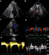
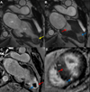
Case report: We present a case of HCM with midventricular hypertrophy and obstruction diagnosed after a CT scan of the abdomen incidentally revealed a filling defect in the left ventricular apex. Transthoracic echocardiography demonstrated mid left ventricular hypertrophy and obstruction, as well as an aneurysmal apex containing a large thrombus. Cardiovascular MRI showed a spade-shaped left ventricle with midcavitary obliteration, an infarcted apex and regions of myocardial fibrosis. Due to the risk of embolization and a relative contraindication to anticoagulation, the patient underwent surgery including thrombectomy, septal myectomy and aneurysmal ligation.
Conclusions: Hypertrophic cardiomyopathy with midventricular hypertrophy leads to cavity obstruction, increased apical wall tension, ischemia and ultimately fibrosis. Over time, patchy apical fibrosis can develop into a confluent scar resembling a transmural myocardial infarction in the left anterior descending coronary artery distribution. Aneurysmal remodeling of the left ventricular apex potentiates thrombus formation and risk of cardioembolism. For these reasons, hypertrophic cardiomyopathy with midventricular obstruction portends a particularly poor prognosis and should be recognized early in the disease process.
Keywords: Aneurysmal apical chamber; Cardiovascular magnetic resonance imaging of hypertrophic cardiomyopathy; Hypertrophic cardiomyopathy; Midcavity obstruction; Midventricular hypertrophy; Paradoxic diastolic jet flow in hypertrophic cardiomyopathy.
Figures


References
-
- Gersh BJ, Maron BJ, Bonow RO, Dearani JA, Fifer MA, et al. 2011 ACCF/AHA guideline for the diagnosis and treatment of hypertrophic cardiomyopathy: a report of the American College of Cardiology Foundation/American Heart Association Task Force on Practice Guidelines. Circulation. 2011;124:e783–e831. - PubMed
-
- Minami Y, Kajimoto K, Terajima Y, Yashiro B, Okayama D, et al. Clinical implications of midventricular obstruction in patients with hypertrophic cardiomyopathy. J Am Coll Cardiol. 2011;57:2346–2355. - PubMed
-
- Duncan K, Shah A, Chaudhry F, Sherrid MV. Hypertrophic cardiomyopathy with massive midventricular hypertrophy, midventricular obstruction and an akinetic apical chamber. Anadolu Kardiyol Derg. 2006;6:279–282. - PubMed
-
- Fighali S, Krajcer Z, Edelman S, Leachman RD. Progression of hypertrophic cardiomyopathy into a hypokinetic left ventricle: higher incidence in patients with midventricular obstruction. J Am Coll Cardiol. 1987;9:288–294. - PubMed
Grants and funding
LinkOut - more resources
Full Text Sources
Other Literature Sources
Miscellaneous
