. 2016 Feb 1;2(2):131-157.
doi: 10.1016/j.jcmgh.2015.11.009.

Loss of MYO5B in mice recapitulates Microvillus Inclusion Disease and reveals an apical trafficking pathway distinct to neonatal duodenum

Affiliations

Loss of MYO5B in mice recapitulates Microvillus Inclusion Disease and reveals an apical trafficking pathway distinct to neonatal duodenum

Victoria G Weis et al. Cell Mol Gastroenterol Hepatol. .

Abstract

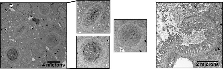
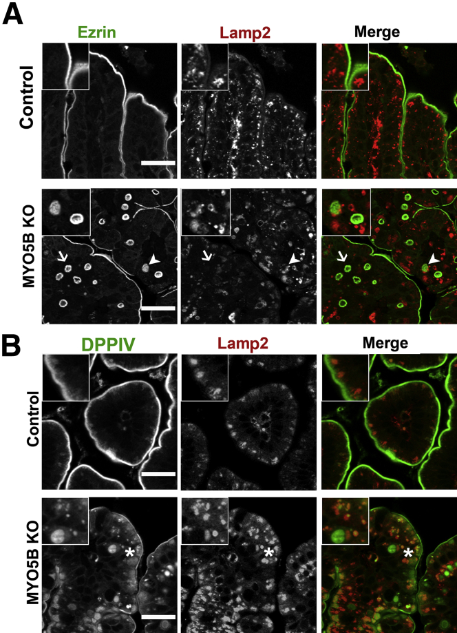
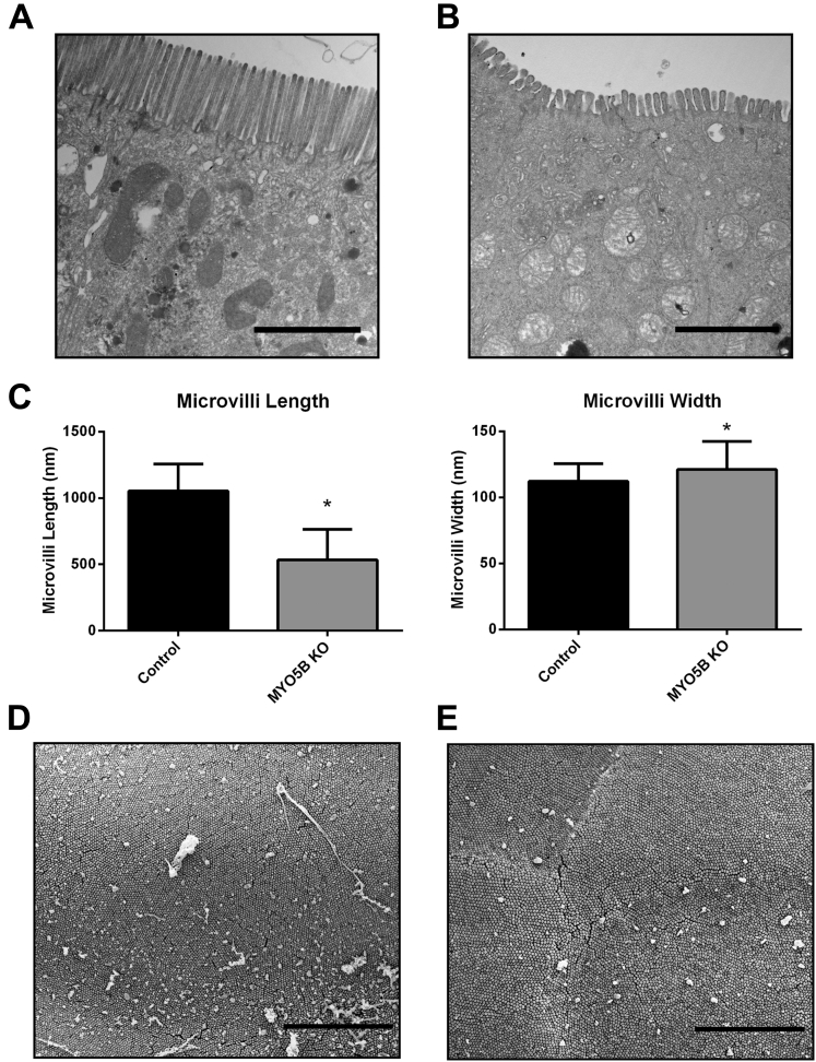
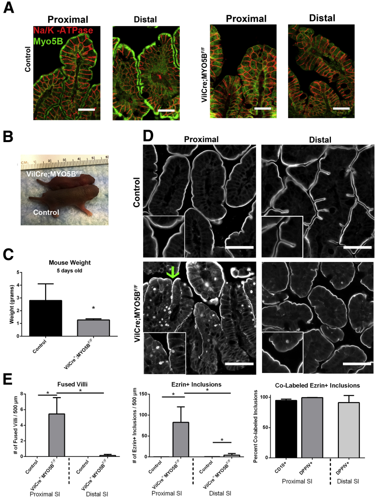
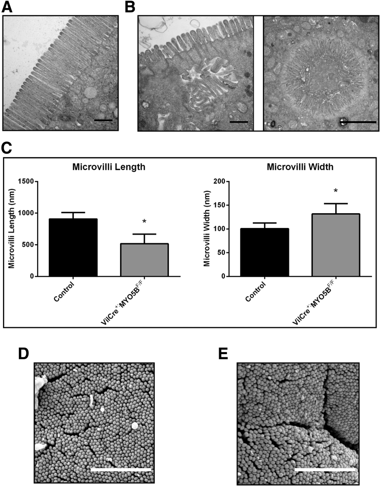
Background and aims: Inactivating mutations in MYO5B cause severe neonatal diarrhea in Microvillus Inclusion Disease. Loss of active MYO5B causes the formation of pathognomonic inclusions and aberrations in brush border enzymes.

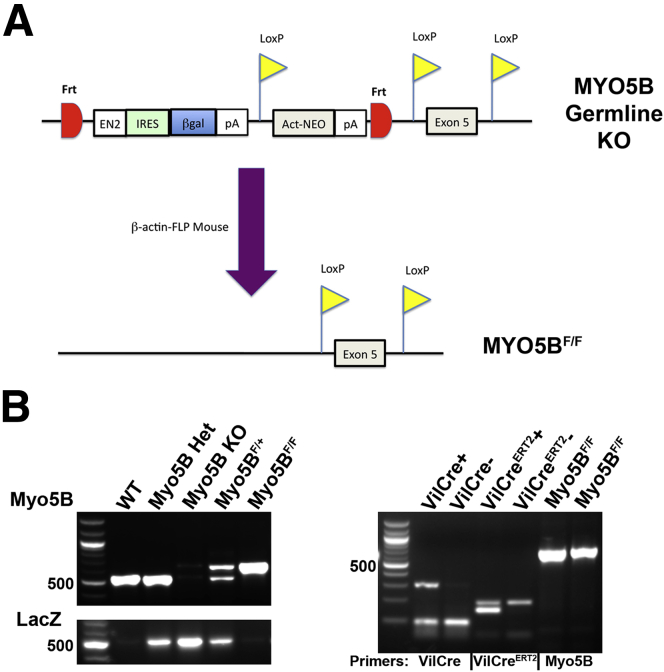
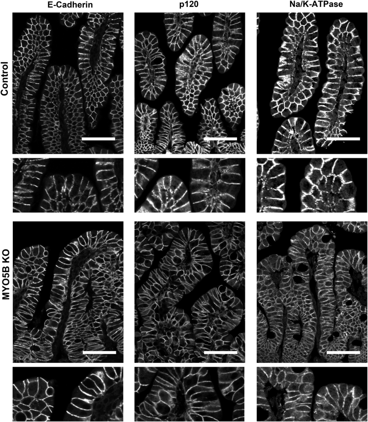
Methods: We developed three mouse models of germline, constitutively intestinal targeted and inducible intestinal targeted deletion of MYO5B. The mice were evaluated for enterocyte cellular morphology.

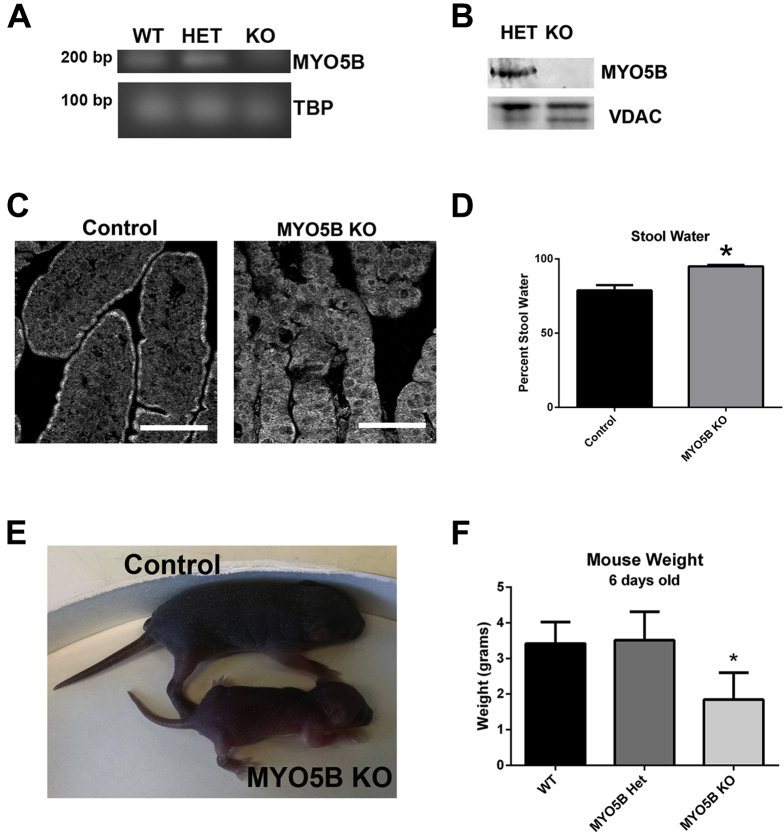
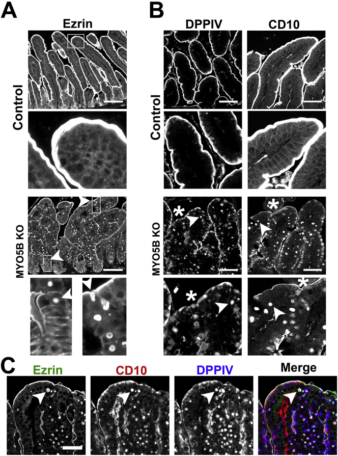
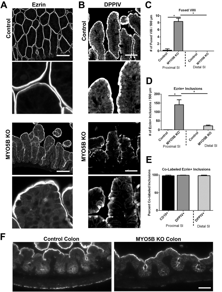
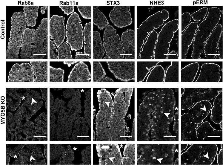
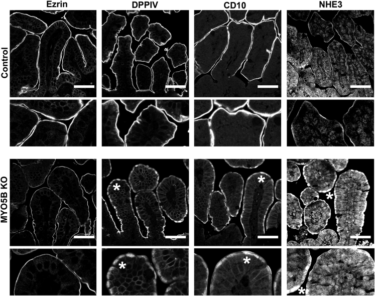
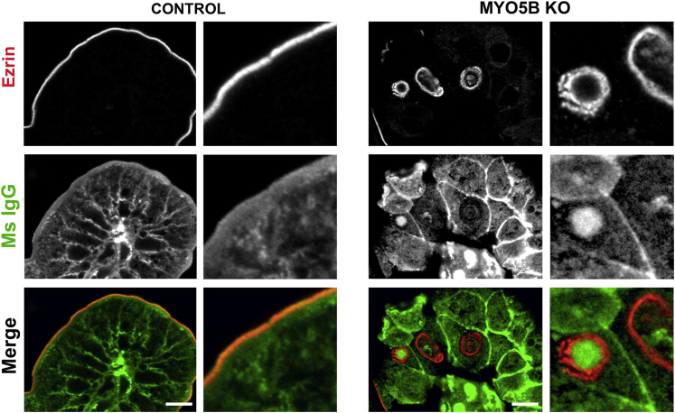
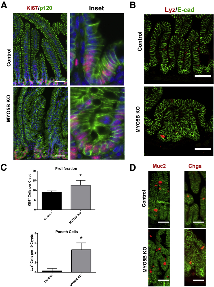
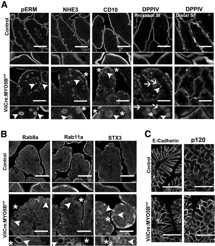
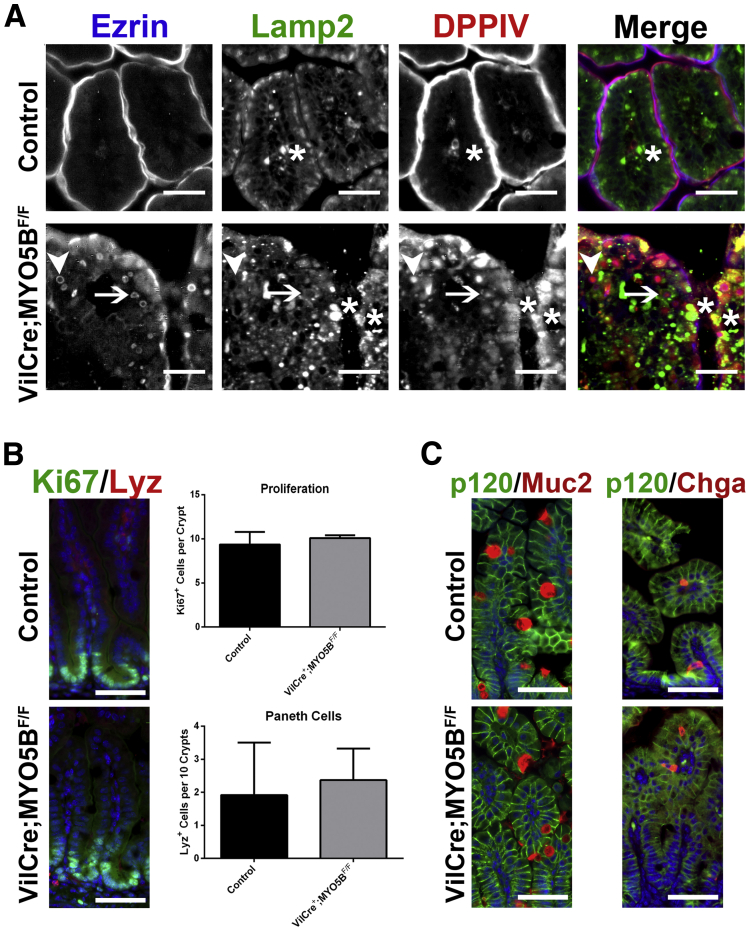
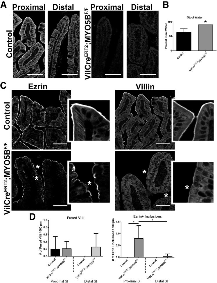
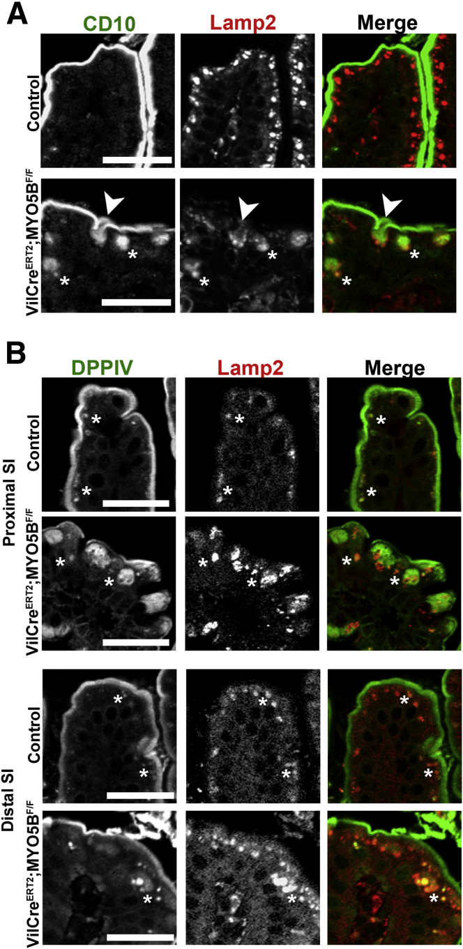
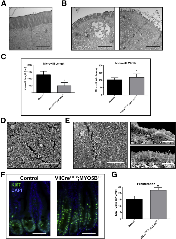
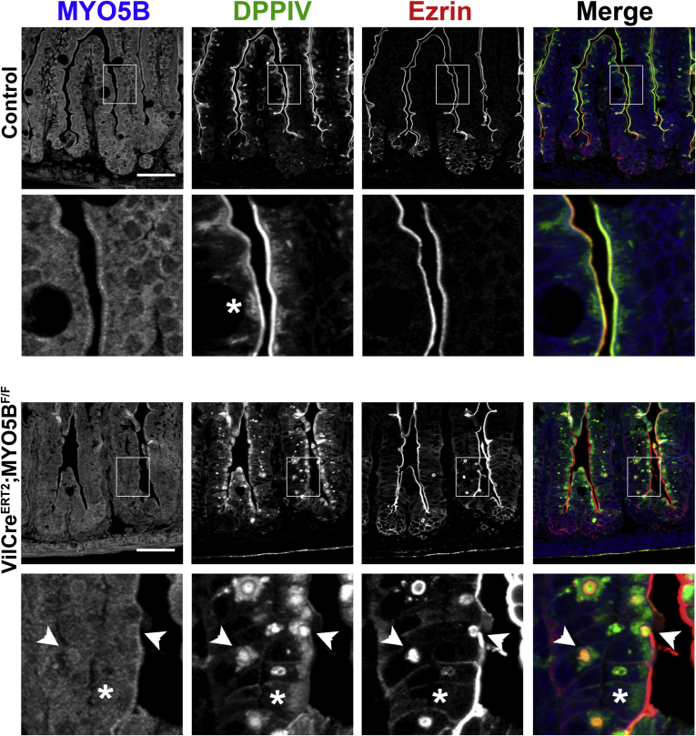
Results: Germline MYO5B KO mice showed early diarrhea and failure to thrive with evident microvillus inclusions and loss of apical transporters in the duodenum. IgG was present within inclusions. Apical transporters were lost and inclusions were present in the duodenum, but were nearly absent in the ileum. VillinCre;MYO5BF/F mice showed similar pathology and morphological changes in duodenal enterocytes. In contrast, when MYO5B KO was induced with tamoxifen treatment at 8 weeks of age, VillinCreERT2;MYO5BF/F mice developed severe diarrhea with loss of duodenal brush border enzymes, but few inclusions were observed in enterocytes. However, if tamoxifen is administered to 2-day-old VillinCreERT2;MYO5BF/F mice, prominent microvillus inclusions were observed.

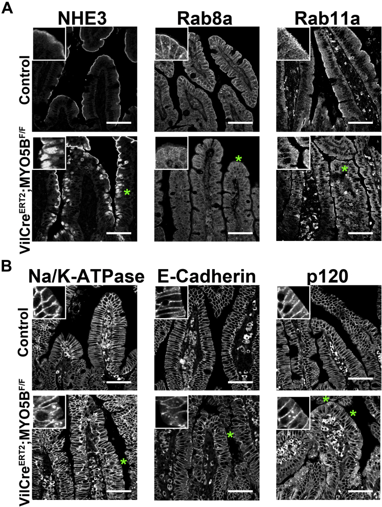
Conclusions: The microvillus inclusions that develop after MYO5B loss reveal the presence of an unrecognized apical membrane trafficking pathway in neonatal duodenal enterocytes. However, the diarrheal pathology after MYO5B loss is due to deficits in transporter presentation at the apical membrane in duodenal enterocytes.

Keywords: Enterocyte trafficking; NHE3; Rab11a; Rab8a; Syntaxin 3; brush border.


Conflict of interest statement

The authors have declared that no conflict of interest exists.

Figures

Figure 1
Figure 1
Generation of germline MYO5B KO and MYO5BF/Fmice. (A) Schematic representation of the knockout first allele for MYO5B from the Knockout Mouse Project is shown. This construct was used to generate germline MYO5B KO mice by early termination of the Myo5B transcription before exon 5. Mice harboring this allele were mated to β-actin–FLP mice to also create MYO5BF/F mice in which LoxP sites flank exon 5. (B) PCR of genomic DNA shows the different PCR product patterns used to identify the genotype of a mouse. The 100-bp DNA Ladder (New England Biolabs, Ipswich, MA) is shown in the left lane of gels to denote PCR product size. The 500-bp marker is indicated.
Figure 2
Figure 2
Germline MYO5B KO mice fail to thrive. (A) PCR of complementary DNA from wild-type (WT), MYO5B Het, and MYO5B KO mouse duodenum confirmed Myo5B messenger RNA loss. Tata-box binding protein (TBP) was used as a loading control. (B) Western blot of duodenum confirms MYO5B protein loss in a MYO5B germline KO mouse as compared with a MYO5B heterozygote mouse. VDAC was used as a loading control. (C) Immunofluorescence for MYO5B in control duodenum shows subapical staining. This subapical staining was lost in MYO5B KO duodenum. Scale bar: 50 μm (D) The percentage of stool constituted by water was calculated in control and MYO5B KO mice (n = 3). MYO5B KO mice had significantly higher water content in stool collected from the colon. *P = .05. (E) Picture of control and MYO5B KO littermates at 5 days old shows the reduced size of MYO5B KO mice. (F) Mice weighed at 6 days old show no difference between WT and MYO5B Het mice. MYO5B KO mice were significantly smaller than control littermates (both WT and Het) (n=3). *P = .05.
Figure 3
Figure 3
Immunofluorescence of MYO5B KO duodenum. (A) Ezrin immunostaining in control mice show a fully formed brush border along the length of single villi. The inset shows no internal ezrin staining. In MYO5B KO duodenum, ezrin labeled internal inclusions in addition to the apical brush border. Left inset: inclusions formed in cells just exiting the crypt (arrowhead). Inclusions also were found forming along the apical membrane (arrowhead, right inset). Ezrin immunostaining also showed fused villi in MYO5B KO duodenum. Scale bar: 100 μm. (B) DPPIV and CD10 both labeled the apical brush border in control mice with no internal structures labeled (inset). In MYO5B KO duodenum, DPPIV and CD10 was collapsed from the apical surface into a diffuse subapical localization (*) as well as localized to internal inclusions (arrowheads). DPPIV also was found localized to small punctate vesicles (DPPIV inset). Scale bar: 50 μm. (C) MYO5B duodenum immunostained for ezrin (green), CD10 (red), and DPPIV (blue) show ezrin-positive inclusions co-labeled with CD10 and DPPIV (arrowhead). Scale bar: 50 μm.
Figure 4
Figure 4
Comparison of distal small intestine and colon. (A) Distal small intestine from control and MYO5B KO mice was immunolabeled for ezrin. In control distal small intestine, ezrin labeled the apical brush border with no intracellular immunoreactivity, as found in the proximal intestine. Distal MYO5B KO small intestine showed only occasional fused villi and few ezrin-positive inclusions along with the apical membrane labeling. Scale bar: 100 μm. (B) In control distal small intestine, DPPIV labeled the apical membrane with a small subapical population. DPPIV localization in MYO5B KO distal small intestine appeared similar to control tissue with a compact subapical localization in addition to the apical membrane. In addition, fewer DPPIV-positive intracellular inclusions were found. Scale bar: 50 μm. (C) The number of fused villi was counted in both the proximal and distal intestine in control and MYO5B KO mice. MYO5B KO proximal intestine had approximately 8 fused villi per 500-μm length of intestine, which was increased significantly from the controls. A decreasing gradient in the number of fused villi was observed in MYO5B KO intestine with only rare fused villi found in the distal small intestine. *P = .05. (D) MYO5B KO duodenum had approximately 140 ezrin-positive inclusions per 500 μm of intestine. Again, the number of inclusions was decreased significantly in MYO5B KO distal small intestine as compared with MYO5B KO duodenum. *P = .05. (E) The percentage of ezrin-positive inclusions that co-labeled with CD10 or DPPIV was quantified. More than 98% of all ezrin-positive inclusions were dual-labeled with ezrin and CD10 or ezrin and DPPIV. No difference in co-labeled inclusions was found between the proximal and distal small intestine. (F) Colon from control and MYO5B KO mice was immunolabeled for ezrin. No microvillus inclusions were observed in MYO5B KO colon. Scale bar: 50 μm.
Figure 5
Figure 5
TEM identification of microvillus inclusions. TEM analysis of MYO5B KO duodenum showed inclusions with fully formed microvilli, confirming the formation of microvillus inclusions. Several microvillus inclusions were visualized within the enterocytes of the duodenum (left four panels). Scale bar: 4 μm. In addition, microvillus inclusion was visualized at the apical membrane with characteristic omega-shaped invaginations of the brush-border microvilli (right panel). Scale bar: 2 μm.
Figure 6
Figure 6
Trafficking of ezrin-positive inclusions and DPPIV. (A) Duodenum from control and MYO5B KO mice were co-stained for ezrin (green) and Lamp2 (red). In control enterocytes, Lamp2-positive lysosomes concentrated just under the apical surface. Upon MYO5B loss, a subset of ezrin-positive inclusions were surrounded closely or cupped by Lamp2-positive lysosomes (arrowheads). However, other ezrin-positive inclusions were not located near Lamp2 (arrows). Scale bars: 25 μm. (B) In controls, the small DPPIV population occasionally found in the subapical region co-localized with Lamp2. *In MYO5B KO duodenum, the subset of DPPIV that mistrafficked to small punctate vesicles was found to co-label with Lamp2. Scale bars: 25 μm.
Figure 7
Figure 7
Disruption of apical trafficking in MYO5B KO duodenum. Control (top row) and MYO5B KO (bottom row) duodenal sections were immunostained for apical proteins (Rab8a, Rab11a, STX3, NHE3, and pERM). Apical trafficking proteins, Rab8a, Rab11a, and STX3, were concentrated subapically in control tissue, with Rab8a also labeling along lateral membranes. *Rab8a and Rab11a were lost from the apical membrane and occasionally localized to inclusions (arrowheads) in MYO5B KO duodenum. STX3 was collapsed into a broad and diffuse subapical region (asterisks and arrowheads), as well as localized to inclusions (arrowheads). The apical exchanger NHE3 was normally located on the apical membrane of enterocytes in the small intestine. Upon MYO5B loss, NHE3 was mislocalized to a diffuse subapical compartment (asterisks) and labels inclusions (arrowheads). In addition, a microvilli structural protein, pERM, was examined to further investigate the status of microvilli in MYO5B KO tissue. pERM in control duodenum labeled only on the apical surface of the villus enterocytes. As with ezrin, in MYO5B KO duodenum, pERM immunostaining identified microvillus inclusions inside enterocytes as well as the formation of microvillus inclusions at the apical surface of enterocytes (arrowheads). Scale bars: 50 μm.
Figure 8
Figure 8
Immunofluorescence staining for basolateral trafficking. Duodenum sections were immunostained for junctional proteins E-cadherin and p120 and the basolateral Na/K-ATPase. All 3 proteins were localized to the basolateral membrane in control duodenum (top row). Similar basolateral localization was observed in MYO5B KO duodenum (bottom row), thus showing no disruption in basolateral trafficking upon MYO5B loss. Scale bars: 50 μm.
Figure 9
Figure 9
TEM and SEM analysis of apical microvilli. (A) Control duodenum was imaged by TEM. Images show long and straight microvilli on the apical surface of enterocytes. Scale bar: 2 μm. (B) TEM imaging of MYO5B KO duodenum showed shortened apical microvilli. Scale bar: 2 μm. (C) The length and width of apical microvilli from control and MYO5B KO duodenum was measured. In MYO5B KO duodenum, microvilli were half the height of control microvilli. In addition, microvilli were slightly wider in MYO5B KO duodenal enterocytes. *P < .05. (D) Control duodenum examined by SEM showed tightly packed microvilli. Scale bar: 5 μm. (E) By SEM, tightly packed microvilli were visualized in MYO5B KO duodenum, showing that packing remained largely unchanged even with shortened microvilli. Scale bar: 5 μm.
Figure 10
Figure 10
Immunofluorescence staining of embryonic duodenum. Embryonic (E18.5) duodenum from control and MYO5B KO littermates was immunostained for apical markers to examine mistrafficking and microvillus inclusion formation. As in neonates, all markers (ezrin, DPPIV, CD10, and NHE3) labeled the apical membrane in control embryonic duodenum (top row). In contrast to neonates, ezrin-positive inclusions were not observed in MYO5B KO duodenum, suggesting that microvillus inclusions were not formed until after birth. However, DPPIV, CD10, and NHE3 staining was collapsed to a subapical localization (asterisk) not found in the control littermates. These findings show apical mistrafficking is present before birth, but microvillus inclusions likely do not form until after birth. Scale bars: 50 μm.
Figure 11
Figure 11
IgG internalization in ezrin-positive inclusions. Mouse IgG internalization was visualized by immunofluorescence staining for mouse IgG (Ms IgG, green) and ezrin (red). To detect endogenous mouse IgG, the Mouse on Mouse block kit could not be used, thus resulting in high background signal from interstitial cells. Nonetheless, no significant internal mouse IgG was observed in control duodenum (left panels). In contrast, in MYO5B KO duodenum, mouse IgG was found inside ezrin-positive inclusions (right panels). Scale bars: 10 μm.
Figure 12
Figure 12
Proliferation and cell lineages in MYO5B KO duodenum. (A) Ki67 was used as a marker of proliferation. Sections were stained for Ki67 (red) and p120 (green). Ki67-positive proliferating cells were confined to the short crypts in control mice. In MYO5B KO mice, an increase in proliferation was observed as well as an expansion of the crypt. 4’,6-diamidino-2-phenylindole (blue). Scale bar: 50 μm. (B) To determine the status of Paneth cells, sections were immunostained for Lysozyme (red) and E cadherin (E-cad) (green). A premature maturation of Paneth cells was observed in MYO5B KO duodenum. Scale bar: 50 μm. (C) The number of Ki67-positive proliferating cells per crypt was quantified for both control and MYO5B KO duodenum. A small but significant increase in proliferation was found in MYO5B KO duodenum. *P = .05. (B) Lysozyme-positive Paneth cells appear earlier in MYO5B KO than in control littermates. Although less than 1 Paneth cell per 10 crypts was found in control duodenum, more than 4 Paneth cells per 10 crypts were counted in MYO5B KO duodenum *P = .05. (D) Tissue sections were co-stained for lineage markers Muc2 or Chga (red) and p120 (green). Goblet cells (Muc2-positive) and enteroendocrine cells (Chga-positive) were present in both control and MYO5B KO tissue. Scale bar: 50 μm.
Figure 13
Figure 13
Characterization of VillinCre;MYO5BF/Fintestine. (A) Proximal and distal small intestine from control and VillinCre;MYO5BF/F mice was immunostained for MYO5B (green) and Na/K-ATPase (red). A clear subapical concentration of MYO5B was observed in control tissue, however, this immunoreactivity was lost in VillinCre;MYO5BF/F intestine (proximal and distal). Of note, no significant changes were observed in Na/K-ATPase expression and localization. Scale bar: 25 μm. (B) An image of a VillinCre;MYO5BF/F mouse and a control littermate mouse show an obvious size difference. (C) Mice weighed at 5 days old showed that VillinCre;MYO5BF/F mice were approximately half the weight of control littermates. *P = .05 (D) Ezrin immunofluorescence showed fused villi (green arrow) and the presence of ezrin-positive inclusions in VillinCre;MYO5BF/F proximal small intestine. However, only occasional inclusions were observed in the VillinCre;MYO5BF/F distal small intestine and, rarely, fused villi (lower right panel). Scale bar: 50 μm. (E) Similar to germline MYO5B KO mice, VillinCre;MYO5BF/F mice showed a gradient of phenotype from proximal to distal small intestine. Approximately 5 fused villi per 500-μm length of proximal intestine was measured, which decreased to only rare fused villi in the distal small intestine. Likewise, the VillinCre;MYO5BF/F small intestine showed significantly fewer inclusions in the distal small intestine compared with proximal. Again, more than 98% of all ezrin-positive inclusions also labeled with CD10 or DPPIV with no significant differences found between the proximal and distal small intestine. *P = .05. SI, small intestine.
Figure 14
Figure 14
Disruption of apical trafficking in VillinCre;MYO5BF/Fduodenum. (A) Control (top row) and VillinCre;MYO5BF/F (bottom row) duodenal sections were immunostained for apical proteins (pERM, NHE3, CD10, and DPPIV). pERM staining in control duodenum showed microvilli located only on the apical surface of the villi enterocytes. In VillinCre;MYO5BF/F duodenum, pERM immunostaining identified microvillus inclusions inside enterocytes as well as the formation of microvillus inclusions at the apical surface of enterocytes (arrowhead). The apical exchanger NHE3 normally was located on the apical membrane of enterocytes in the small intestine. Upon MYO5B loss, NHE3 was mislocalized to a diffuse subapical compartment (asterisk) and labeled in inclusions (arrowhead). CD10 and DPPIV both labeled the apical brush border in control mice. In VillinCre;MYO5BF/F duodenum, CD10 and DPPIV were relocated into a diffuse subapical localization (asterisk) as well as into internal inclusions (arrowheads). DPPIV also was found localized to small punctate vesicles (arrows). In VillinCre;MYO5BF/F distal small intestine (last column), although some DPPIV subapical accumulation was observed, no apparent overall difference was noted in comparison with control. Scale bar: 50 μm. (B) Apical trafficking proteins, Rab8a, Rab11a, and STX3, were concentrated subapically in control tissue, with Rab8a also labeling along lateral membranes. Rab8a and Rab11a were lost from the apical membrane with a small accumulation in the subapical regions (asterisk), and occasionally localized to inclusions (arrowheads) in VillinCre;MYO5BF/F duodenum. STX3 was collapsed into a diffuse subapical region (asterisk) as well as localized to inclusions (arrowheads). Scale bar: 50 μm. (C) Control and VillinCre;MYO5BF/F duodenum were immunostained for E-cadherin and p120. In both controls and VillinCre;MYO5BF/F duodenum, E-cadherin and p120 labeled the basolateral membranes with no obvious differences between them. Scale bar: 50 μm. SI, small intestine.
Figure 15
Figure 15
Localization of lysosomes and characterization of cellular make-up in villi. (A) Duodenum from control and VillinCre;MYO5BF/F duodenum was triple-stained for ezrin (blue), Lamp2 (green), and DPPIV (red). In control enterocytes, Lamp2-positive lysosomes concentrated just under the ezrin and DPPIV-positive apical surface. In VillinCre;MYO5BF/F duodenum, a subset of ezrin-positive inclusions were surrounded closely or cupped by Lamp2-positive lysosomes (arrow). However, other ezrin-positive inclusions were not located near Lamp2 (arrowhead). DPPIV internalized into small vesicles co-localized with Lamp2 (asterisks). Scale bar: 25 μm. (B) Control and VillinCre;MYO5BF/F duodenum was analyzed for proliferating cells (Ki67 in green) and the early maturation of Paneth cells (lysozyme in red). No significant differences were found between the 2 mouse strains. 4′,6-diamidino-2-phenylindole (blue). Scale bar: 50 μm. (C) Sections were immunostained for Muc2 (red, left panel) and Chga (red, right panel) with p120 (green). Similar to the germline MYO5B KO, no significant difference was observed between the control littermates and VillinCre;MYO5BF/F duodenums. 4′,6-diamidino-2-phenylindole (blue). Scale bar: 50 μm.
Figure 16
Figure 16
TEM and SEM analysis of apical microvilli in VillinCre;MYO5BF/Fmice. (A) Control duodenum was imaged by TEM. Images showed long and straight microvilli on the apical surface of enterocytes. Scale bar: 500 nm. (B) TEM imaging of VillinCre;MYO5BF/F duodenum showed shortened apical microvilli and the presence of internal inclusions with obvious microvilli. Scale bar: 500 nm (left panel). TEM analysis also showed internal microvillus inclusions with fully formed microvilli. Scale bar: 2 μm (right panel). (C) The length and width of apical microvilli from control and VillinCre;MYO5BF/F duodenum was measured. In VillinCre;MYO5BF/F duodenum, microvilli were significantly shorter than control microvilli. In addition, microvilli also were wider in VillinCre;MYO5BF/F duodenal enterocytes. *P < .05. (D) Control duodenum examined by SEM showed tightly packed microvilli. Scale bar: 2 μm. (E) By SEM, tightly packed microvilli were visualized in VillinCre;MYO5BF/F duodenum, showing that packing remained largely unchanged even with shortened microvilli. Scale bar: 2 μm.
Figure 17
Figure 17
Characterization of VillinCreERT2;MYO5BF/Fmouse small intestine in 8-week-old mice induced with tamoxifen. Eight-week-old VillinCreERT2;MYO5BF/F mice were treated with a single 2 mg intraperitoneal dose of tamoxifen to induce intestine KO of MYO5B. (A) Proximal and distal small intestine from control and VillinCreERT2;MYO5BF/F mice were immunostained for MYO5B. A clear subapical concentration of MYO5B was observed in control tissue, however, MYO5B immunofluorescence was lost in VillinCreERT2;MYO5BF/F mice in both proximal and distal intestine. Scale bar: 50 μm. (B) The percentage of stool constituted by water was determined in control and VillinCreERT2;MYO5BF/F mice. VillinCreERT2;MYO5BF/F mice had significantly higher water content in stool collected from the colon (90% stool water). *P = .05. (C) Ezrin immunostaining in control mice showed a fully formed brush border along the length of the villi. Inset: a strong sharp band of ezrin-positive brush border. In VillinCreERT2;MYO5BF/F duodenum, ezrin labeled a thin band along the brush border, suggesting a shortening or loss of apical microvilli (asterisk). Of note, only occasional ezrin-positive inclusions were observed in VillinCreERT2;MYO5BF/F mouse intestines. Similarly, villin labeled a bright and wide brush border in control duodenums. In VillinCreERT2;MYO5BF/F duodenums, the apical villin stain was much thinner and even absent in some locations (asterisk), with only rare inclusions observed. Scale bar: 50 μm. (D) Fused villi and the total number of ezrin-positive inclusions were quantified. No difference was observed in the number of fused villi in control and VillinCreERT2;MYO5BF/F proximal and distal small intestine. There was a significant increase in the number of ezrin-positive inclusions as compared with control in the proximal intestine. However, the total number of inclusions was only 0.75 ezrin-positive inclusions per 500 μm of tissue in VillinCreERT2;MYO5BF/F duodenums. *P = .05. SI, small intestine.
Figure 18
Figure 18
Internalization and trafficking of CD10 and DPPIV to lysosomes. (A) Duodenum from control and VillinCreERT2;MYO5BF/F mice were co-stained for CD10 (green) and Lamp2 (red). In control enterocytes, Lamp2-positive lysosomes were located just under the CD10-labeled apical surface. Upon induced MYO5B loss, a subset of CD10 was internalized (asterisks and arrow). Lamp2 was found surrounding CD10 as it was internalized from the apical surface (arrow), and, in addition, Lamp2 co-localized with internal CD10 populations (asterisks). Scale bar: 25 μm. (B) Proximal and distal small intestine from controls and VillinCreERT2;MYO5BF/F mice were immunolabeled for DPPIV (green) and Lamp2 (red). In both proximal and distal small intestine controls, DPPIV predominately resided at the brush border, but a small DPPIV population was found in the subapical region co-localized with Lamp2 (asterisks). In VillinCreERT2;MYO5BF/F duodenum, DPPIV was collapsed into a diffuse subapical region as well as to small punctate vesicles (asterisks). The DPPIV-labeled vesicles, along with a portion of the diffuse DPPIV staining, co-labeled with Lamp2 (asterisks). In VillinCreERT2;MYO5BF/F distal small intestine, most DPPIV was retained on the apical membrane with a small subset of internalized DPPIV found in the small vesicles (asterisk). As in the duodenum, this internalized DPPIV population co-labeled with Lamp2 (asterisk). Scale bar: 25 μm. SI, small intestine.
Figure 19
Figure 19
Disruption of apical and basolateral trafficking in VillinCreERT2;MYO5BF/Fmice. (A) Control and VillinCreERT2;MYO5BF/F duodenum was stained for an apical exchanger protein (NHE3) and for apical trafficking proteins (Rab8a and Rab11a). The normal apical localization of NHE3 (top left panel) found in control duodenum was collapsed to a diffuse pattern in the cytoplasm in VillinCreERT2;MYO5BF/F duodenum (green asterisk). Correspondingly, the normal concentrated subapical localization of Rab8a and Rab11a was disrupted in VillinCreERT2;MYO5BF/F duodenum (green asterisks). In VillinCreERT2;MYO5BF/F duodenum, Rab8a and Rab11a was prominently lost from the subapical region and diverted to a very diffuse cytoplasmic localization. Scale bar: 50 μm. (B) Basolateral trafficking was analyzed by immunostains for Na/K-ATPase, E-cadherin, and p120 in control and VillinCreERT2;MYO5BF/F duodenum. In control duodenum, all 3 proteins were tightly localized to the basolateral membrane of enterocytes. Although some normal basolateral localization was retained in VillinCreERT2;MYO5BF/F duodenum, aberrant small internal punctate vesicles also were observed for Na/K-ATPase, E-cadherin, and p120 (green asterisks and insets), suggesting that loss of MYO5B disrupts basolateral trafficking. Scale bar: 50 μm.
Figure 20
Figure 20
Proliferation and EM analysis of VillinCreERT2;MYO5BF/Fduodenum. (A) TEM images of control duodenum showed long and straight microvilli on the apical surface of enterocytes. Scale bar: 2 μm. (B) By TEM analysis, VillinCreERT2;MYO5BF/F duodenum showed varying aberrations in the apical microvilli. Frequent areas of shortened apical microvilli (left panel) were observed as well as patches of apical microvilli denudation (right panel). Scale bar: 2 μm. (C) The length and width of apical microvilli from control and VillinCreERT2;MYO5BF/F duodenal enterocytes was quantified. In VillinCreERT2;MYO5BF/F duodenal enterocytes, microvilli that were observed were significantly shorter than control microvilli. In addition, apical microvilli were also wider in VillinCreERT2;MYO5BF/F duodenal enterocytes. *P < .05. (D) Control duodenum examined by SEM showed tightly packed microvilli. Scale bar: 2 μm. (E) By SEM, areas of tightly packed microvilli were visualized in VillinCreERT2;MYO5BF/F duodenum, as were areas of shortened microvilli. Scale bar: 2 μm (left panel). In addition, SEM analysis of some cross-sectioned enterocytes further confirmed areas of short microvilli (top right panel; scale bar: 1 μm) as well as showed aberrant microvilli morphology such as a linked appearance (bottom right panel; scale bar: 1 μm). (F) Proliferation in control and VillinCreERT2;MYO5BF/F duodenum was examined via Ki67 (green) and 4′,6-diamidino-2-phenylindole (DAPI) (blue) immunofluorescence. Proliferation (noted by Ki67 immunoreactivity) was restricted to the crypts in both control and VillinCreERT2;MYO5BF/F duodenum. Scale bar: 50 μm. (G) The number of Ki67-positive cells per crypt was quantified for comparison. A significant increase in proliferation was seen in VillinCreERT2;MYO5BF/F duodenal crypts compared with control duodenal crypts. *P = .05.
Figure 21
Figure 21
Neonatal induction of MYO5B loss in VillinCreERT2;MYO5BF/Fmice. Tamoxifen-induced MYO5B loss in 2-day-old littermates of controls (top row and second inset row) and VillinCreERT2;MYO5BF/F mice (third row and bottom inset row) was examined to analyze the formation of microvillus inclusions in neonates as compared with adult VillinCreERT2;MYO5BF/F mice. Sections of duodenum were triple-stained for MYO5B (blue), DPPIV (green), and ezrin (red). In controls, MYO5B localized to the subapical region just below the DPPIV and ezrin-positive co-labeled brush border. Some diffuse internal DPPIV is observed in controls (asterisk). In VillinCreERT2;MYO5BF/F neonatal duodenum, the prominent MYO5B subapical localization was lost in the bottom portion of villi. Of note, MYO5B was not completely lost from the upper half of villi. DPPIV was lost from the apical membrane and collapsed into a prominent, diffuse subapical region (asterisk). Furthermore, in contrast to adult VillinCreERT2;MYO5BF/F mice, VillinCreERT2;MYO5BF/F neonates formed numerous internal ezrin-positive inclusions in the duodenum (arrowheads) that co-labeled with DPPIV. These inclusions resemble the microvillus inclusions found in in MYO5B KO and VillinCre;MYO5BF/F neonatal mice. Scale bar: 50 μm.

References

    1. Erickson R.P., Larson-Thome K., Valenzuela R.K. Navajo microvillous inclusion disease is due to a mutation in MYO5B. Am J Med Genet A. 2008;146A:3117–3119. - PubMed
    1. Muller T., Hess M.W., Schiefermeier N. MYO5B mutations cause microvillus inclusion disease and disrupt epithelial cell polarity. Nat Genet. 2008;40:1163–1165. - PubMed
    1. Ruemmele F.M., Muller T., Schiefermeier N. Loss-of-function of MYO5B is the main cause of microvillus inclusion disease: 15 novel mutations and a CaCo-2 RNAi cell model. Hum Mutat. 2010;31:544–551. - PubMed
    1. Davidson G.P., Cutz E., Hamilton J.R. Familial enteropathy: a syndrome of protracted diarrhea from birth, failure to thrive, and hypoplastic villus atrophy. Gastroenterology. 1978;75:783–790. - PubMed
    1. Ruemmele F.M., Schmitz J., Goulet O. Microvillous inclusion disease (microvillous atrophy) Orphanet J Rare Dis. 2006;1:22. - PMC - PubMed