. 2016 Feb 1;2(2):70-83.
doi: 10.1016/j.trecan.2015.12.006.

The Impact of DNA Methylation in Hematopoietic Malignancies

Affiliations

The Impact of DNA Methylation in Hematopoietic Malignancies

Maria Guillamot et al. Trends Cancer. .

Abstract

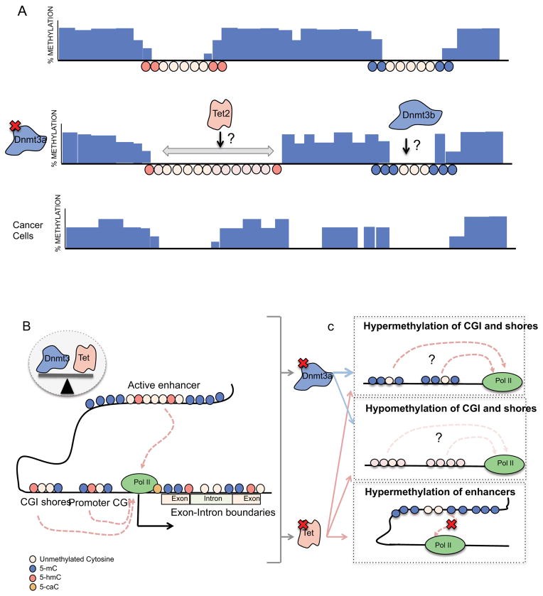
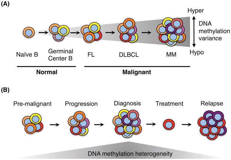
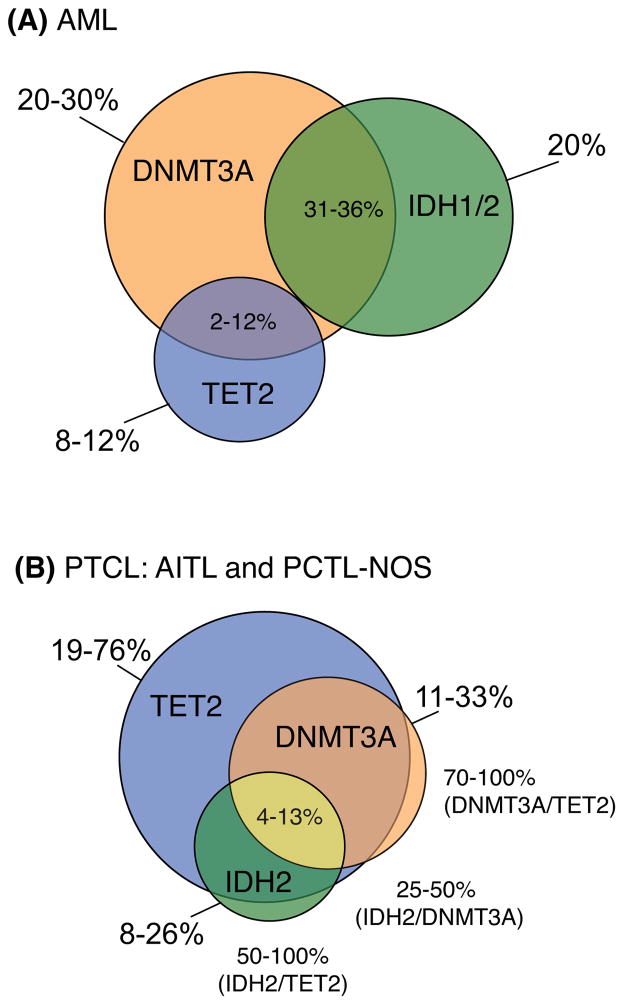
Aberrant DNA methylation is a characteristic feature of cancer including blood malignancies. Mutations in the DNA methylation regulators DNMT3A, TET1/2 and IDH1/2 are recurrent in leukemia and lymphoma. Specific and distinct DNA methylation patterns characterize subtypes of AML and lymphoma. Regulatory regions such as promoter CpG islands, CpG shores and enhancers show changes in methylation during transformation. However, the reported poor correlation between changes in methylation and gene expression in many mouse models and human studies reflects the complexity in the precise molecular mechanism for why aberrant DNA methylation promotes malignancies. This review will summarize current concepts regarding the mechanisms behind aberrant DNA methylation in hematopoietic malignancy and discuss its importance in cancer prognosis, tumor heterogeneity and relapse.

Keywords: DNA methylation; epigenetics; leukemia; lymphoma.


Figures

Figure 1
Figure 1. DNA methylation dynamics in normal and malignant hematopoiesis
A) Mammalian cells show 70–80% of global methylation in their genome. Large hypomethylated regions called “canyons” are enriched in H3K4me3 and/or H3K27me3. Edges of the canyons, especially those enriched in H3K4me3 show 5hmC marks. DNMT3A has been shown to play an important role in the maintenance of canyons. Dmnt3-null HSCs show normal global DNA methylated levels but alterations at canyon edges. Canyons occupying active chromatin (H3K4me3) usually expand and are enriched in hematopoietic stem cells genes (Meis1, Hox) that are known to be dysregulated in leukemia. Quiescent canyons (H3K27me3) do not expand with DNMT3A loss and often shrink suggesting that DNMT3B activity or other mechanisms drive the hypermethylation phenotype. DNMT3A mutant AML cells show global hypomethylation and newly formed hypomethylated valleys in their genome compared to normal cells. B) Hypomethylated areas of the genome are enriched in active regulatory elements such us enhancers and promoters. Hypomethylated CGIs and shores are enriched in active and poised genes, while hypermethylation of CGIs is related to gene silencing. Proper balance between DNMT3 and TET activity determine the DNA methylation status of CpGs. 5mC and 5hmC in genes bodies correlate to active transcription and exon splicing while 5caC interacts with RNA Pol II during elongation. C) DNMT3A mutant HSC and AML cells, display both differentially hypomethylated and hypermethylated CGIs. Hypomethylated CGIs are enriched in genes related to hematologic malignancies like Prdm16, Stat1, Ccnd1, Myc, Mn1, Msi2, Men1, Erg and Runx1. Changes in gene expression do not correlate with differentially methylated CGIs. TET2 mutant CMML patients and Tet2-null HSCs show a global increase in DNA methylation. In an AML mouse model (AML1-ETO) Tet2 depletion promotes hypermethylation of enhancers related to tumor suppressor genes. The activity of DNMT3 and TET proteins at the same genomic sites may suggest that these loci are tightly controlled by the balance of DNA methylation. The reported poor correlation between changes in methylation and gene expression in both mouse models and human samples may reflect non-coding and transcription independent roles for DNA methylation.
Figure 2
Figure 2. DNA methylation heterogeneity in mature B cell lymphoma
A) DNA methylation heterogeneity increases in germinal center B cells most likely as a result of sub-clonal expansion during an immune response. B-cell lymphomas display increased intra-tumor and inter-tumor methylation heterogeneity, ranging from hypo- to hyper-methylation when compared to normal B cells. MM shows the highest degree of DNA methylation heterogeneity compared to DLBCL and FL. B) Increased DNA methylation heterogeneity is associated with disease progression. A higher level of heterogeneity at diagnosis increases the risk of relapse. Relapsed tumors often display lower methylation heterogeneity than at diagnosis suggesting clonal evolution. A higher degree of DNA methylation heterogeneity at diagnosis may increase the probability that treatment-resistant clones can survive to promote relapse.
Figure 3
Figure 3. Co-mutation in regulators of cytosine methylation in AML and PTCL
A) In de-novo AML patients, mutation in DNMT3A is more frequent than IDH1/2 or TET2 and co-occurs with both enzymes independently, given that TET2 and IDH mutations are mutually exclusive. Mutation frequencies are shown per gene outside the venn diagram and in co-occurrence with DNMT3A within overlapping segments. B) Subsets of PTCL, in particular AITL and PCTL-NOS, have a high prevalence of TET2 mutation (>70%) that co-occurs 73–100% of the time with DNMT3A mutation. IDH1 is never seen to be mutated in these patients however IDH2 mutations readily co-occur with TET2 mutation and DNMT3A showing that co-mutation in all three genes is a distinct feature of T-cell lymphoma. Mutation frequencies are shown per gene and in co-occurrence with each other outside the venn diagram, or together in co-occurrence with TET2 mutations within the overlapping segment. Frequencies are representative of recent sequencing studies in AML [45, 63, 107, 138] and PTCL [31, 80, 98, 99, 102].

Similar articles

Cited by

References

    1. Ji H, et al. Comprehensive methylome map of lineage commitment from haematopoietic progenitors. Nature. 2010;467(7313):338–42. - PMC - PubMed
    1. Bock C, et al. DNA methylation dynamics during in vivo differentiation of blood and skin stem cells. Mol Cell. 2012;47(4):633–47. - PMC - PubMed
    1. Cedar H, Bergman Y. Epigenetics of haematopoietic cell development. Nat Rev Immunol. 2011;11(7):478–88. - PubMed
    1. Figueroa ME, et al. DNA methylation signatures identify biologically distinct subtypes in acute myeloid leukemia. Cancer Cell. 2010;17(1):13–27. - PMC - PubMed
    1. Borgel J, et al. Targets and dynamics of promoter DNA methylation during early mouse development. Nat Genet. 2010;42(12):1093–100. - PubMed

LinkOut - more resources