Clinical Trial
. 2016 Jun;24(6):1135-1149.
doi: 10.1038/mt.2016.63. Epub 2016 Mar 29.

GD2-specific CAR T Cells Undergo Potent Activation and Deletion Following Antigen Encounter but can be Protected From Activation-induced Cell Death by PD-1 Blockade

Affiliations
Clinical Trial

GD2-specific CAR T Cells Undergo Potent Activation and Deletion Following Antigen Encounter but can be Protected From Activation-induced Cell Death by PD-1 Blockade

Tessa Gargett et al. Mol Ther. 2016 Jun.

Abstract

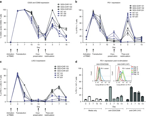
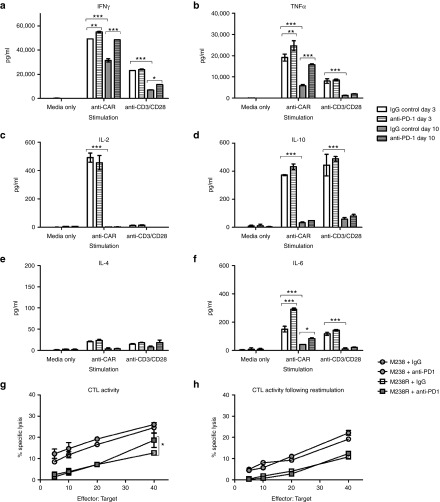
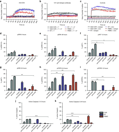
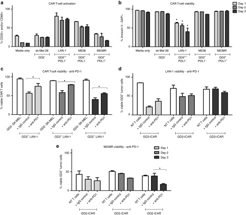
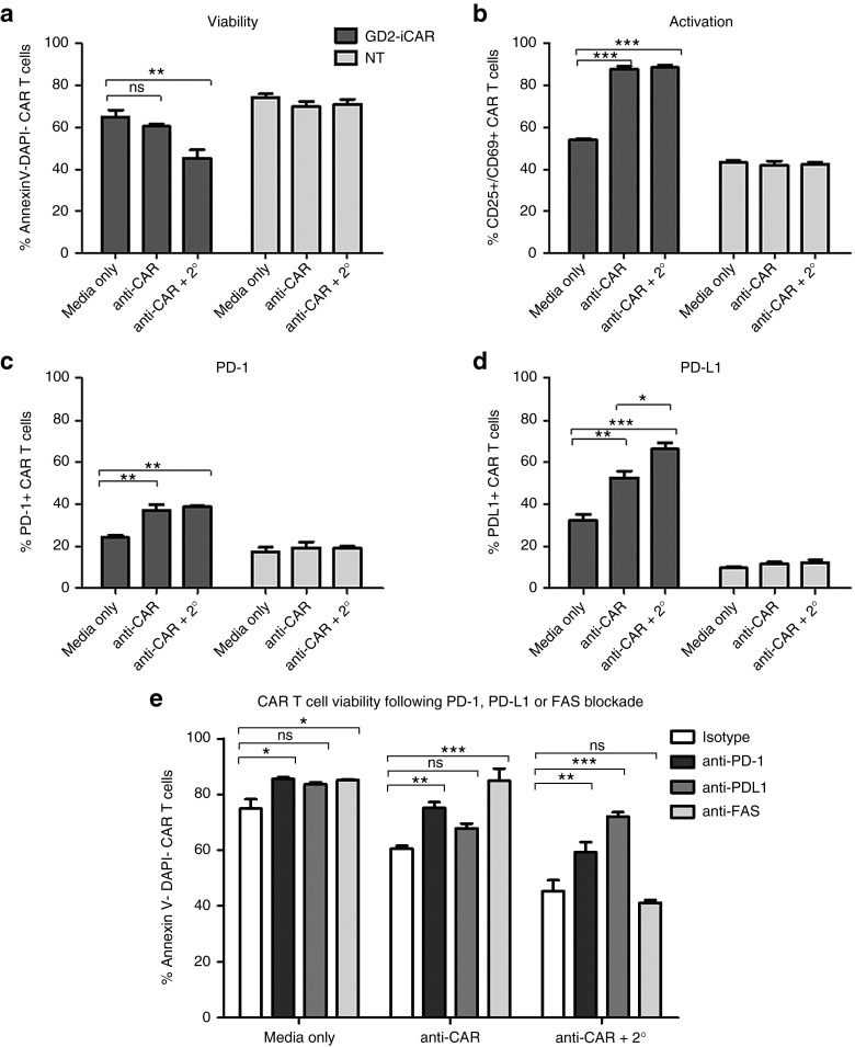
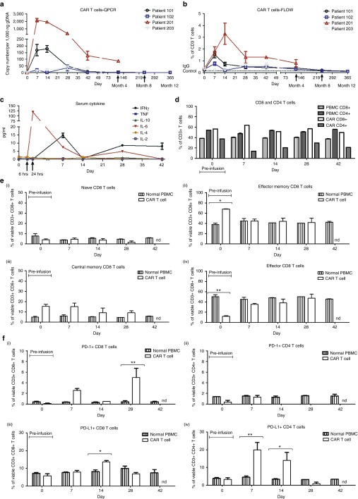
Chimeric antigen receptor (CAR) T cells have shown great promise in the treatment of hematologic malignancies but more variable results in the treatment of solid tumors and the persistence and expansion of CAR T cells within patients has been identified as a key correlate of antitumor efficacy. Lack of immunological "space", functional exhaustion, and deletion have all been proposed as mechanisms that hamper CAR T-cell persistence. Here we describe the events following activation of third-generation CAR T cells specific for GD2. CAR T cells had highly potent immediate effector functions without evidence of functional exhaustion in vitro, although reduced cytokine production reversible by PD-1 blockade was observed after longer-term culture. Significant activation-induced cell death (AICD) of CAR T cells was observed after repeated antigen stimulation, and PD-1 blockade enhanced both CAR T-cell survival and promoted killing of PD-L1(+) tumor cell lines. Finally, we assessed CAR T-cell persistence in patients enrolled in the CARPETS phase 1 clinical trial of GD2-specific CAR T cells in the treatment of metastatic melanoma. Together, these data suggest that deletion also occurs in vivo and that PD-1-targeted combination therapy approaches may be useful to augment CAR T-cell efficacy and persistence in patients.


Figures

Figure 1
Figure 1
Markers of activation and exhaustion on GD2-iCAR and paired nontransduced T cells in vitro. Patient peripheral blood mononuclear cells were activated, transduced with GD2-iCAR retroviral vector and expanded for 14 days before cryopreservation. Chimeric antigen receptor (CAR) T cells and nontransduced (NT) T-cell controls were manufactured from four patients with metastatic melanoma. Cells were then thawed and restimulated. (a) CD25 and CD69 expression during culture. (b) PD-1 expression during culture. (c) LAG-3 expression during culture. (d) Difference in PD-1 expression after restimulation with 1A7 or -anti-CD3/CD28 antibodies. Representative data are shown for patient 101. All samples were analyzed in duplicate, with graphs displaying mean ± standard error of the mean. The gating strategy is shown in Supplementary Figure S1.
Figure 2
Figure 2
Functional capacity of GD2-iCAR T cells in vitro with or without PD-1 blockade. Thawed chimeric antigen receptor (CAR) T cells were stimulated via CD3/CD28 or CAR in the absence of exogenous cytokine and in the absence (white) or presence (gray) of 20 µg/ml anti-PD-1 blocking antibody. Supernatants were collected after 3 days (day 3 plain bars) stimulation or 3 days after a restimulation on day 7 (Day 10, striped bars) and diluted 1/10 prior to analysis for cytokine secretion. (a) IFNγ. (b) TNFα. (c) IL-2. (d) IL-10. (e) IL-4. (f) IL-6. (g) CAR T-cell killing of GD2+ PD-L1- (M238, circles) and PD-L1 + (M238R, squares) melanoma cell lines ± 20 µg/ml anti-PD-1. Cells were thawed, rested overnight and then cultured with 51Cr-labeled tumor lines for 6 hours. (h) CAR T-cell killing of melanoma cell lines on day 7 after 3 days of stimulation. Cells were thawed, rested overnight and then stimulated with anti-CAR for 3 days and rested for 4 days before culture with 51Cr-labeled tumor lines for 6 hours. Flow samples were analyzed in duplicate and 51Cr assays in triplicate. Representative data are shown for patient 101, and graph display mean ± standard error of the mean. Cytokine secretion for nontransduced controls are shown in Supplementary Figure S2.
Figure 3
Figure 3
Comparison of downstream signaling events following chimeric antigen receptor (CAR) or CD3/CD28 stimulation. Calcium flux after stimulation of thawed CAR T cells with primary (1°) antibodies specific for (a) the CAR (1A7) or (b) the CD3 (OKT3) receptors, with or without cross-linking by antimouse IgG secondary (2°) antibodies. (c) Ionomycin and isotype control and a secondary antibody were used as positive and negative controls, respectively. Anti-CD28 and anti-OX40 antibodies were also used in conjunction with anti-CD3 but did not induce markedly different calcium flux compared to anti-CD3 alone (data not shown). CAR T cells, nontransduced (NT) T cells and peripheral blood mononuclear cells (PBMC) were stimulated via CAR or CD3/CD28 receptors and analyzed for signaling molecule phosphorylation. (d) and (e) pERK+ T cells at 6 and 48 hours. (f) and (g) pAKT+ T cells 12 and 24 hours. (h) pSTAT5+ T cells at 24 hours. (i) pSTAT6+ T cells at 12 hours. (j) and (k) active caspase 3 in T cells at 12 and 24 hours. CAR T cells and NT T cells and PBMC were tested in duplicate. Representative data are shown for patient 102, and graphs display mean ± standard error of the mean. The gating strategy is shown in Supplementary Figure S3.
Figure 4
Figure 4
Activation-induced cell death (AICD) after repeated antigen stimulation of GD2-iCAR T cells with GD2+ tumor cells. Thawed chimeric antigen receptor (CAR) T cells were passaged daily onto tumor cells, and cells were collected at days 1, 2, and 3 for analysis of activation (CD25 and CD69) and cell death (Annexin V and DAPI) by flow cytometry. SK-Mel cells provide a GD2- control and nontransduced (NT) T cells provided controls lacking CAR expression. Anti-PD-1was included in some cultures. CAR T-cell activation (a) and viability (b) after repeated stimulation with tumor cell lines. (c) CAR T-cell death after repeated stimulation by GD2hi neuroblastoma cell lines (LAN-1), with or without anti-PD-1. (d) CAR T-cell killing of GD2hi PD-L1- neuroblastoma cells (LAN-1), with or without anti-PD-1. (e) CART-cell killing of GD2+PD-L1+ melanoma cells (M238R), with or without anti-PD-1. Repetitive antigen stress-test assays were performed in duplicate. Representative data are shown for patient 101, and graphs display mean ± standard error of the mean. The gating strategy is shown in Supplementary Figure S4.
Figure 5
Figure 5
PD-1/PD-L1-dependent activation-induced cell death (AICD) after activation of GD2-iCAR T cells. (a) PD-L1 expression on thawed chimeric antigen receptor (CAR) T cells after stimulation in vitro. (b) Immune phenotype of CAR PD-L1+ and PD-L1- T cells at day 3 poststimulation. Immune phenotype defined as: Naive (CD45RA+CCR7+), effector memory (Tem; ––CD45RACCR7), central memory (Tcm; CD45RACCR7+), effector (CD45RA+CCR7), activated (CD25+/CD69+), proliferating (CFSE low). (c) CAR T-cell viability after repetitive stimulation with LAN-1. (d) GD2+PD-L1- M238 tumor cell viability and (e) GD2++PD-L1- LAN-1 tumor cell viability and (f) GD2+PD-L1+ M238R tumor cell viability after stress-test assay with PD-L1 blockade (anti-PD-L1). Repetitive antigen stress-test assays and flow cytometry analyses were performed in duplicate. Representative data are shown for patient 101, and graphs display mean ± stanadard error of the mean. The gating strategy is shown in Supplementary Figure S5.
Figure 6
Figure 6
Chimeric antigen receptor (CAR) cross-linking also induces activation-induced cell death. (a) Viability, (b) activation, (c) PD-1, and (d) PD-L1 expression after CAR cross-linking with 1A7 and secondary antibody (2°) of thawed CAR T cells and nontransduced (NT) controls. (e) GD2-iCAR T-cell AICD in the presence of PD-1-, PD-L1-, or FAS-blocking antibodies after CAR cross-linking. Cross-linking assays and flow cytometry analyses were performed in duplicate. Representative data are shown for patient 101, and graphs display mean ± stanadard error of the mean. The gating strategy is shown in Supplementary Figure S5.
Figure 7
Figure 7
Chimeric antigen receptor (CAR) T-cell persistence and immune phenotype in patients receiving third-generation GD2-specific CAR T cells for the treatment of V600 BRAF-mutant metastatic melanoma. (a) Flow cytometry using 1A7 to detect GD2-iCAR-expressing T cells in peripheral blood. (b) Quantitative polymerase chain reaction (QPCR) to detect GD2-iCAR transgene-positive cells in peripheral blood. (c) Patient 102 serum cytokine levels. (d) Patient 102 CD4 and CD8 CAR T-cell subsets for peripheral blood mononuclear cells, the GD2-iCAR T-cell product and circulating GD2-iCAR T cells detected from day 7 postadministration. (e) Patient 102 CD8+ T-cell immune phenotype and (f) PD1/PD-L1 expression on peripheral blood normal T cells and 1A7+ GD2-iCAR T cells. Experiments were performed in duplicate (flow cytometry) or triplicate (QPCR), with graphs displaying mean ± standard error of the mean. Gating strategy and data for Patients 101, 201 and 203 are shown in Supplementary Figure S6.

References

    1. Porter, DL, Levine, BL, Kalos, M, Bagg, A and June, CH (2011). Chimeric antigen receptor-modified T cells in chronic lymphoid leukemia. N Engl J Med 365: 725–733. - PMC - PubMed
    1. Grupp, SA, Kalos, M, Barrett, D, Aplenc, R, Porter, DL, Rheingold, SR et al. (2013). Chimeric antigen receptor-modified T cells for acute lymphoid leukemia. N Engl J Med 368: 1509–1518. - PMC - PubMed
    1. Kershaw, MH, Westwood, JA, Parker, LL, Wang, G, Eshhar, Z, Mavroukakis, SA et al. (2006). A phase I study on adoptive immunotherapy using gene-modified T cells for ovarian cancer. Clin Cancer Res 12(20 Pt 1): 6106–6115. - PMC - PubMed
    1. Louis, CU, Savoldo, B, Dotti, G, Pule, M, Yvon, E, Myers, GD et al. (2011). Antitumor activity and long-term fate of chimeric antigen receptor-positive T cells in patients with neuroblastoma. Blood 118: 6050–6056. - PMC - PubMed
    1. Lamers, CH, Sleijfer, S, van Steenbergen, S, van Elzakker, P, van Krimpen, B, Groot, C et al. (2013). Treatment of metastatic renal cell carcinoma with CAIX CAR-engineered T cells: clinical evaluation and management of on-target toxicity. Mol Ther 21: 904–912. - PMC - PubMed

Publication types