

Functions and importance of mycobacterial extracellular vesicles
- PMID: 27020292
- PMCID: PMC4879809
- DOI: 10.1007/s00253-016-7484-x
Functions and importance of mycobacterial extracellular vesicles
Abstract
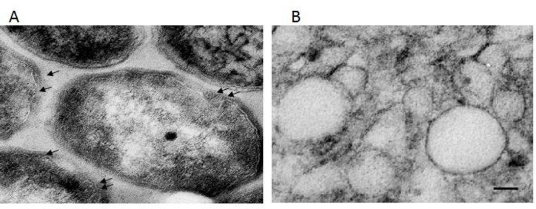
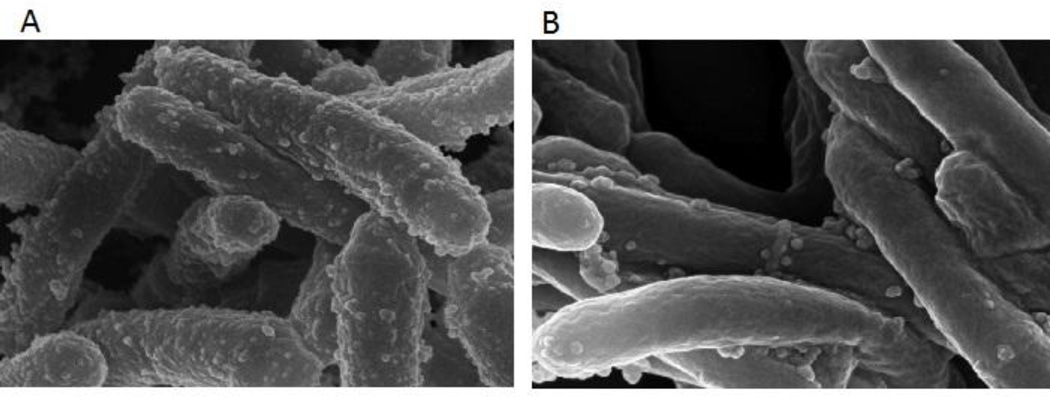
The release of cellular factors by means of extracellular vesicles (EVs) is conserved in archaea, bacteria, and eukaryotes. EVs are released by growing bacteria as part of their interaction with their environment and, for pathogenic bacteria, constitute an important component of their interactions with the host. While EVs released by gram-negative bacteria have been extensively studied, the vesicles released by thick cell wall microorganisms like mycobacteria were recognized only recently and are less well understood. Nonetheless, studies of mycobacterial EVs have already suggested roles in pathogenesis, opening exciting new avenues of research aimed at understanding their biogenesis and potential use in antitubercular strategies. In this minireview, we discuss the discovery of mycobacterial vesicles, the current understanding of their nature, content, regulation, and possible functions, as well as their potential therapeutic applications.
Keywords: Iron; Lipoproteins; Mycobacterium; Siderophores; Vaccines; Vesicles.
Conflict of interest statement
Figures
References
-
- Basaraba RJ, Bielefeldt-Ohmann H, Eschelbach EK, Reisenhauer C, Tolnay AE, Taraba LC, Shanley CA, Smith EA, Bedwell CL, Chlipala EA, Orme IM. Increased expression of host iron-binding proteins precedes iron accumulation and calcification of primary lung lesions in experimental tuberculosis in the guinea pig. Tuberculosis (Edinb) 2008;88:69–79. - PMC - PubMed
-
- Cairo G, Recalcati S, Mantovani A, Locati M. Iron trafficking and metabolism in macrophages: contribution to the polarized phenotype. Trends in immunology. 2011;32:241–247. - PubMed
-
- Ciofu O, Beveridge TJ, Kadurugamuwa J, Walther-Rasmussen J, Hoiby N. Chromosomal beta-lactamase is packaged into membrane vesicles and secreted from Pseudomonas aeruginosa. J Antimicrob Chemother. 2000;45:9–13. - PubMed
-
- Daffe M. The cell envelope of tubercle bacilli. Tuberculosis (Edinb) 2015;95(Suppl 1):S155–S158. - PubMed
Publication types
MeSH terms
Substances
Grants and funding
LinkOut - more resources
Full Text Sources
Other Literature Sources
