Fluorescence guided surgery and tracer-dose, fact or fiction?
- PMID: 27020580
- PMCID: PMC4969335
- DOI: 10.1007/s00259-016-3372-y
Fluorescence guided surgery and tracer-dose, fact or fiction?
Abstract
Introduction: Fluorescence guidance is an upcoming methodology to improve surgical accuracy. Challenging herein is the identification of the minimum dose at which the tracer can be detected with a clinical-grade fluorescence camera. Using a hybrid tracer such as indocyanine green (ICG)-(99m)Tc-nanocolloid, it has become possible to determine the accumulation of tracer and correlate this to intraoperative fluorescence-based identification rates. In the current study, we determined the lower detection limit of tracer at which intraoperative fluorescence guidance was still feasible.
Methods: Size exclusion chromatography (SEC) provided a laboratory set-up to analyze the chemical content and to simulate the migratory behavior of ICG-nanocolloid in tissue. Tracer accumulation and intraoperative fluorescence detection findings were derived from a retrospective analysis of 20 head-and-neck melanoma patients, 40 penile and 20 prostate cancer patients scheduled for sentinel node (SN) biopsy using ICG-(99m)Tc-nanocolloid. In these patients, following tracer injection, single photon emission computed tomography fused with computed tomography (SPECT/CT) was used to identify the SN(s). The percentage injected dose (% ID), the amount of ICG (in nmol), and the concentration of ICG in the SNs (in μM) was assessed for SNs detected on SPECT/CT and correlated with the intraoperative fluorescence imaging findings.
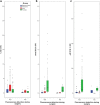
Results: SEC determined that in the hybrid tracer formulation, 41 % (standard deviation: 12 %) of ICG was present in nanocolloid-bound form. In the SNs detected using fluorescence guidance a median of 0.88 % ID was present, compared to a median of 0.25 % ID in the non-fluorescent SNs (p-value < 0.001). The % ID values could be correlated to the amount ICG in a SN (range: 0.003-10.8 nmol) and the concentration of ICG in a SN (range: 0.006-64.6 μM).
Discussion: The ability to provide intraoperative fluorescence guidance is dependent on the amount and concentration of the fluorescent dye accumulated in the lesion(s) of interest. Our findings indicate that intraoperative fluorescence detection with ICG is possible above a μM concentration.
Keywords: Fluorescence-guided surgery; Microdosing; Multimodal; SPECT/CT; Sentinel node.
Figures


Similar articles
-
Multispectral Fluorescence Imaging During Robot-assisted Laparoscopic Sentinel Node Biopsy: A First Step Towards a Fluorescence-based Anatomic Roadmap.Eur Urol. 2017 Jul;72(1):110-117. doi: 10.1016/j.eururo.2016.06.012. Epub 2016 Jun 23. Eur Urol. 2017. PMID: 27345689
-
Hybrid Indocyanine Green-99mTc-nanocolloid for Single-photon Emission Computed Tomography and Combined Radio- and Fluorescence-guided Sentinel Node Biopsy in Penile Cancer: Results of 740 Inguinal Basins Assessed at a Single Institution.Eur Urol. 2020 Dec;78(6):865-872. doi: 10.1016/j.eururo.2020.09.007. Epub 2020 Sep 17. Eur Urol. 2020. PMID: 32950298
-
A hybrid radioactive and fluorescent tracer for sentinel node biopsy in penile carcinoma as a potential replacement for blue dye.Eur Urol. 2014 Mar;65(3):600-9. doi: 10.1016/j.eururo.2013.11.014. Epub 2013 Nov 26. Eur Urol. 2014. PMID: 24355132
-
[Sentinel node in melanoma and breast cancer. Current considerations].Rev Esp Med Nucl Imagen Mol. 2015 Jan-Feb;34(1):30-44. doi: 10.1016/j.remn.2014.09.006. Epub 2014 Nov 7. Rev Esp Med Nucl Imagen Mol. 2015. PMID: 25455506 Review. Spanish.
-
Fluorescent-guided surgery for sentinel lymph node detection in gastric cancer and carcinoembryonic antigen targeted fluorescent-guided surgery in colorectal and pancreatic cancer.J Surg Oncol. 2018 Aug;118(2):315-323. doi: 10.1002/jso.25139. Epub 2018 Sep 14. J Surg Oncol. 2018. PMID: 30216455 Free PMC article. Review.
Cited by
-
Intraoperative near-infrared imaging can identify canine mammary tumors, a spontaneously occurring, large animal model of human breast cancer.PLoS One. 2020 Jun 17;15(6):e0234791. doi: 10.1371/journal.pone.0234791. eCollection 2020. PLoS One. 2020. PMID: 32555698 Free PMC article.
-
Robot-assisted laparoscopic surgery using DROP-IN radioguidance: first-in-human translation.Eur J Nucl Med Mol Imaging. 2019 Jan;46(1):49-53. doi: 10.1007/s00259-018-4095-z. Epub 2018 Jul 27. Eur J Nucl Med Mol Imaging. 2019. PMID: 30054696 Free PMC article.
-
Multi-wavelength fluorescence imaging with a da Vinci Firefly-a technical look behind the scenes.J Robot Surg. 2021 Oct;15(5):751-760. doi: 10.1007/s11701-020-01170-8. Epub 2020 Nov 11. J Robot Surg. 2021. PMID: 33179201 Free PMC article.
-
Click Chemistry in the Design and Production of Hybrid Tracers.ACS Omega. 2019 Jul 22;4(7):12438-12448. doi: 10.1021/acsomega.9b01484. eCollection 2019 Jul 31. ACS Omega. 2019. PMID: 31460363 Free PMC article.
-
Click-on fluorescence detectors: using robotic surgical instruments to characterize molecular tissue aspects.J Robot Surg. 2023 Feb;17(1):131-140. doi: 10.1007/s11701-022-01382-0. Epub 2022 Apr 9. J Robot Surg. 2023. PMID: 35397108 Free PMC article.
References
-
- ICG Fluorescence Imaging and Navigation Surgery - Mitsuo Kusano, Norihiro Kokudo, Masakazu Toi, Masaki Kaibori ISBN 978-4-431-55528-5
-
- Vermeeren L, Muller SH, Meinhardt W, Valdés Olmos RA. Optimizing the colloid particle concentration for improved preoperative and intraoperative image-guided detection of sentinel nodes in prostate cancer. Eur J Nucl Med Mol Imaging. 2010;37(7):1328–34. doi: 10.1007/s00259-010-1410-8. - DOI - PubMed
-
- Manny TB, Patel M, Hemal AK. Fluorescence-enhanced robotic radical prostatectomy using real-time lymphangiography and tissue marking with percutaneous injection of unconjugated indocyanine green: the initial clinical experience in 50 patients. Eur Urol. 2014;65(6):1162–8. doi: 10.1016/j.eururo.2013.11.017. - DOI - PubMed
MeSH terms
Substances
LinkOut - more resources
Full Text Sources
Other Literature Sources

