. 2016 Aug;23(8):2586-95.
doi: 10.1245/s10434-016-5186-3. Epub 2016 Mar 28.

(Near-Infrared) Fluorescence-Guided Surgery Under Ambient Light Conditions: A Next Step to Embedment of the Technology in Clinical Routine

Affiliations

(Near-Infrared) Fluorescence-Guided Surgery Under Ambient Light Conditions: A Next Step to Embedment of the Technology in Clinical Routine

Nynke S van den Berg et al. Ann Surg Oncol. 2016 Aug.

Abstract

Background and purpose: In open surgery procedures, after temporarily dimming the lights in the operation theatre, the Photo Dynamic Eye (PDE) fluorescence camera has, amongst others, been used for fluorescence-guided sentinel node (SN) biopsy procedures. To improve the clinical utility and logistics of fluorescence-guided surgery, we developed and evaluated a prototype modified PDE (m-PDE) fluorescence camera system.

Methods: The m-PDE works under ambient light conditions and includes a white light mode and a pseudo-green-colored fluorescence mode (including a gray-scaled anatomical background). Twenty-seven patients scheduled for SN biopsy for (head and neck) melanoma (n = 16), oral cavity (n = 6), or penile (n = 5) cancer were included. The number and location of SNs were determined following an indocyanine green-(99m)Tc-nanocolloid injection and preoperative imaging. Intraoperatively, fluorescence guidance was used to visualize the SNs. The m-PDE and conventional PDE were compared head-to-head in a phantom study, and in seven patients. In the remaining 20 patients, only the m-PDE was evaluated.

Results: Phantom study: The m-PDE was superior over the conventional PDE, with a detection sensitivity of 1.20 × 10(-11) M (vs. 3.08 × 10(-9) M) ICG in human serum albumin. In the head-to-head clinical comparison (n = 7), the m-PDE was also superior: (i) SN visualization: 100 versus 81.4 %; (ii) transcutaneous SN visualization: 40.7 versus 22.2 %; and (iii) lymphatic duct visualization: 7.4 versus 0 %. Findings were further underlined in the 20 additionally included patients.

Conclusion: The m-PDE enhanced fluorescence imaging properties compared with its predecessor, and provides a next step towards routine integration of real-time fluorescence guidance in open surgery.


Figures
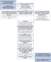
Fig. 1
Fig. 1
Workflow for sentinel node localization and excision. Following preoperative image analysis by the surgeon to virtually determine the location of the SNs (1), blue dye can be injected (2). Prior to incision a portable gamma camera (Sentinella; Oncovision, Valencia, Spain), a gamma probe (Neoprobe; Johnson & Johnson Medical, Hamburg, Germany), and the fluorescence camera (c-PDE or m-PDE; Hamamatsu Photonics K.K., Hamamatsu, Japan) are use to determine the location of the SNs (3). After incision (4) the SN is pursued via gamma tracing, after which alternating attempts were made to visualize the SN via fluorescence imaging and, when applicable, blue-dye visualization (5). After identification of the SN, the node was excised, after which the wound bed was checked for the presence of residual radioactivity/remaining fluorescence activity at the site of a previously excised SN. Additionally excised nodes were considered part of a cluster of multiple adjacent SNs (6). Following completion of SN biopsy via the combined radio- and fluorescence-guided (and, when applicable, blue dye) approach, the wound-bed was palpated for the presence of suspicious non-radioactive, non-fluorescent and, when applicable, non-blue-dye-stained lymph nodes (8). Thereafter the wound bed was closed (9). SN sentinel node, PDE Photo Dynamic Eye
Fig. 2
Fig. 2
Determination of the sensitivity of the m-PDE and c-PDE fluorescence camera systems for ICG–HSA. (a) Fluorescence intensity curve of the various steps of the dilution range measured with the IVIS Spectrum. (b) Visual fluorescence images obtained with the IVIS Spectrum, c-PDE, and m-PDE when measured in full darkness, with all lights in the operating room turned on (satellite lamps, plenum, and surrounding lights), and with the satellite lamps directly lighting the sterile field turned off, but the plenum and surrounding lights on. (c) Light spectrum of the lamps present in the operating room. The light blue area shows the area in which ICG emits its light. (d) Absorption and emission spectrum of 1.50 × 10−9 M ICG–HSA. ICG indocyanine green, HSA human serum albumin, PDE Photo Dynamic Eye
Fig. 3
Fig. 3
Fluorescence-guided sentinel node excision in a patient with a melanoma of the neck. (a) Preoperative imaging. Left Static lymphoscintigram acquired 2 h after hybrid tracer injection showing only the IS. Middle Following fusion of the acquired SPECT and CT images, a 3D volume rendering was generated showing the injection site, as well as an SN in level IV (white arrow) and a supraclavicular SN. Right Axial fused SPECT/CT (left) and CT (right) slice showing the SN in level IV being part of a cluster (indicated because no clear node could be identified on the CT, only a strand of tissue). (b) After re-excision of the melanoma scar, the SN cluster in level IV was pursued via fluorescence imaging using the m-PDE fluorescence camera. The timeline shows fluorescence-guided excision of this cluster of SNs. Switching between the fluorescence and white light image allowed the surgeon to work under continuous fluorescence guidance. A total of three fluorescent (and radioactive) SNs were removed from the area where the hotspot was seen on SPECT/CT imaging. IS injection site, SN sentinel node, SPECT/CT single photon emission computed tomography combined with computed tomography, 3D three-dimensional

References

    1. Crane LM, Themelis G, Arts HJ, Buddingh KT, Brouwers AH, Ntziachristos V, et al. Intraoperative near-infrared fluorescence imaging for sentinel lymph node detection in vulvar cancer: first clinical results. Gynecol Oncol. 2011;120(2):291–295. doi: 10.1016/j.ygyno.2010.10.009. - DOI - PubMed
    1. Mieog JS, Troyan SL, Hutteman M, Donohoe KJ, van der Vorst JR, Stockdale A, et al. Toward optimization of imaging system and lymphatic tracer for near-infrared fluorescent sentinel lymph node mapping in breast cancer. Ann Surg Oncol. 2011;18(9):2483–2491. doi: 10.1245/s10434-011-1566-x. - DOI - PMC - PubMed
    1. Kusano M, Kokudo N, Toi M, Kaibori M (eds). ICG fluorescence imaging and navigation surgery. New York: Springer; 2016.
    1. KleinJan GH, van den Berg NS, Brouwer OR, de Jong J, Acar C, Wit EM, et al. Optimisation of fluorescence guidance during robot-assisted laparoscopic sentinel node biopsy for prostate cancer. Eur Urol. 2014;66(6):991–998. doi: 10.1016/j.eururo.2014.07.014. - DOI - PubMed
    1. Brouwer OR, Klop WM, Buckle T, Vermeeren L, van den Brekel MW, Balm AJ, et al. Feasibility of sentinel node biopsy in head and neck melanoma using a hybrid radioactive and fluorescent tracer. Ann Surg Oncol. 2012;19(6):1988–1994. doi: 10.1245/s10434-011-2180-7. - DOI - PMC - PubMed

Publication types

MeSH terms

Substances

LinkOut - more resources