. 2016 Aug;229(2):314-25.
doi: 10.1111/joa.12465. Epub 2016 Mar 29.

Morphogenesis of myocardial trabeculae in the mouse embryo

Affiliations

Morphogenesis of myocardial trabeculae in the mouse embryo

Gabriella Captur et al. J Anat. 2016 Aug.

Abstract

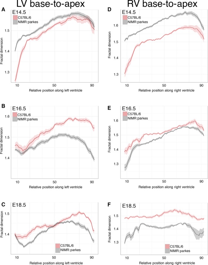
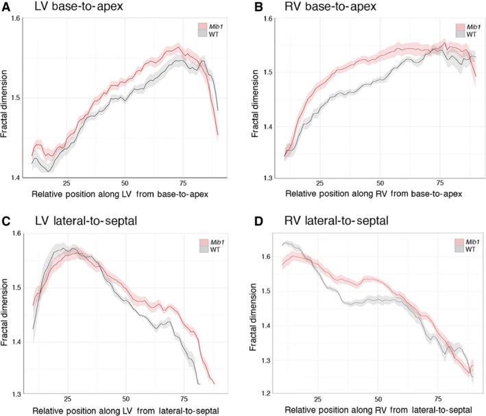
Formation of trabeculae in the embryonic heart and the remodelling that occurs prior to birth is a conspicuous, but poorly understood, feature of vertebrate cardiogenesis. Mutations disrupting trabecular development in the mouse are frequently embryonic lethal, testifying to the importance of the trabeculae, and aberrant trabecular structure is associated with several human cardiac pathologies. Here, trabecular architecture in the developing mouse embryo has been analysed using high-resolution episcopic microscopy (HREM) and three-dimensional (3D) modelling. This study shows that at all stages from mid-gestation to birth, the ventricular trabeculae comprise a complex meshwork of myocardial strands. Such an arrangement defies conventional methods of measurement, and an approach based upon fractal algorithms has been used to provide an objective measure of trabecular complexity. The extent of trabeculation as it changes along the length of left and right ventricles has been quantified, and the changes that occur from formation of the four-chambered heart until shortly before birth have been mapped. This approach not only measures qualitative features evident from visual inspection of 3D models, but also detects subtle, consistent and regionally localised differences that distinguish each ventricle and its developmental stage. Finally, the combination of HREM imaging and fractal analysis has been applied to analyse changes in embryonic heart structure in a genetic mouse model in which trabeculation is deranged. It is shown that myocardial deletion of the Notch pathway component Mib1 (Mib1(flox/flox) ; cTnT-cre) results in a complex array of abnormalities affecting trabeculae and other parts of the heart.

Keywords: cardiac embryology; cardiogenesis; developmental biology; non-compaction cardiomyopathy.


Figures

Figure 1
Figure 1
Three‐dimensional trabecular architecture in the early mouse embryo heart. (A) Thoracic region of an E9.5 embryo (ventral view), reconstructed by volume rendering from HREM data. The ballooning left and right ventricular chambers (LV and RV) are clearly visible through the thin pericardium, as is the outflow tract (OT). The plane of erosion for (B) is shown (red box). Scale bar: 200 μm. (B) Erosion from the dorsal side reveals a mesh‐like network of trabeculae throughout the common ventricular chamber, which contrasts with the smooth luminal surface of the outflow tract (yellow arrow). The trabeculae appear raised and more closely interconnected at the site of the future IVS (yellow box). Scale bar: 200 μm. (C and D) Three‐dimensional models of the heart at E10.5 and E11.5, respectively. Ventro‐inferior erosion through the ventricular chambers towards the atrioventricular valve reveals progressive coalescence of trabeculae to form the base of the IVS (boxed). Scale bar: 200 μm.
Figure 2
Figure 2
Changes in trabecular morphology during embryo development. Three‐dimensional models from HREM data for isolated hearts at successive stages of embryonic development. Models have been eroded in the short axis to provide views of the trabecular meshwork in the apex of the ventricles. Erosion in the plane of the IVS provides views of the left and right ventricular (LV and RV) chambers. Scale bars: 200 μm (E10.5); 500 μm (E18.5).
Figure 3
Figure 3
Trabeculae support the developing papillary muscles. Three‐dimensional models digitally eroded along the indicated planes (yellow) to view papillary muscles (P) of the LV at E14.5, E16.5 and E18.5 (views from the right; regions enlarged are shown in red). The trabecular mesh merges to form the roots of the developing papillary muscles, and this intimate arrangement is maintained throughout embryonic development. Scale bar: 500 μm (E18.5).
Figure 4
Figure 4
Fractal‐based quantification of trabecular complexity during normal development. The FD was calculated from HREM image data obtained with the outbred mouse strain, NIMR:Parkes. Fractal analysis is a method of quantifying complex geometric patterns in biological structures. The resulting FD is a unitless measure index of how completely the object fills space, increasing with increased structural complexity. FD was measured using a box‐counting method on consecutive HREM slices of the embryo hearts in two orientations: base‐to‐apex and lateral‐to‐septal. (A) Base‐to‐apex profile of LV and RV trabecular development (pink and grey, respectively) at stages E14.5 (LV: n = 15; RV: n = 11); E16.5 (LV: n = 13; RV: n = 11) and E18.5 (LV: n = 14; RV: n = 12). The fractal signatures of the morphological RV and LV are relatively similar at E14.5 (P = 0.951), but their patterns diverge as development proceeds, and by E18.5 are markedly different (P < 0.0001). (B) The LV and RV chambers from E16.5 (n = 23) and E18.5 hearts (n = 20) were digitally resliced in an orthogonal plane, from lateral‐to‐septal surface. At E16.5 and E18.5 note the differing lateral‐to‐septal patterns of FD between LV and RV (pink and grey, respectively) and the consistently higher FD value in the lateral wall of the RV compared with the equivalent position in the LV (P < 0.0001 both stages). (Solid lines: mean FD value; shaded ribbons: 95% confidence interval. Only FD values for slices from 10% to 90% of Base‐to‐apex or lateral‐to‐septal axis are shown.)
Figure 5
Figure 5
Strain differences in trabeculation detected by fractal analysis. Comparison of base‐to‐apex fractal profile of LV and RV trabeculae between the outbred strain, NIMR:Parkes (grey) and the inbred strain, C57BL/6 (pink). (A–C) LV data: E14.5 (n = 28); E16.5 (n = 21); E18.5 (n = 24). (D–F) RV data: E14.5 (n = 19); E16.5 (n = 18); E18.5 (n = 18). Also see Fig. S3. (Solid lines: mean FD value; shaded ribbons: 95% confidence interval. Only FD values for slices from 10% to 90% of base‐to‐apex are shown.)
Figure 6
Figure 6
Morphological abnormalities in the Mib1 mutant heart. Three‐dimensional volume renderings of E16.5 Mib1 mutant and wild‐type, sibling hearts. Models are eroded from the anterior (A and C) to give a four‐chamber view, and from the dorsal side (B and D) at the level of the developing aortic valve. Regions highlighted are shown (red boxes). Note the grossly aberrant trabecular arrangement, thin ventricular wall and ventricular septal defect (arrow) in the Mib1 mutant (C). Wild‐type left and right atria have normal pectinate muscle morphology, while there is near‐complete effacement in Mib1 mutants (compare A with C, highlighted). The developing aortic valve in the Mib1 mutant is abnormal, in this case the left and non‐coronary leaflets (highlight D, asterisks) lying well above a reduced sized right leaflet. Compare with the normal trifoliate (left, right and non‐coronary) leaflet arrangement (highlight B, labels L, R and N, respectively). Scale bar: 500 μm (E16.5).
Figure 7
Figure 7
Fractal analysis of Mib1 mutant hearts. Base‐to‐apex and lateral‐to‐septal fractal dimension (FD) plots comparing E16.5 Mib1 mutant (Mib1 flox/flox; cTnT‐cre) hearts (pink) with wild‐type littermates (grey). Note that for both LV and RV, fractal analysis detects significant and consistent patterns of variation between mutant and control hearts. Also see Fig. S5. (Solid lines, mean; shaded ribbons, 95% confidence intervals).

References

    1. Axel L (2004) Papillary muscles do not attach directly to the solid heart wall. Circulation 109, 3145–3148. - PubMed
    1. Captur G, Lopes LR, Patel V, et al. (2014) Abnormal cardiac formation in hypertrophic cardiomyopathy: fractal analysis of trabeculae and preclinical gene expression. Circ Cardiovasc Genet 7, 241–248. - PubMed
    1. Captur G, Karperien AL, Li C, et al. (2015) Fractal frontiers in CMR: an invitation toward clinical implementation. J Cardiovasc Magn Reson 19, 1–10. - PMC - PubMed
    1. Carlhäll CJ, Bolger A (2010) Passing strange: flow in the failing ventricle. Circ Heart Fail 3, 326–331. - PubMed
    1. Filipoiu FM(2014) Atlas of Heart Anatomy and Development. Springer‐Verlag London: Springer Science & Business Media.

Publication types