Review
. 2016 Sep:150:149-65.
doi: 10.1016/j.exer.2016.03.018. Epub 2016 Mar 26.

Retinal remodeling in human retinitis pigmentosa

Affiliations
Review

Retinal remodeling in human retinitis pigmentosa

B W Jones et al. Exp Eye Res. 2016 Sep.

Abstract

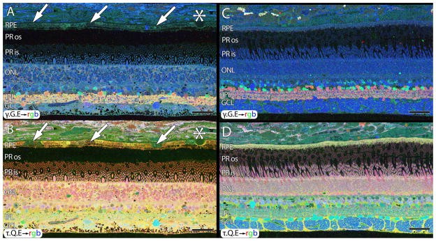
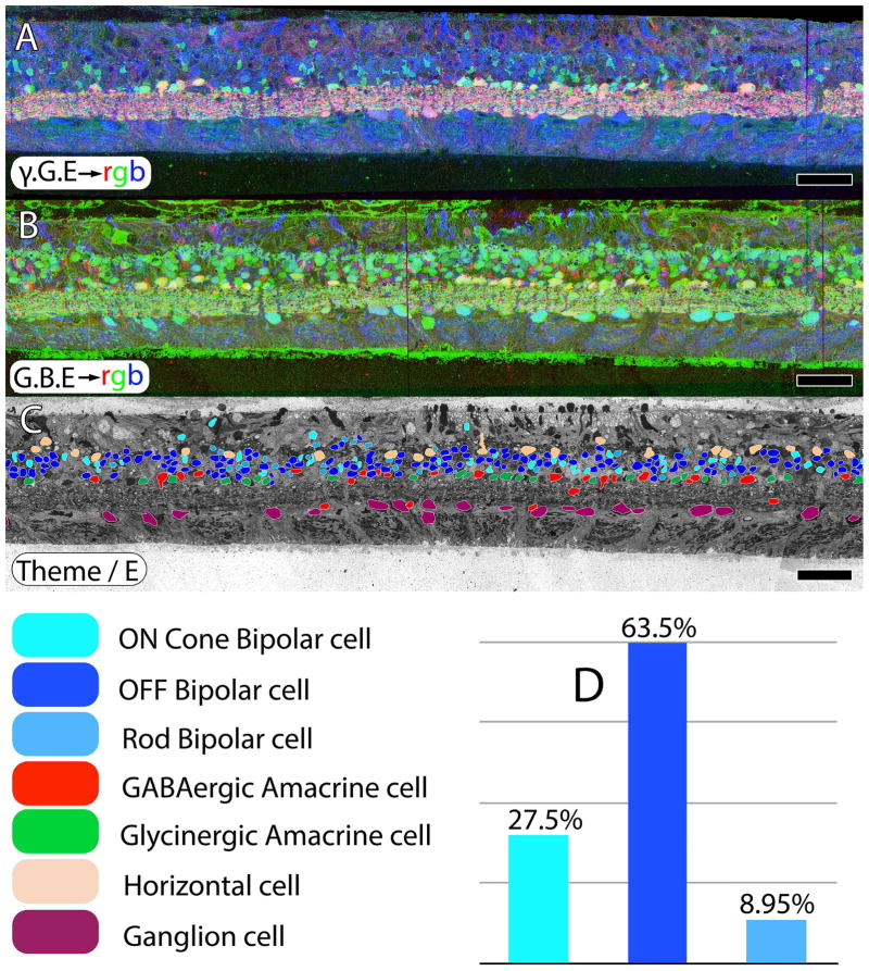
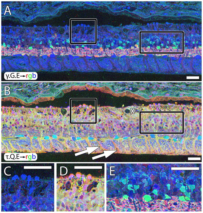
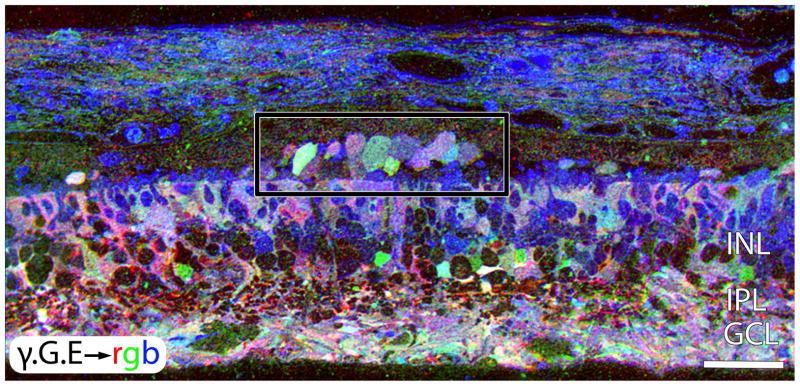
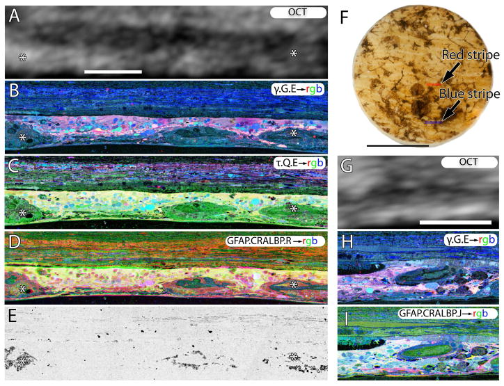
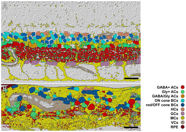
Retinitis Pigmentosa (RP) in the human is a progressive, currently irreversible neural degenerative disease usually caused by gene defects that disrupt the function or architecture of the photoreceptors. While RP can initially be a disease of photoreceptors, there is increasing evidence that the inner retina becomes progressively disorganized as the outer retina degenerates. These alterations have been extensively described in animal models, but remodeling in humans has not been as well characterized. This study, using computational molecular phenotyping (CMP) seeks to advance our understanding of the retinal remodeling process in humans. We describe cone mediated preservation of overall topology, retinal reprogramming in the earliest stages of the disease in retinal bipolar cells, and alterations in both small molecule and protein signatures of neurons and glia. Furthermore, while Müller glia appear to be some of the last cells left in the degenerate retina, they are also one of the first cell classes in the neural retina to respond to stress which may reveal mechanisms related to remodeling and cell death in other retinal cell classes. Also fundamentally important is the finding that retinal network topologies are altered. Our results suggest interventions that presume substantial preservation of the neural retina will likely fail in late stages of the disease. Even early intervention offers no guarantee that the interventions will be immune to progressive remodeling. Fundamental work in the biology and mechanisms of disease progression are needed to support vision rescue strategies.

Keywords: Amacrine cell; Bipolar cell; Computational molecular phenotyping (CMP); Cone photoreceptor; Ganglion cell; Müller cell; Neural remodeling; Retina; Retinal pigment epithelium (RPE); Retinal remodeling; Retinitis pigmentosa (RP); Rod photoreceptor.


Figures

Figure 1
Figure 1
CMP of 77 year old normal human male (A , B) and primate (C,D). A. γ.G.E → rgb mapping of human peripheral retina from a 77 year old male. Inhomogeneity in the small molecule signals of the RPE indicated by arrows in B. Small druse, asterisk. Mild elevation of GABA in the Müller cells. B. τ.Q.E rgb mapping reveals Müller cells in yellow. Mild postmortem elevations of glutamine and taurine in Müller cells. RPE heterogeneity (arrows). C. Normal macaque retina, γ.G.E → rgb mapping. No RPE heterogeneity. No elevation of GABA in Müller cells. D. Normal macaque retina, τ.G.E → rgb mapping. No RPE heterogeneity. No abnormal elevation of glutamine or taurine in Müller cells. Scale = 40 μm.
Figure 2
Figure 2
Fundoscopic images of a 67 year old normal human retina (HS1) in A and a fundoscopic image from an RP donor (RP1), then aged 58 when imaged in 1991 in B, 20 years prior to death. The fundoscopic image of RP1 demonstrates a pale fundus, optic nerve atrophy, and vessel attenuation. This retina provided the tissue for sample RP1.
Figure 3
Figure 3
Backlit extended depth-of-focus view of sample RP2 retina, visualizing pigmented bone spicules invading the neural retina. Spicules appear to fill the spaces between cells and function as highways or facilitated pathways for migration of other cell classes that ultimately alter the topology throughout the retina. Scale = 200 μm.
Figure 4
Figure 4
A. Hypertrophic blood vessel adjacent to the inner limiting membrane invested by pigmented RPE processes.Scale = 15 μm B. RPE pigment granules deep within the neural retina in the former ganglion cell layer, Scale = 3 μm. C. A remnant RPE cell invading the neural retina. Scale = 9 μm. D. An inset from C showing detail of the pigment granules and intracellular debris, pigment granules surrounding vascular elements. Scale = 2 μm.
Figure 5
Figure 5
CMP and excitation mapping in human sample RP2. A. γ.G.E → rgb mapping. B. G.AGB.E→ mapping. The AGB signal is induced by activating iGluRs with 25 μM kainic acid. C. Classified retinal neurons, revealing ON cone bipolar, OFF bipolar, rod bipolar, GABAergic amacrine, glycinergic amacrine, horizontal and ganglion cells. Retinal bipolar cell classes in normal retina should approximate 33% each for ON, OFF and rod bipolar cell classes. However, in retinitis pigmentosa, excitation mapping reveals extensive class switching of rod bipolar cells from ON to OFF subclasses as manifested by iGluR functional display D. Scale = 60 μm.
Figure 6
Figure 6
Sparse cone sparing in sample RP2. A. γ.G.E → rgb mapping showing high blue glutamate signals in a cluster of cones (square) and isolated cones projecting into the subretinal space (arrows). B. τ.Q.E rgb mapping showing characteristic magenta cone and yellow-gold Müller cell signatures. C. Square Inset from A. D. Square inset from B. E. Rectangle inset from in A showing excessive glycine+ neuritogenesis in the outer retina. Asterisk in B demonstrates onset of Müller glial seal. All scales = 42 μm.
Figure 7
Figure 7
γ .G.E → mapping in a peripheral region of retina from Sample RP2. This region is devoid of remnant rods or cones. The area in the rectangle shows GABAergic and glycinergic amacrine cells emigrating into the remnant sub-retinal space. Scale = 42 μm.
Figure 8
Figure 8
Retina from late phase III remodeling in sample RP1. A, OCT of the area of retina subserved by the red stripe, registered to the histology shown in B-E. B. γ.G.E → rgb mapping, showing nearly complete loss of bipolar and ganglion cells. C. tau.Q.E→ rgb mapping revealing that Müller cells in yellow now dominate the retina. D. GFAP.CRALBP.R→rgb mapping. E. Phase contrast image showing pigment granules around vascular elements and in the neural retina. F. Backlit image of the 3mm biopsy punch. G. OCT of the area of retina subserved by the blue stripe, registered to the histology shown in H and I. H.γ G.E → rgb mapping. I. GFAP. CRALBP. J.→ rgb mapping. Scales A-E, G-I = 100 μm. Scale, F = 2 mm.
Figure 9
Figure 9
ISODATA clustering of retinal cell classes. A. In normal primate retina lamination is precise (outer plexiform layer, OPL; inner nuclear layer, INL; inner plexiform layer IPL, ganglion cell layer GCL, optif fiber layer, OFL, inner limiting membrane ILM). The IPL is ≈ 40 μm thick B. Sample RP3.(republished from Jones et. al. 2003) showing remodeling clearly disrupting normal lamination of the retina. The IPL and GCL are essentially gone. Scale = 40 μm.
Figure 10
Figure 10
A near horizontal section through the Müller cell end feet revealing metabolic instability. A. T.J.E.→ rgb. B. GS.T.Q.→ rgb. Scale, = 200 μm.
Figure 11
Figure 11
A two regions of human RP retina, stacked, probed and registered in serial sections for CRALBP, GS, GFAP in A, taurine, glutamine and glutamate in B. The isolated GS channel is shown in C. All three panels demonstrate inhomogeneous levels of GS expression occurring in human RP in response to retinal degeneration and subsequent retinal remodeling. Before all photoreceptors die off, GS increases in concentration only to disappear in regions where Müller cells are engaging in seal formation. Scale = 200 μm.

References

    1. Aguilar J, Humanes-Valera D, Alonso-Calvino E, Yague JG, Moxon KA, Oliviero A, Foffani G. Spinal cord injury immediately changes the state of the brain. J Neurosci. 2010;30:7528–7537. - PMC - PubMed
    1. Aleman TS, Cideciyan AV, Sumaroka A, Schwartz SB, Roman AJ, Windsor EA, Steinberg JD, Branham K, Othman M, Swaroop A, Jacobson SG. Inner retinal abnormalities in X-linked retinitis pigmentosa with RPGR mutations. Invest Ophthalmol Vis Sci. 2007;48:4759–4765. - PMC - PubMed
    1. Allikmets R. Simple and complex ABCR: genetic predisposition to retinal disease. American journal of human genetics. 2000;67:793–799. - PMC - PubMed
    1. Allikmets R, Shroyer NF, Singh N, Seddon JM, Lewis RA, Bernstein PS, Peiffer A, Zabriskie NA, Li Y, Hutchinson A, Dean M, Lupski JR, Leppert M. Mutation of the Stargardt disease gene (ABCR) in age-related macular degeneration. Science. 1997a;277:1805–1807. - PubMed
    1. Allikmets R, Singh N, Sun H, Shroyer NF, Hutchinson A, Chidambaram A, Gerrard B, Baird L, Stauffer D, Peiffer A, Rattner A, Smallwood P, Li Y, Anderson KL, Lewis RA, Nathans J, Leppert M, Dean M, Lupski JR. A photoreceptor cell-specific ATP-binding transporter gene (ABCR) is mutated in recessive Stargardt macular dystrophy. Nature genetics. 1997b;15:236–246. - PubMed

Publication types

MeSH terms