Review
. 2016 Jul:71:19-25.
doi: 10.1016/j.jaut.2016.03.007. Epub 2016 Mar 24.

Antigen presentation events during the initiation of autoimmune diabetes in the NOD mouse

Affiliations
Review

Antigen presentation events during the initiation of autoimmune diabetes in the NOD mouse

Stephen T Ferris et al. J Autoimmun. 2016 Jul.

Abstract

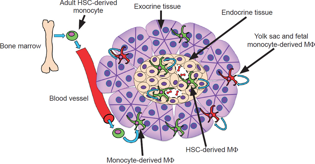
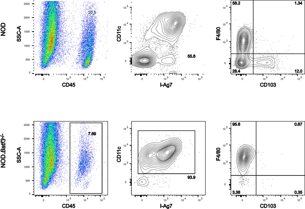
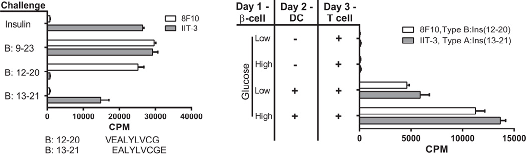
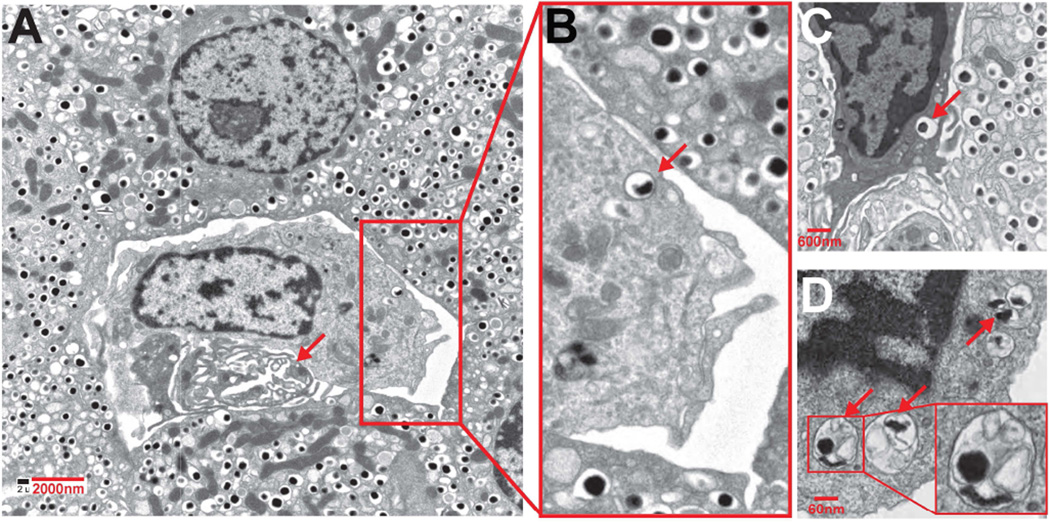
This is a brief summary of our studies of NOD autoimmune diabetes examining the events during the initial stage of the process. Our focus has been on antigen presentation events and the antigen presenting cells (APC) inside islets. Islets of non-diabetic mice contain resident macrophages that are developmentally distinct from those in the inter-acinar stroma. The autoimmune process starts with the entrance of CD4+ T cells together with a burst of a subset of dendritic cells (DC) bearing CD103. The CD103+ DC develop under the influence of the Batf3 transcription factor. Batf3 deficient mice do not develop diabetes and their islets are uninfiltrated throughout life. Thus, the CD103+ DC are necessary for the progression of autoimmune diabetes. The major CD4+ T cell response in NOD are the T cells directed to insulin. In particular, the non-conventional 12-20 segment of the insulin B chain is presented by the class II MHC molecule I-A(g7) and elicits pathogenic CD4+ T cells. We discuss that the diabetic process requires the CD103+ DC, the CD4+ T cells to insulin peptides, and NOD specific I-Ag(7) MHC-II allele. Finally, our initial studies indicate that beta cells transfer insulin containing vesicles to the local APC in a contact-dependent reaction. Live images of beta cells interactions with the APC and electron micrographs of islet APCs also show the transfer of granules.

Keywords: Antigen presenting cells; Antigen processing; Autoimmune diabetes; Non-obese diabetic; T cells.


Figures

Fig. 1
Fig. 1
The multiple origins of pancreatic macrophages and their maintenance. The illustration depicts the origin of islet-resident macrophages (adult HSC derived, shown in green) and their self-maintenance by in situ proliferation. These show basal M1 features. In contrast, the exocrine pancreatic (stroma) macrophages are composed of two subsets: one in continual replacement by circulating monocytes (adult HSC derived) and not self-maintained and the second derived from yolk sac and fetal liver monocytes (shown in red) and selfmaintained. Half the interacinar macrophages expressed CD206 and CD301 and were preferentially situated among pancreatic ducts. The second set derives from circulating monocytes and did not express CD206 or CD301. All macrophages in the interacinar stroma have M2 features. From Calderon et al. [2] with permission.
Fig. 2
Fig. 2
Islet APC composition differs between the NOD and NOD.Batf3−/− mice. Scatter plots represent the flow cytometry of dispersed islets from two NOD (top) or NOD.Batf3−/− (bottom) mice at 12 wks of age. Each gate is predicated upon the panel to the left, e.g. CD11c+ MHCII+ gate displays cells from the CD45+ gate. The NOD shows two APCS, the F4/80+ macrophage and the CD103+ DC, whereas the NOD.Batf3−/− mouse is missing the CD103+ cells.
Fig. 3
Fig. 3
Beta cells transfer of insulin granules to DC. The characterization of the two CD4+ T cells reactive to insulin is shown in the left panel. T cells were incubated with splenic DCs isolated from FMS-like tyrosine kinase 3 ligand (FLT-3L) treated mice. The antigens were either insulin or the B:9–23 peptide, each at 10 µM. 8F10 is a T cell hybridoma that reacts with peptides B:9–23 or B:12–20 (sequence shown below the graph), but not with insulin or B:13–21. The IIT-3 is a T cell hybridoma that reacts with insulin and peptides B:9–23 and B:13–21, but not B:12–20. The panel on the right shows the reactivity of the insulin T cells to beta cell derived antigen. Beta cells are incubated with or without DC, after which CD4+ T cell hybridomas reactive to insulin peptides are added. The bars represent the degree of activation of the T cells as measured by IL-2 production and the proliferation of the IL-2 dependent CTLL-2 cell line as read by counts per minute (CPM). Experiments were performed in the presence of 5mM (Low) or 25mM (High) glucose. From Vomund, et al. with permission from the Proceedings of the National Academy of Science [3].
Fig. 4
Fig. 4
Electron micrographs of islets showing phagocytes containing vesicles with the morphology of dense core granules. A represents an islet from an 8-wkold female NOD; B–D are islets taken from NOD.Rag1−/− mice at 14 wk of age. The arrow in A indicates a vessel. In the enlarged area, one of the dense-core granules is indicated by an arrow. In panel C a phagocyte is shown in between beta cells, and an arrow points to a dense-core granule. Panel D shows a portion of a phagocyte with endocytosed material in the form of vesicles containing an electron-dense core, with others containing amorphous content. From Vomund. et al. with permission from the Proceedings of the National Academy of Science [3]

References

    1. Ferris ST, Carrero JA, Mohan JF, Calderon B, Murphy KM, Unanue ER. A minor subset of Batf3-dependent antigen presenting cells in islets of Langerhans is essential for the development of autoimmune diabetes. Immunity. 2014;41:657–669. - PMC - PubMed
    1. Calderon B, Carrero JA, Ferris ST, Sojka DK, Moore L, Epelman S, et al. The pancreas anatomy conditions the origin and properties of resident macrophages. J Exp Med. 2015;212:1497–1512. - PMC - PubMed
    1. Vomund AN, Zinselmeyer BH, Hughes J, Calderon B, Valderrama C, Ferris ST, et al. CV325 Beta cells transfer vesicles containing insulin to phagocytes for presentation to T cells. Proc Natl Acad Sci USA. 2015;112:5496–5502. - PMC - PubMed
    1. Carrero JA, Calderon B, Towfic F, Artyomov MN, Unanue ER. Defining the transcriptional and cellular landscape of type 1 diabetes in the NOD mouse. PLoS One. 2013;8:e59701. - PMC - PubMed
    1. Calderon B, Carrero JA, Unanue ER. The central role of antigen presentation in islets of Langerhans in autoimmune diabetes. Curr Opin Immunol. 2014;26:32–40. - PMC - PubMed

Publication types

MeSH terms