

Magnifying Endoscopy with Narrow Band Imaging of Early Gastric Cancer: Correlation with Histopathology and Mucin Phenotype
- PMID: 27021504
- PMCID: PMC4933412
- DOI: 10.5009/gnl15364
Magnifying Endoscopy with Narrow Band Imaging of Early Gastric Cancer: Correlation with Histopathology and Mucin Phenotype
Abstract
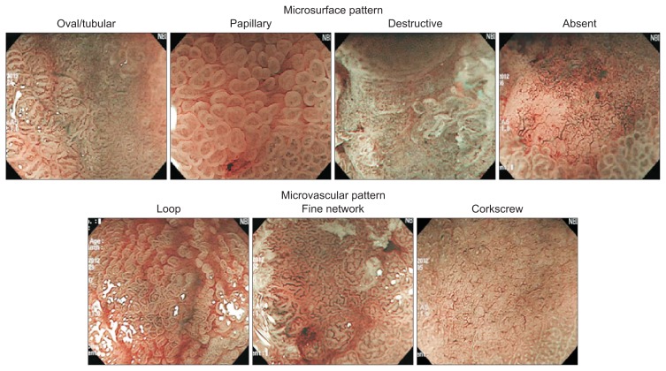
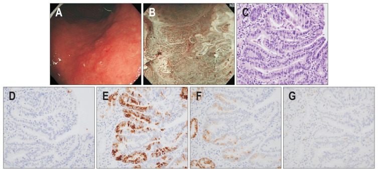
Background/aims: Magnifying endoscopy with narrow band imaging (ME-NBI) is a useful modality for the detailed visualization of microsurface (MS) and microvascular (MV) structures in the gastrointestinal tract. This study aimed to determine whether the MS and MV patterns in ME-NBI differ according to the histologic type, invasion depth, and mucin phenotype of early gastric cancers (EGCs).
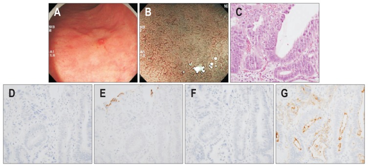
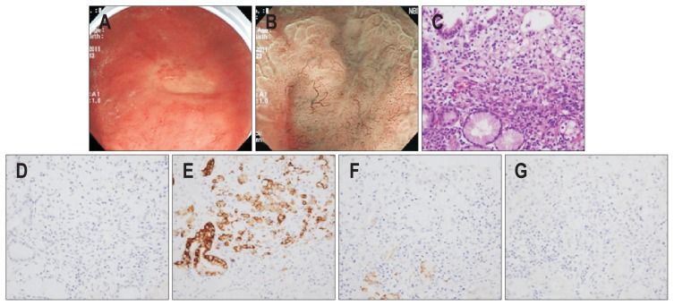
Methods: The MS and MV patterns of 160 lesions in 160 patients with EGC who underwent ME-NBI before endoscopic or surgical resection were prospectively collected and analyzed. EGCs were categorized as either differentiated or undifferentiated and as either mucosal or submucosal, and their mucin phenotypes were determined via immunohistochemistry of the tumor specimens.
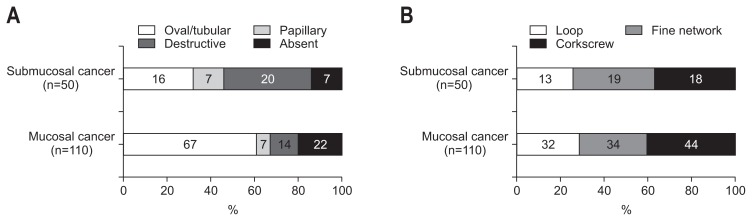
Results: Differentiated tumors mainly displayed an oval and/or tubular MS pattern and a fine network or loop MV pattern, whereas undifferentiated tumors mainly displayed an absent MS pattern and a corkscrew MV pattern. The destructive MS pattern was associated with submucosal invasion, and this association was more prominent in the differentiated tumors than in the undifferentiated tumors. MUC5AC expression was increased in lesions with either a papillary or absent MS pattern and a corkscrew MV pattern, whereas MUC6 expression was increased in lesions with a papillary MS pattern and a loop MV pattern. CD10 expression was more frequent in lesions with a fine network MV pattern.
Conclusions: ME-NBI can be useful for predicting the histopathology and mucin phenotype of EGCs.
Keywords: Magnifying endoscopy; Mucins; Narrow band imaging; Stomach neoplasms.
Figures
Similar articles
-
Diagnosis of early gastric cancer using narrow band imaging and acetic acid.World J Gastroenterol. 2015 Jan 28;21(4):1268-74. doi: 10.3748/wjg.v21.i4.1268. World J Gastroenterol. 2015. PMID: 25632201 Free PMC article.
-
Correlation between White Globe Appearance and Clinicopathologic Characteristics in Early Gastric Cancer.Gut Liver. 2025 Jan 15;19(1):50-58. doi: 10.5009/gnl240097. Epub 2024 Oct 8. Gut Liver. 2025. PMID: 39376041 Free PMC article.
-
Magnifying endoscopy of gastric epithelial dysplasia based on the morphologic characteristics.World J Gastroenterol. 2014 Nov 14;20(42):15771-9. doi: 10.3748/wjg.v20.i42.15771. World J Gastroenterol. 2014. PMID: 25400462 Free PMC article.
-
[NBI magnifying endoscopic classification using crystal violet staining].Nihon Rinsho. 2008 May;66(5):1023-7. Nihon Rinsho. 2008. PMID: 18464526 Review. Japanese.
-
Detection and characterization of early gastric cancer for curative endoscopic submucosal dissection.Dig Endosc. 2013 Mar;25 Suppl 1:44-54. doi: 10.1111/den.12004. Epub 2013 Jan 24. Dig Endosc. 2013. PMID: 23362939 Review.
Cited by
-
Image-Enhanced Endoscopy and Its Corresponding Histopathology in the Stomach.Gut Liver. 2021 May 15;15(3):329-337. doi: 10.5009/gnl19392. Gut Liver. 2021. PMID: 32200589 Free PMC article. Review.
-
Comparison of clinical and pathological features between early-stage gastric-type and intestinal-type differentiated adenocarcinoma: a retrospective study.BMC Gastroenterol. 2023 Mar 28;23(1):92. doi: 10.1186/s12876-023-02733-3. BMC Gastroenterol. 2023. PMID: 36977979 Free PMC article.
-
Endoscopic submucosal dissection for early gastric cancer: It is time to consider the quality of its outcomes.World J Gastroenterol. 2023 Nov 21;29(43):5800-5803. doi: 10.3748/wjg.v29.i43.5800. World J Gastroenterol. 2023. PMID: 38074917 Free PMC article.
-
Magnifying endoscopy with narrow-band imaging for gastric heterotopic pancreas.Endosc Int Open. 2018 Mar;6(3):E369-E375. doi: 10.1055/s-0044-101350. Epub 2018 Mar 7. Endosc Int Open. 2018. PMID: 29527560 Free PMC article.
-
Narrow-Band Imaging: Clinical Application in Gastrointestinal Endoscopy.GE Port J Gastroenterol. 2018 Dec;26(1):40-53. doi: 10.1159/000487470. Epub 2018 Mar 27. GE Port J Gastroenterol. 2018. PMID: 30675503 Free PMC article. Review.
References
-
- Yagi K, Nakamura A, Sekine A, Umezu H. Magnifying endoscopy with narrow band imaging for early differentiated gastric adenocarcinoma. Dig Endosc. 2008;20:115–122. doi: 10.1111/j.1443-1661.2008.00788.x. - DOI
Publication types
MeSH terms
Substances
LinkOut - more resources
Full Text Sources
Other Literature Sources
Medical
