. 2016 Sep;30(9):1913-6.
doi: 10.1038/leu.2016.62. Epub 2016 Mar 8.

Generation of the Fip1l1-Pdgfra fusion gene using CRISPR/Cas genome editing

Affiliations

Generation of the Fip1l1-Pdgfra fusion gene using CRISPR/Cas genome editing

M Vanden Bempt et al. Leukemia. 2016 Sep.
No abstract available


Figures

Figure 1
Figure 1
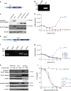
Use of CRISPR/Cas genome editing to generate Fip1l1–Pdgfra fusions. (a) Structure of PDGFRα and the FIP1L1-PDGFRα fusion protein. Formation of the fusion leads to disruption of the JM domain between two tryptophan (W) residues in PDGFRα (TM=transmembrane domain, JM=juxtamembrane domain). (b) Representation of the Fip1l1 and Pdgfra mouse genes. Exons are indicated by vertical bars. Red arrows indicate the location of the gRNA target sites in mouse Fip1l1 and Pdgfra. Black arrows indicate homologous sequences of breakpoints found in patients. (c) Efficiency of the individual gRNAs targeting Fip1l1 and Pdgfra in Ba/F3 cells as determined by Illumina next-generation sequencing. (d) Growth curve showing the transforming capacities of Ba/F3 cells harboring an endogenous FP1 fusion. Ba/F3 cells harboring a FP2–FP6 fusion had similar transformation rates (data not shown). Electroporation of only 1 gRNA targeting Fip1l1 or Pdgfra could not transform the Ba/F3 cells. Cas9 only refers to a vector containing Cas9 without a gRNA sequence. (e) PCR to detect six different gene fusions in Ba/F3 cells electroporated with a vector containing Cas9 and gRNA sequences targeting Fip1l1 and Pdgfra. Cas9 only refers to a vector containing Cas9 without a gRNA sequence. (f) Western blot showing six different Fip1l1–Pdgfrα fusion proteins expressed in Ba/F3 cells. Different breakpoints in Fip1l1 lead to different molecular weights. (g) Sequencing trace showing a fusion between Fip1l1 and Pdgfra in Ba/F3 single cell clones of two different fusion genes (FP1 and FP2). (h) FISH on Ba/F3 cells electroporated with empty vector or with vectors containing Fip1l1–Pdgfra gRNAs. The white arrow indicates loss of one copy of Chic2, a gene in the deleted region between Fip1l1 and Pdgfra.
Figure 2
Figure 2
Study of the properties of the Fip1l1–Pdgfrα fusion protein. (a) Structure of the FP7 fusion. Break points are located in Fip1l1 exon 1 and Pdgfra exon 12 (W=tryptophan residue, TM=transmembrane domain, JM=juxtamembrane domain). (b) PCR to detect the FP7 fusion in Ba/F3 cells. (c) Western blot showing expression of the FP7 fusion protein in Ba/F3 cells after electroporation of gRNAs targeting Fip1l1 exon 1 and Pdgfra exon 12. (d) Growth curve showing the transforming capacities of Ba/F3 cells harboring an endogenous FP7 fusion. (e) Structure of the FP9 fusion. Break points are located in Fip1l1 exon 9 and Pdgfra exon 11 (W=tryptophan residue, TM=transmembrane domain, JM=juxtamembrane domain). (f) PCR to detect four different FP9 fusions with four different gRNAs targeting the upstream region of Pdgfra exon 11. (g) Growth curve of Ba/F3 cells harboring an endogenous FP1 or FP9 fusion. (h) Western blot showing the effects of Imatinib on the Fip1l1–Pdgfrα fusion protein. Ba/F3 cells harboring an endogenous FP1 fusion (FP1) and Ba/F3 cells expressing a human FIP1L1–PDGFRA cDNA (FP retro) were treated for 90 min with 0 or 100 nM Imatinib. On the blots showing expression of (p-)Fip1l1–Pdgfra, a 1/10 dilution of FP retro is shown. Difference in molecular weight of Fip1l1–Pdgfrα is due to differences in molecular weight between the mouse and human fusion proteins. (i) Dose response curve for Imatinib. EC50 values are represented by 95% confidence intervals.

References

    1. Cools J, DeAngelo DJ, Gotlib J, Stover EH, Legare RD, Cortes J et al. A tyrosine kinase created by fusion of the PDGFRA and FIP1L1 genes as a therapeutic target of imatinib in idiopathic hypereosinophilic syndrome. N Engl J Med 2003; 348: 1201–1214. - PubMed
    1. Tefferi A, Vardiman JW. Classification and diagnosis of myeloproliferative neoplasms: the 2008 World Health Organization criteria and point-of-care diagnostic algorithms. Leukemia 2008; 22: 14–22. - PubMed
    1. Jovanovic JV, Score J, Waghorn K, Cilloni D, Gottardi E, Metzgeroth G et al. Low-dose imatinib mesylate leads to rapid induction of major molecular responses and achievement of complete molecular remission in FIP1L1-PDGFRA-positive chronic eosinophilic leukemia. Blood 2007; 109: 4635–4640. - PubMed
    1. Lierman E, Michaux L, Beullens E, Pierre P, Marynen P, Cools J et al. FIP1L1-PDGFRalpha D842V, a novel panresistant mutant, emerging after treatment of FIP1L1-PDGFRalpha T674I eosinophilic leukemia with single agent sorafenib. Leukemia 2009; 23: 845–851. - PubMed
    1. von Bubnoff N, Sandherr M, Schlimok G, Andreesen R, Peschel C, Duyster J. Myeloid blast crisis evolving during imatinib treatment of an FIP1L1-PDGFR alpha-positive chronic myeloproliferative disease with prominent eosinophilia. Leukemia 2005; 19: 286–287. - PubMed

Publication types

MeSH terms

Substances