Review
. 2016 Jun 15;56(6):310-6.
doi: 10.2176/nmc.ra.2016-0006. Epub 2016 Mar 28.

Vascular Anatomy of the Cauda Equina and Its Implication on the Vascular Lesions in the Caudal Spinal Structure

Affiliations
Review

Vascular Anatomy of the Cauda Equina and Its Implication on the Vascular Lesions in the Caudal Spinal Structure

Katsunari Namba. Neurol Med Chir (Tokyo). .

Abstract

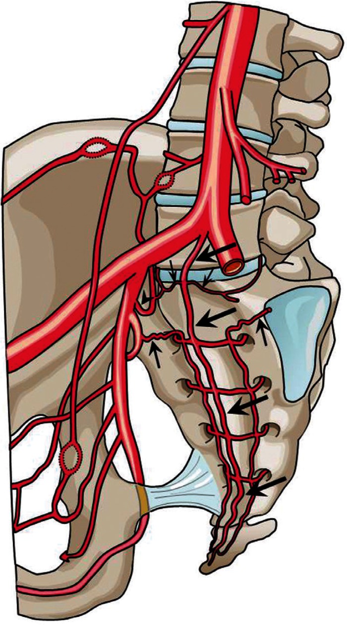
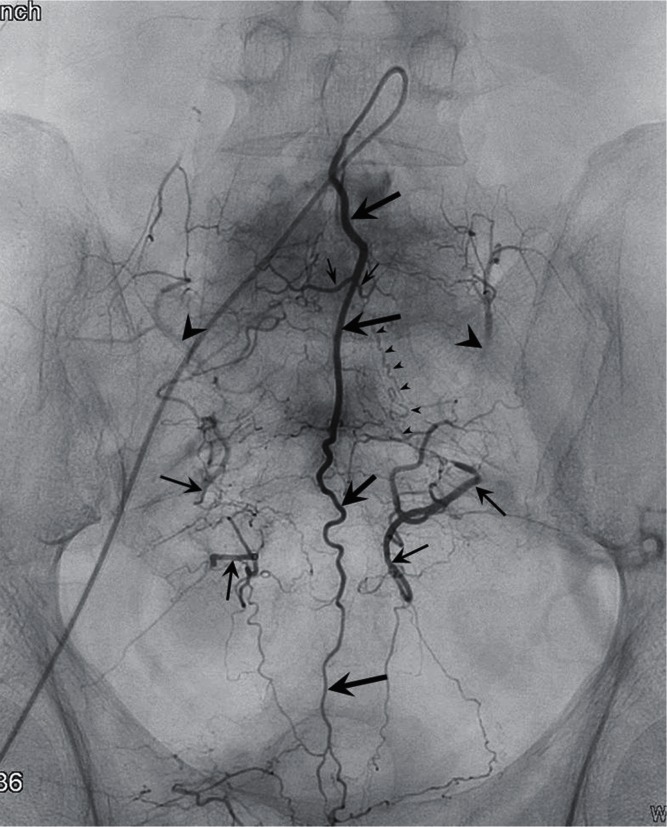
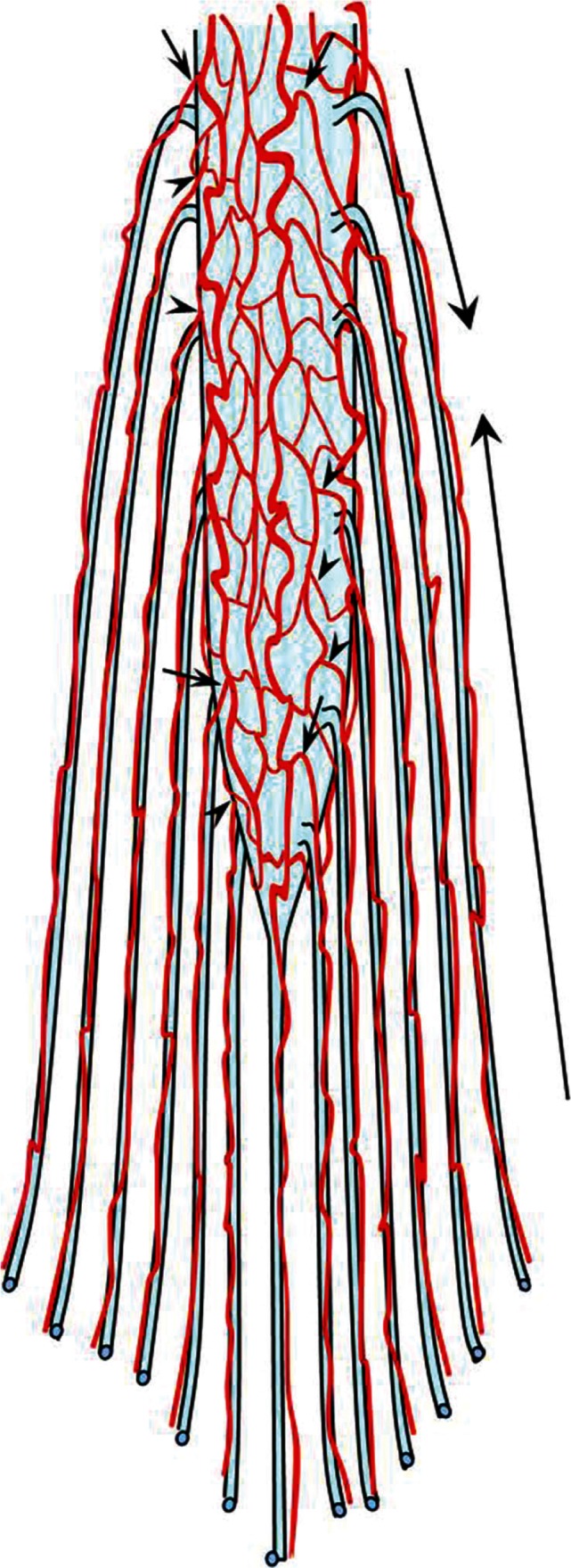
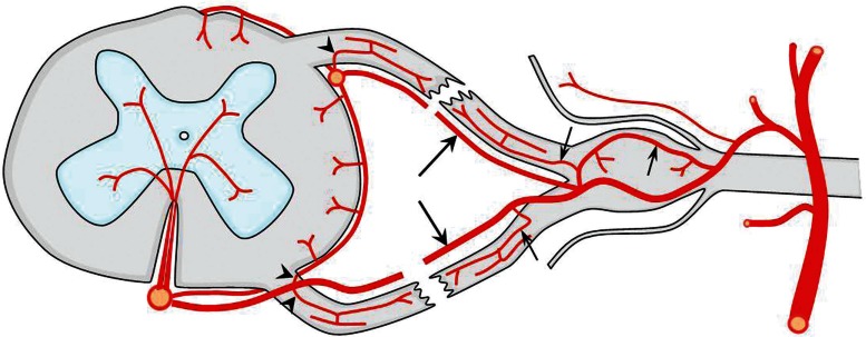
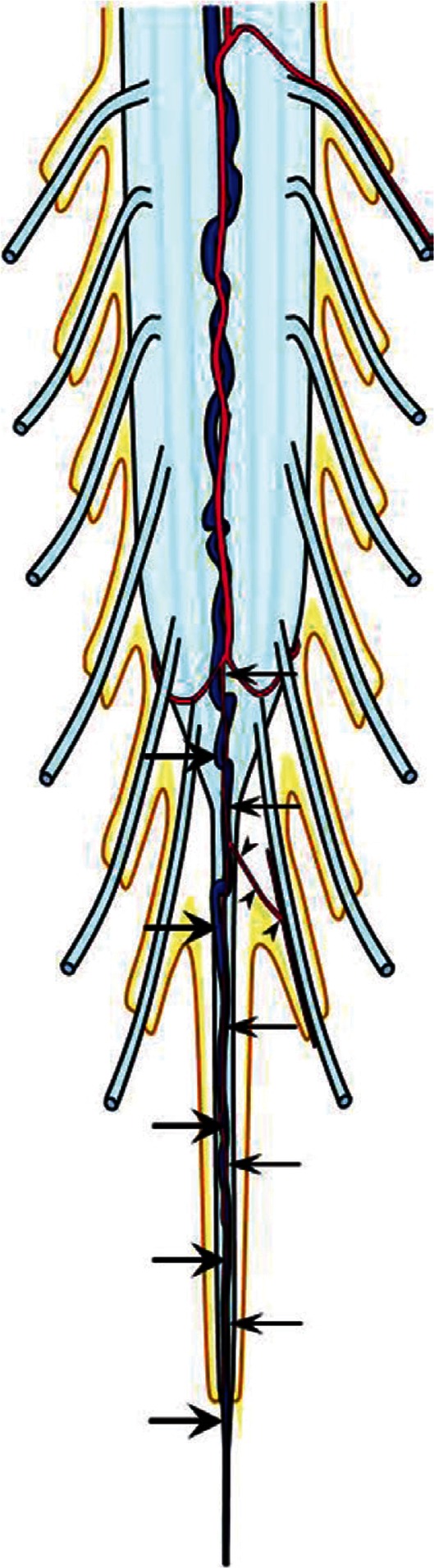
The cauda equina is composed of the lumbosacral and the coccygeal nerve roots and the filum terminale. In the embryonic period, discrepancy in development between the termination of the spinal cord and the spinal column results in elongation of the nerve roots as well as the filum terminale in this region. Although the vascular anatomy of the caudal spinal structure shares many common features with the other metameric levels, this elongation forms the basis of the characteristic vascular anatomy in this region. With the evolution of the high quality imaging techniques, vascular lesions in the cauda equina are being diagnosed more frequently than ever before. Albeit the demand for accurate knowledge of the vascular anatomy in this region, descriptions are often fragmented and not easily accessible. In this review, the author attempted to organize the existing knowledge of the vascular anatomy in the cauda equina and its implication on the vascular lesions in this region. Also reviewed is the clinically relevant embryological development of the cauda equina.


Conflict of interest statement

Conflicts of Interest Disclosure

None.

Figures

Fig. 1
Fig. 1
A schema showing the arterial supply to the lumbosacral and coccygeal spinal structures. The middle sacral artery (large arrows) gives rise to a pair of the lowest lumbar artery (small arrows), which is the L5 segmental artery. A branch of the iliiolumbar artery (arrowhead), may also supply the L5 region. The size of this vessel is inversely related to the development of the lowest lumbar artery. The spinal structures within the sacral vertebral body are usually supplied by the branches of the lateral sacral arteries (arrows), which course medial to the anterior sacral foramen. The branches of the sacral artery form numerous anastomoses with the lateral branches of the medial sacral artery.
Fig. 2
Fig. 2
An angiogram of the medial sacral artery (large arrows) demonstrates the arterial network of the lumbosacral and coccygeal region. A pair of the lowest lumbar artery is rising from the medial sacral artery (small arrows). Retrograde opacification of the iliolumbar arteries (arrowheads), lateral sacral arteries (arrows) are seen bilaterally. Note the opacification of a pathologic S1 radicular artery (small arrowheads) supplying an arteriovenous fistula in a higher level (not shown) that originates from the lateral sacral artery.
Fig. 3
Fig. 3
A schema showing the arterial anatomy of the cauda equina and conus medullaris. On the right-hand side, the arteries of the anterior roots are shown, whereas on the left-hand side, the arteries of the posterior roots are shown. Note the multiple anastomoses among the radicular artery and the spinal cord arteries (arrows) or the vasa corona (arrowheads). The long arrows on the right signify that the proximal one-third of the root is supplied from the radicular artery arising from the arterial plexus of the spinal cord and the distal two-thirds from the segmental radicular artery.
Fig. 4
Fig. 4
A schema illustrating all the possible arterial structures of the spinal nerve roots that may be applicable to all segmental levels. The radiculomedullary and radiculopial arteries (arrows) course along their respective nerve roots to supply the spinal cord. The segmental arteries give rise to the distal radicular arteries to supply the distal part of the roots (small arrows). The anterior and posterior spinal arteries or the vasa corona give rise to the radicular arteries in the proximal nerve roots (arrowheads).
Fig. 5
Fig. 5
A schema of the vascular anatomy of the filum terminale. The artery of the filum terminale originates from the termination of the anterior spinal artery (arrows). The filum artery may give rise to arterioles which supply the coccygeal nerve roots adherent to the filum terminale (arrowheads). The vein of the filum terminale exists constantly on the ventral aspect of the filum, behind the artery (large arrows). The vein is continuous from the anterior spinal vein to the sacral dural sac, where it traverses the dura to join the sacral venous plexus.

References

    1. Parke WW, Gammell K, Rothman RH: Arterial vascularization of the cauda equina. J Bone Joint Surg Am 63: 53– 62, 1981. - PubMed
    1. Crock HV, Yamagishi M, Crock MC: The Conus Medullaris and Cauda Equina in Man. An Atlas of the Arteries and Veins. Wien-New York, Springer-Verlag, 1986.
    1. Djindjian M, Ribeiro A, Ortega E, Gaston A, Poirier J: The normal vascularization of the intradural filum terminale in man. Surg Radiol Anat 10: 201– 209, 1988. - PubMed
    1. Sadler TW: Langman’s Medical Embryology, ed 11 Baltimore, Lippincott Williams & Wilkins, 2010, pp 67– 70
    1. Saitsu H, Shiota K: Involvement of the axially condensed tail bud mesenchyme in normal and abnormal human posterior neural tube development. Congenit Anom (Kyoto) 48: 1– 6, 2008. - PubMed

MeSH terms