Review
. 2016 Apr;19(4):542-53.
doi: 10.1038/nn.4273.

Modeling ALS with motor neurons derived from human induced pluripotent stem cells

Affiliations
Review

Modeling ALS with motor neurons derived from human induced pluripotent stem cells

Samuel Sances et al. Nat Neurosci. 2016 Apr.

Abstract

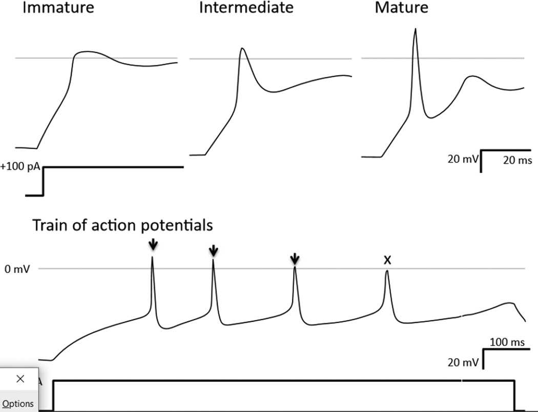
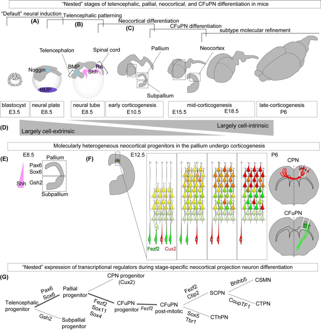
Directing the differentiation of induced pluripotent stem cells into motor neurons has allowed investigators to develop new models of amyotrophic lateral sclerosis (ALS). However, techniques vary between laboratories and the cells do not appear to mature into fully functional adult motor neurons. Here we discuss common developmental principles of both lower and upper motor neuron development that have led to specific derivation techniques. We then suggest how these motor neurons may be matured further either through direct expression or administration of specific factors or coculture approaches with other tissues. Ultimately, through a greater understanding of motor neuron biology, it will be possible to establish more reliable models of ALS. These in turn will have a greater chance of validating new drugs that may be effective for the disease.


Figures

Figure 1
Figure 1
Emulating MN Developmental Signaling In Vitro. Developmental stages of human MNs (a) are reproduced in vitro (b) through the use of small molecule and recombinant signaling molecules. (i) Blastocyst containing pluripotent stem cells derived from the inner cell mass (blue) is generated in vitro from adult somatic tissue through reprograming into iPSC cultures. During gastrulation, Wnt-dependent primitive streak formation (ii) is simulated using a GSK3 inhibitor (CHIR99021). Neural ectoderm that emerges during neurulaiton (iii) is directed through the use of dual-SMAD inhibitors SB431542 and LDN193189 (SB, LDN). Retinoic Acid (RA) (iv) is produced by neighboring somites (not shown) that act as caudalizing molecules that specify a hindbrain and anterior spinal cord fate (iv’). (v) Sonic Hedgehog (SHH) is released from the ventral notochord, causing a gradient that induces MN fate in the ventral portion of the spinal cord. (v’) This is reproduced in vitro with small molecule (smSHH) or recombinant SHH signaling agonists. (vi) MN progenitors depend on trophic support to connect axon projections to target muscles and develop into functioning LMNs. (vi’) Neurotrophic factors (NTFs) such as GDNF, BDNF, CNTF and others are used in vitro to provide maturation and survival signaling.
Figure 2
Figure 2
Comparison of Published LMN Differentiation Protocols. 12 iPSC to LMN protocols compared with respect to time (days in vitro). End of experiment (Assay) based on last data presented. Fibroblast growth factor 2 (FGF2), brain-derived neurotropic factor (BDNF), glial cell line-derived neurotropic factor (GDNF), cilliary neurotropic factor (CNTF), retinoic acid (RA), retinoic acid receptor (RAR), sonic hedgehog (SHH), purmorphamine (PMN). Yellow column lists summary of results, unique MN subtype markers observed, and approximate percent yield of MNs based on reported percent cells expressing HB9 or Islet-1. Induced action potentials (iAP), spontaneous action potentials (sAP), Caudal spinal cord associated Hox family member C (HoxC9), Forkhead Box P1, P2 (FoxP1), (FoxP2) LIM Homeobox 3 (LHX3), LIM Homeobox 1/2 (LHX1/2), Paired Mesoderm Homeobox 2B (PHOX2B).
Figure 3
Figure 3
Induced Action Potentials Evolve Over Time. (a) Current injection (100pA) whole-cell recordings of human iPSC-derived MNs (hPSC-MNs) over time in culture. Example action potential recordings show maturity over time in vitro as depolarization and hyperpolarization events occur more rapidly. (b) More mature hPSC-MNs in vitro display trains of action potentials (black arrows) with an abortive event (x). Action potentials displayed as change in membrane voltage (mv) over time (ms).
Figure 4
Figure 4
Co-Culture of Neuromuscular Circuit. (a). hPSC-astrocytes (b) taken with permission from Sareen et. al 2014. hPSC-myofibers (c) taken with permission from Hosoyama 2014. These examples of published iPSC-derived cell types could comprise of (d) a conceptualized neuronmuscular circuit. Other cell types such as myelinating schwann cells (dotted lines) and terminal schwann cells (not shown) have not yet been generated from iPSCs and may be required for functional NMJ formation in vitro. Neurofiliment Heavy chain (NFH) and specific NFH epitope SMI32, Islet-1 (Isl1), Glial fibrillary acidic protein (GFAP), Neurofiliment Heavy chain Smooth Muscle Actin (SMA).
Figure 5
Figure 5
Classification of Diverse Neocortical Projection Neurons. Neocortical projection neurons can be subdivided into broad classes, types, and subtypes largely based on their axonal projections. (Figure adapted from Greig et al., Nat Rev Neurosci, 2013.) Illustrations are of the mouse brain.
Figure 6
Figure 6
Cell-extrinsic and Cell-intrinsic Factors Regulate the Development of CFuPN within Sequential, “Nested” Stages of Differentiation. (a) “Default” neural and rostral differentiation occurs by repression of alternate signaling pathways induced by multiple morphogens (e.g., Noggin inhibits BMP signaling during neural plate formation at ~E3.5–E6.5 in mice; cortical progenitors require low or absent expression of caudalizing retinoids (RA) and ventralizing Shh at ~E6.5–E8.5). (b) The dorsal aspect of the telencephalon is called the pallium, which gives rise to the neocortex. In contrast, the ventral telencephalon is called the subpallium. The delineation of these two telencephalic progenitor domains occurs between ~E8.5 and ~E10.5. (c) During corticogenesis, beginning at ~E10.5 in mice, multiple diverse cortical projection neuron classes, types, and subtypes are sequentially generated from cortical progenitors. These projection neurons become refined with continued maturation through post-natal ages. (d) Early stages of corticofugal projection neuron (CFuPN) differentiation are largely mediated by cell-extrinsic factors, whereas later stages of are largely mediated by cell-intrinsic factors. (e) Following Shh-mediated dorsal-ventral patterning of the telencephalon, cortical and ventral identities are reinforced by transcriptional regulation (Pax6 and Sox6 in the pallium; Gsh2 in the ventral areas). (f) Early cortical progenitors give rise to more definitive (neo)cortical progenitors, which generate projection neuron subtypes at ~E10.5. CFuPN populate the deep layers of the cortex. Later-born CPN populate both deep and superficial layers of cortex. Molecular distinction of CPN and CFuPN occurs with continued maturation (represented by transition from yellow, dual-marker expression to red or green single-marker expression). (g) “Nested” expression of distinct transcriptional regulators at distinct developmental stages promotes stepwise CFuPN and thus UMN differentiation.

References

    1. Takahashi K, et al. Induction of pluripotent stem cells from adult human fibroblasts by defined factors. Cell. 2007;131:861–872. - PubMed
    1. Yu J, et al. Induced pluripotent stem cell lines derived from human somatic cells. Science. 2007;318:1917–1920. - PubMed
    1. Svendsen CN. Back to the future: how human induced pluripotent stem cells will transform regenerative medicine. Human molecular genetics. 2013;22:R32–R38. - PMC - PubMed
    1. Sandoe J, Eggan K. Opportunities and challenges of pluripotent stem cell neurodegenerative disease models. Nature neuroscience. 2013;16:780–789. - PubMed
    1. Ericson J, Thor S, Edlund T, Jessell TM, Yamada T. Early stages of motor neuron differentiation revealed by expression of homeobox gene Islet-1. Science. 1992;256:1555–1560. - PubMed

Publication types

MeSH terms