. 2016 May 27;291(22):11736-50.
doi: 10.1074/jbc.M116.721084. Epub 2016 Mar 28.

Do Src Kinase and Caveolin Interact Directly with Na,K-ATPase?

Affiliations

Do Src Kinase and Caveolin Interact Directly with Na,K-ATPase?

Eliyahu Yosef et al. J Biol Chem. .

Abstract

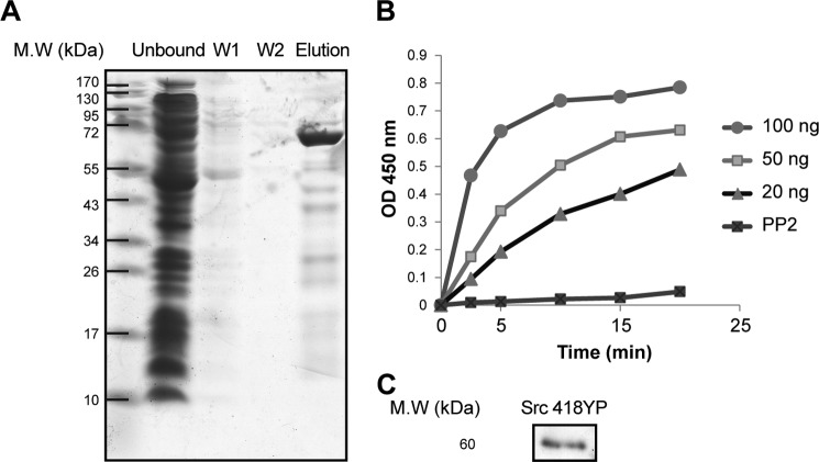
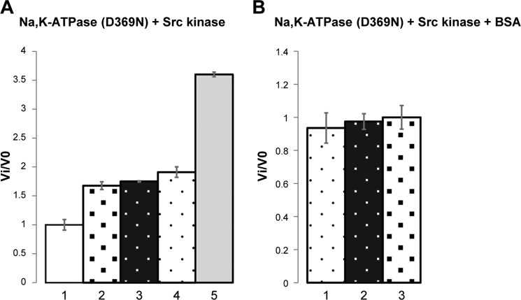
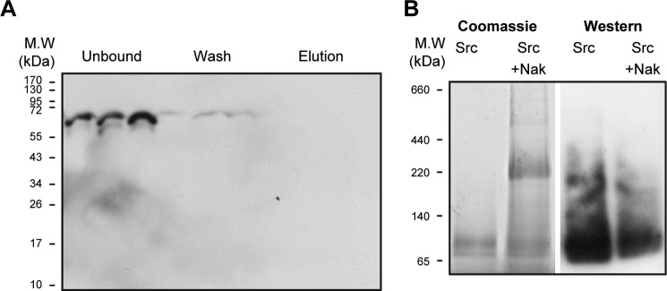
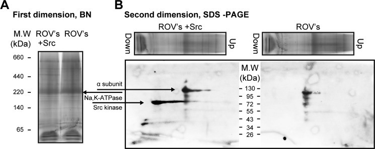
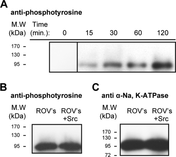
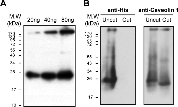
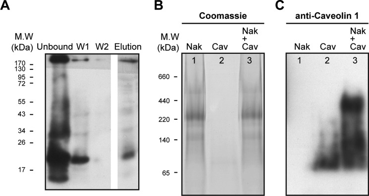
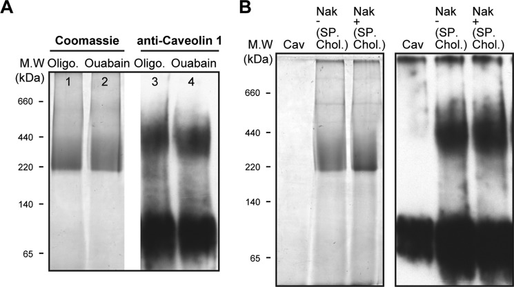
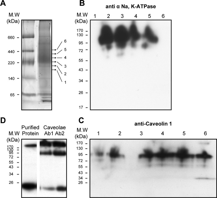
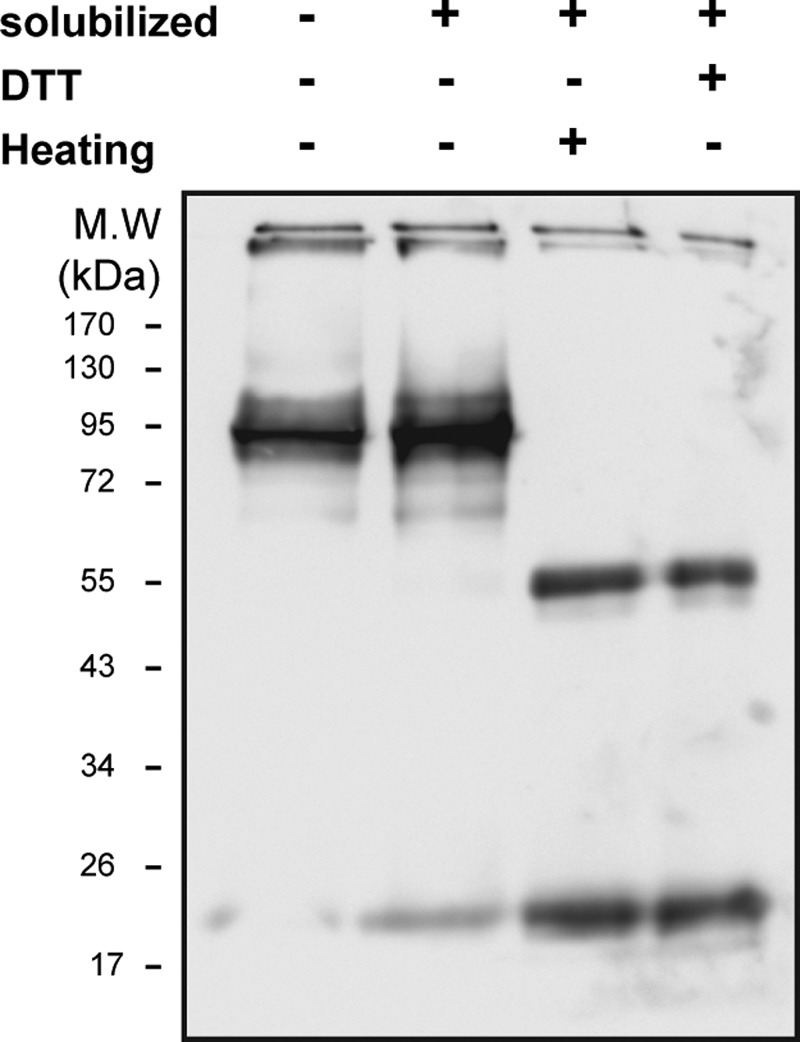
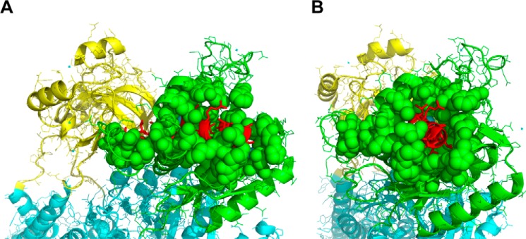
Much evidence points to a role of Na,K-ATPase in ouabain-dependent signal transduction. Based on experiments with different cell lines and native tissue membranes, a current hypothesis postulates direct interactions between the Na,K-ATPase and Src kinase (non-receptor tyrosine kinase). Na,K-ATPase is proposed to bind Src kinase and inhibit its activity, whereas ouabain, the specific Na,K-ATPase inhibitor, binds and stabilizes the E2 conformation, thus exposing the Src kinase domain and its active site Tyr-418 for activation. Ouabain-dependent signaling is thought to be mediated within caveolae by a complex consisting of Na,K-ATPase, caveolin, and Src kinase. In the current work, we have looked for direct interactions utilizing purified recombinant Na,K-ATPase (human α1β1FXYD1 or porcine α1D369Nβ1FXYD1) and purified human Src kinase and human caveolin 1 or interactions between these proteins in native membrane vesicles isolated from rabbit kidney. By several independent criteria and techniques, no stable interactions were detected between Na,K-ATPase and purified Src kinase. Na,K-ATPase was found to be a substrate for Src kinase phosphorylation at Tyr-144. Clear evidence for a direct interaction between purified human Na,K-ATPase and human caveolin was obtained, albeit with a low molar stoichiometry (1:15-30 caveolin 1/Na,K-ATPase). In native renal membranes, a specific caveolin 14-5 oligomer (95 kDa) was found to be in direct interaction with Na,K-ATPase. We inferred that a small fraction of the renal Na,K-ATPase molecules is in a ∼1:1 complex with a caveolin 14-5 oligomer. Thus, overall, whereas a direct caveolin 1/Na,K-ATPase interaction is confirmed, the lack of direct Src kinase/Na,K-ATPase binding requires reassessment of the mechanism of ouabain-dependent signaling.

Keywords: Na+/K+-ATPase; Src; caveolin; protein-protein interaction; signaling.


Figures

FIGURE 1.
FIGURE 1.
Purification and properties of human recombinant Src tyrosine kinase. SDS gel separation showing different Src kinase purification steps (right to left): unbound, wash 1 (W1), wash 2 (W2), and elution fractions (n = 6). B, Src kinase activity with 20, 50, and 100 ng of Src kinase. With 20 ng of Src kinase, the rate of color development was linear over 20 min. The linear regression line, y = 0.0239x + 0.0416 (r2 = 0.97), gave a calculated activity of 1.195 ± 0.322 A450 nm/min/μg Src (n = 6). C, immunoblot of 20 ng of purified Src kinase with phosphorylated Tyr-418 antibody (n = 2).
FIGURE 2.
FIGURE 2.
Effects of purified Na,K-ATPase (α1D369Nβ1FXYD1) and BSA on Src kinase activity. The specific Src kinase activity of the control sample was 1.195 A450 nm/min/μg. In the different conditions, Src kinase activity is presented as the ratio Vi/V0. A, purified porcine Na,K-ATPase (α1D369Nβ1FXYD1) was mixed with Src kinase at a molar ratio of 3:1 (Na,K-ATPase/Src). The Na,K-ATPase was pre-equilibrated with ouabain or oligomycin + 100 mm NaCl, 2 mm ATP, and 2 mm MgCl2 for 30 min at room temperature to stabilize E2-P·ouabain or E1–3Na·oligomycin or after denaturation (heating for 80 °C for 1 h). Where indicated, BSA was added at 1% (w/v) (∼150 μm). The values ± S.E. represent averages from 3 experiments/condition. A, column 1, Src kinase; column 2, Src kinase/Na,K-ATPase/ouabain; column 3, Src kinase/Na,K-ATPase/oligomycin; column 4, Src kinase/BSA; column 5, Src kinase/denatured Na,K-ATPase. B, column 1, Src kinase/BSA; column 2, Src kinase/BSA/Na,K-ATPase/oligomycin; column 3, Src kinase/BSA/Na,K-ATPase/ouabain. A, p values for the differences in columns 3, 4, and 5 versus control in column 1 (control) are in the range from 0.001 to 0.003. The p value for the difference between the values in column 5 (denatured Na,K-ATPase) and columns 3 and 4 (native Na,K-ATPase) is 0.0001.
FIGURE 3.
FIGURE 3.
Tests for a direct interaction between purified human Src kinase and purified human Na,K-ATPase. A, pull-down assay. The Na,K-ATPase (1.5 μg diluted 10-fold to reduce imidazole to 20 mm) was incubated with TEV-treated Src kinase at molar ratios of 1:2, 1:4, and 1:8 (Na,K-ATPase/Src) as described under “Methods.” Unbound, wash, and elution fractions were loaded on an SDS gel. Western blot was probed with anti-Src (n = 2). B, BN gel separation. Lanes 1 and 2, Coomassie stain. Lanes 3 and 4, Western blots probed with anti-Src. Lane 1, Src (8 μg). Lane 2, Src (8 μg) premixed with purified human Na,K-ATPase (α1β1FXYD1-NaK) (10 μg). Lane 3, Src (8 μg). Lane 4, Src plus Na,K-ATPase (α1β1FXYD1-NaK) (10 μg) (n = 4).
FIGURE 4.
FIGURE 4.
Blue native gel. A test for direct interaction between purified human Src kinase and native renal Na,K-ATPase is shown. A, blue native gel, first dimension, Coomassie-stained. Lane 1, marker proteins; lane 2, native rabbit kidney membranes, ROV (100 μg) plus Src (8 μg); lane 3, ROV (100 μg) (n = 4). B, SDS-PAGE, second dimension, Western blotting. Lane 2, probed with anti-α and anti-Src; lane 3, probed with anti-α (n = 2).
FIGURE 5.
FIGURE 5.
Phosphorylation of Na,K-ATPase α subunit by Src kinase. A, time-dependent phosphorylation of purified human Na,K-ATPase (α1β1FXYD1) by purified Src kinase condition. 2 μg of Src kinase were mixed with 15 μg of Na,K-ATPase with the addition of 1 mm ouabain, 2 mm ATP, 2 mm MgCl, 100 mm NaCl, and 50 mm Tris, pH 7.00. The sample was rotated at room temperature, and 2 μl were taken at each time point, mixed in gel buffer, and loaded on the gel (n = 3). B, right-side out rabbit kidney vesicles. Lane 2, untreated ROVs. Lane 3, after incubation of 10 μg of unmasked ROVs in a medium containing 1 mm ouabain, 2 mm ATP, 2 mm MgCl, 100 mm NaCl, and 50 mm Tris, pH 7.00, 2 μg of Src kinase were added. The sample was rotated at room temperature, and 2 μl were taken at each time point, mixed with gel buffer, and loaded on the gel. Western blots were probed with anti-phophotyrosine (n = 3). C, as in B but probed with Na,K-ATPase anti-α antibody (n = 3).
FIGURE 6.
FIGURE 6.
Purification and TEV cleavage of caveolin. A, Western blotting of 20, 40, and 80 ng of purified caveolin, probed with anti-caveolin 1 (n = 3). B, caveolin 1 before and after TEV treatment (see “Methods”). Western blotting was probed with anti-His (left) or anti-caveolin 1 (right)(n = 2).
FIGURE 7.
FIGURE 7.
Tests for a direct interaction between purified human caveolin 1 and purified human Na,K-ATPase. A, pull-down assay. Western blotting of unbound, wash I and II, and elution fractions probed with anti-caveolin 1 (see “Methods”) (n = 2). B and C, blue native gel. B, Coomassie-stained. C, Western blot probed with anti-caveolin 1. Lane 1, markers; lane 2, 5 μg of purified human Na,K-ATPase plus purified caveolin (3 μg), a molar ratio of 3:1 (caveolin/Na,K-ATPase). C, as in lanes 1–3 of B, probed with anti-caveolin 1 antibody (n = 6).
FIGURE 8.
FIGURE 8.
Interaction of purified Na,K-ATPase-caveolin 1 is not altered by the protein conformation or presence of sphingomyelin/cholesterol. A, lane 1, markers; lanes 1 and 2, Coomassie-stained; lane 3, 5 μg of purified human Na,K-ATPase preincubated with 100 μg of oligomycin, 1 mm ATP, 1 mm MgCl2, 100 mm NaCl at room temperature for 30 min, was mixed with 3 μg of purified caveolin; lane 4, purified human Na,K-ATPase preincubated with 2 mm ouabain, 1 mm ATP, 1 mm MgCl2, 100 mm NaCl at room temperature for 30 min was mixed with 3 μg of purified caveolin; lanes 3 and 4, like lanes 2 and 3, Western blots probed with anti-caveolin 1 (n = 4). B, lanes 1–4, Coomassie-stained. Lane 1, markers; lane 2, caveolin 1 (3 μg); lane 3, 5 μg of purified Na,K-ATPase prepared in standard conditions plus caveolin 1 (3 μg); lane 4, 5 μg of purified Na,K-ATPase prepared with sphingomyelin/cholesterol plus (3 μg) caveolin 1 that also was eluted with sphingomyelin and cholesterol; lanes 5–7, as in lanes 2–4, Western blots using anti-caveolin 1 (n = 4).
FIGURE 9.
FIGURE 9.
Blue native gel separation of Na,K-ATPase with bound caveolin in rabbit renal caveolae membranes. A, BN gel. Lane 1, markers; lane 2, caveolae membranes (100 μg), Coomassie stain. Arrows, points of cutting the gel into six 1-mm slices below and above the main band at ∼250 kDa (n = 2). B, SDS-PAGE; Western blotting of second dimension separation of six gel pieces probed with Na,K-ATPase anti-α antibody (n = 2). C, SDS-PAGE; Western blotting of second dimension separation of six gel pieces probed with anti-caveolin 1 antibody (n = 2). D, SDS-PAGE; Western blotting. Lane 1, purified recombinant caveolin shows caveolin bands at 26 kDa and between stacking and separating gels. Lanes 2 and 3, native rabbit renal caveolin, lane 2 probed with anti-caveolin 1 (Ab1, BD Transduction, catalogue no. 610080) and lane 3 probed with anti-caveolin 1 (Ab2, Abcam, catalogue no. ab2910) (n = 2).
FIGURE 10.
FIGURE 10.
Dissociation of the 95-kDa caveolin4–5 oligomer to monomers and dimers. Shown are 10 μg of ROV samples solubilized at different conditions; Western blotting with anti-caveolin antibody. Lane 1, ROVs with no treatment; lane 2, ROVs solubilized with 5 mg/ml urea and 4 mg/ml DDM incubated for 30 min at room temperature; lane 3, ROVs solubilized with 5 mg/ml urea and 4 mg/ml DDM heated for 30 min at 65 °C; lane 4, ROVs solubilized with 5 mg/ml urea and 4 mg/ml DDM incubated with 10 mm DTT for 15 min at room temperature and then with 20 mm iodoacetamide for 15 min (n = 4).
FIGURE 11.
FIGURE 11.
NaKtide sequence is largely buried in the N domain of the α subunit. Shown is the crystal structure of the pig Na,K-ATPase in the E1 state (Protein Data Bank code 3WGV). The N domain is colored in green, and the A domain is colored in yellow. NaKtide is in the N domain, colored in red. In A and B, the N domain is seen from different angles.

Similar articles

Cited by

References

    1. Kometiani P., Li J., Gnudi L., Kahn B. B., Askari A., and Xie Z. (1998) Multiple signal transduction pathways link Na+/K+-ATPase to growth-related genes in cardiac myocytes: the roles of Ras and mitogen-activated protein kinases. J. Biol. Chem. 273, 15249–15256 - PubMed
    1. Haas M., Wang H., Tian J., and Xie Z. (2002) Src-mediated inter-receptor cross-talk between the Na+/K+-ATPase and the epidermal growth factor receptor relays the signal from ouabain to mitogen-activated protein kinases. J. Biol. Chem. 277, 18694–18702 - PubMed
    1. Haas M., Askari A., and Xie Z. (2000) Involvement of Src and epidermal growth factor receptor in the signal-transducing function of Na+/K+-ATPase. J. Biol. Chem. 275, 27832–27837 - PubMed
    1. Aperia A. (2007) New roles for an old enzyme: Na,K-ATPase emerges as an interesting drug target. J. Intern. Med. 261, 44–52 - PubMed
    1. Schoner W. (2002) Endogenous cardiac glycosides, a new class of steroid hormones. Eur. J. Biochem. 269, 2440–2448 - PubMed

Associated data

LinkOut - more resources