. 2016 Mar 28;212(7):845-60.
doi: 10.1083/jcb.201508028.

G3BP-Caprin1-USP10 complexes mediate stress granule condensation and associate with 40S subunits

Affiliations

G3BP-Caprin1-USP10 complexes mediate stress granule condensation and associate with 40S subunits

Nancy Kedersha et al. J Cell Biol. .

Erratum in

Abstract

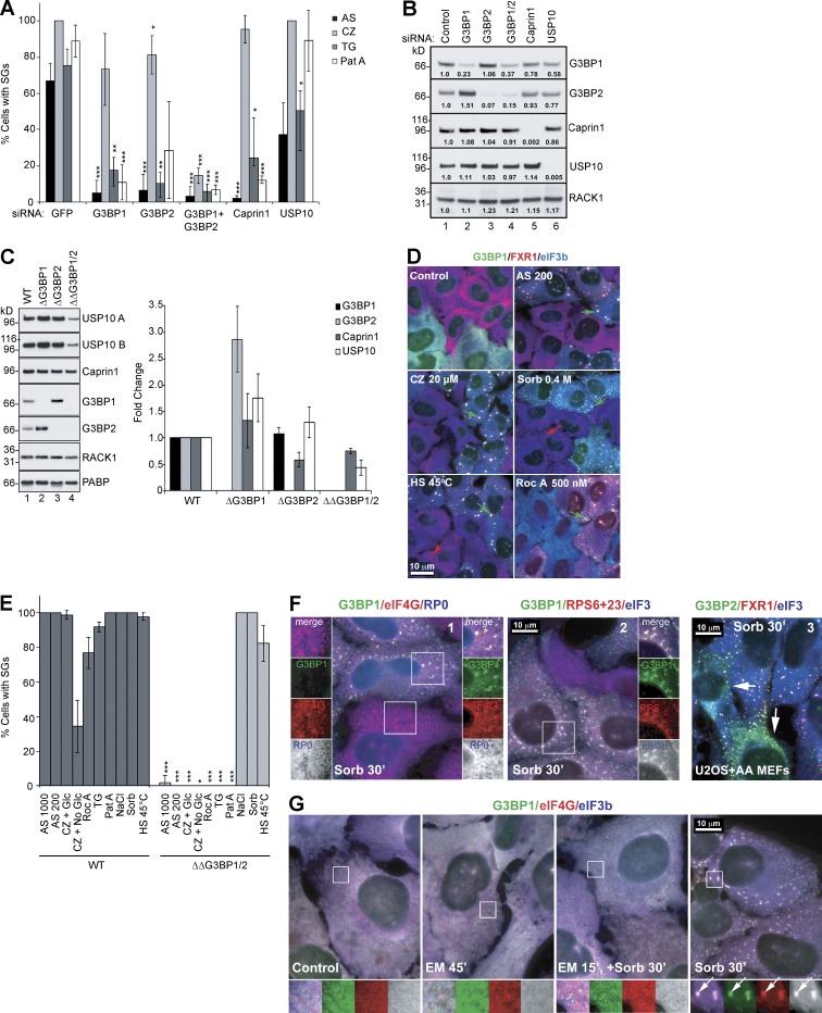
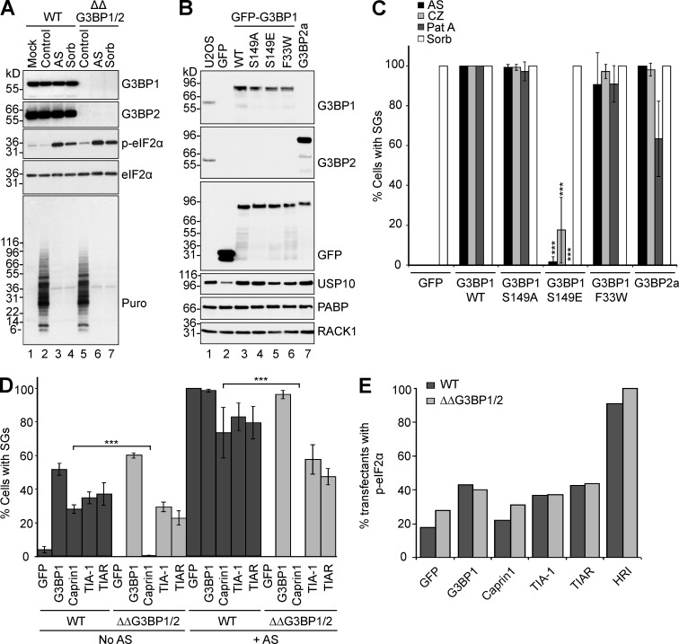
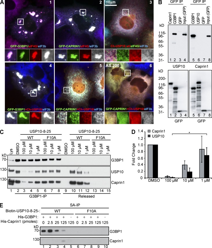
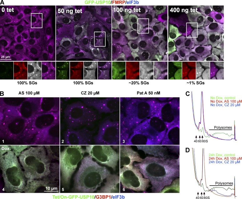
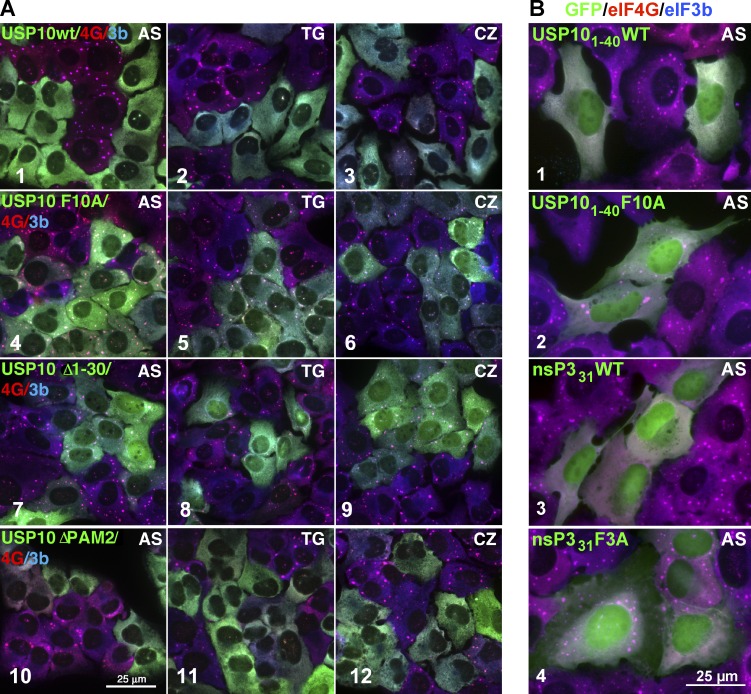
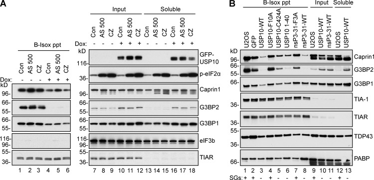
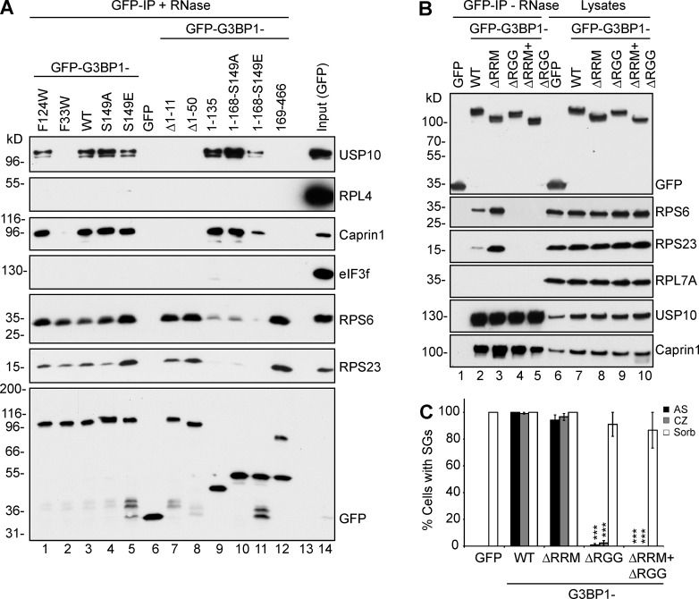
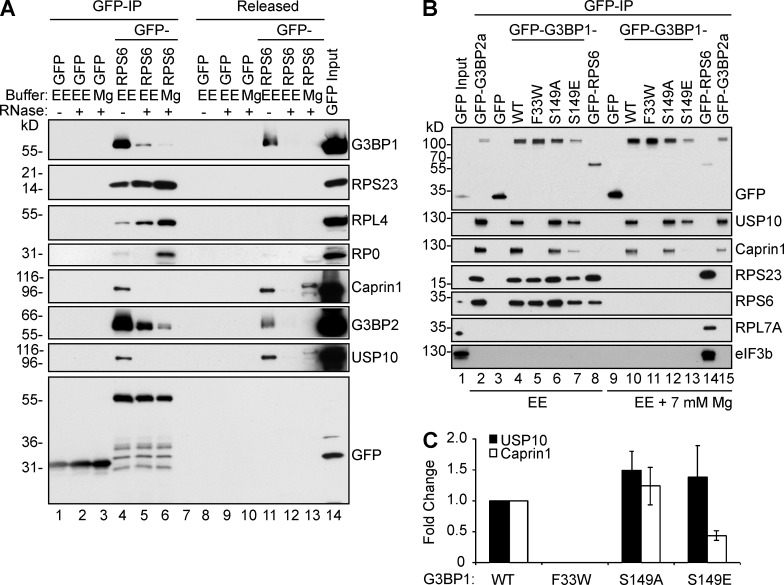
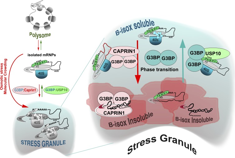
Mammalian stress granules (SGs) contain stalled translation preinitiation complexes that are assembled into discrete granules by specific RNA-binding proteins such as G3BP. We now show that cells lacking both G3BP1 and G3BP2 cannot form SGs in response to eukaryotic initiation factor 2α phosphorylation or eIF4A inhibition, but are still SG-competent when challenged with severe heat or osmotic stress. Rescue experiments using G3BP1 mutants show that phosphomimetic G3BP1-S149E fails to rescue SG formation, whereas G3BP1-F33W, a mutant unable to bind G3BP partner proteins Caprin1 or USP10, rescues SG formation. Caprin1/USP10 binding to G3BP is mutually exclusive: Caprin binding promotes, but USP10 binding inhibits, SG formation. G3BP interacts with 40S ribosomal subunits through its RGG motif, which is also required for G3BP-mediated SG formation. We propose that G3BP mediates the condensation of SGs by shifting between two different states that are controlled by the phosphorylation of S149 and by binding to Caprin1 or USP10.


Figures

Figure 1.
Figure 1.
Role of G3BP and partners in SG assembly. (A) U2OS-WT cells treated with indicated siRNAs were stressed for 1 h with 100 µM AS, 20 µM CZ, 1.0 µM TG, or 50 nM Pat A and then stained for SG markers eIF4G/eIF3b and scored. Data shown are mean ± SEM and are analyzed using the unpaired t test. *, P < 0.05; **, P < 0.01; ***, P < 0.005. siGFP control versus target siRNA treatments, n = 3. (B) Western blot analysis of siRNA-treated U2OS-WT cells. Proteins were quantified from blots using densitometry and normalized to RACK1, n = 3. Fold increase/decrease versus control is indicated. Mr (kD) are shown. (C) Western blot analysis of ΔG3BP1, ΔG3BP2, or ΔΔG3BP1/2 cell lysates, blotted for G3BP1, G3BP2, USP10 (A and B indicate different antibodies), Caprin1, RACK1, and PABP. Mr (kD) are shown. Proteins were quantified from blots using densitometry and normalized to RACK1. Fold change of WT versus KO was plotted. Data shown are mean ± SEM; no statistical analysis was performed, n = 4. (D) SGs in U2OS-WT and ΔΔG3BP1/2 cells, cocultured and treated as indicated, and then stained for G3BP1 (green), FXR1 (red), and eIF3b (blue). SGs in U2OS-WT cells (green arrows) and in ΔΔG3BP1/2 cells (red arrows) are indicated. Bar, 10 µm. (E) SG quantification in U2OS-WT cells (dark bars) and ΔΔG3BP1/2 (light bars), treated as indicated and scored for SGs using eIF4G and eIF3 as markers. Data shown are mean ± SEM and analyzed using the unpaired t test, WT versus ΔΔG3BP1/2. *, P < 0.05; ***, P < 0.005; n = 3. (F) Sorbitol-treated cocultured U2OS-WT (green) and ΔΔG3BP1/2 cells (1 and 2), stained as indicated. 3, sorbitol-treated cocultured mouse eIF2α-S51A MEFs (AA MEFs) and U2OS-WT, stained for G3BP2 (green), FXR1 (red), and eIF3b (blue). SGs in AA MEFs are indicated by white arrows. Insets zoomed 1.3× with separated colors. (G) U2OS-WT cells were untreated (a), EM treated for 45 s (b), EM alone for 15 s and then 0.4 M sorbitol for 30 s (c), or sorbitol alone for 30 s (d). Cells were stained for G3BP1 (green), eIF4G (red), and eIF3b (blue). Insets zoomed 2.3× with separated colors. Arrows indicate SGs. Bars, 10 µm.
Figure 2.
Figure 2.
G3BP is dispensable for translational arrest, required for SG formation, and regulated by S149. (A) U2OS-WT or ΔΔG3BP1/2 cells were untreated or exposed to 500 µM AS (1 h) or 0.4 M sorbitol (30 min) and lysed in SDS and resolved using SDS-PAGE/Western blotting. Blots were probed as indicated. Mr (kD) are shown. (B) ΔΔG3BP1/2 cells stably expressing the indicated constructs, analyzed by Western blot and probed as indicated. Mr (kD) are shown. (C) ΔΔG3BP1/2 cells stably expressing the indicated proteins, treated as indicated, stained, and scored for SGs and quantified. Data shown are mean ± SEM and analyzed using the unpaired t test, WT versus G3BP1-S149E. ***, P < 0.005; n = 3. (D) U2OS-WT (dark bars) or ΔΔG3BP1/2 cells (light bars) were transiently transfected with indicated plasmids, untreated or exposed to 500 µM AS for 1 h, and then stained and scored for SGs. Data shown are mean ± SEM and analyzed using the unpaired t test. ***, P < 0.005; n = 3. (E) U2OS-WT (dark bars) or ΔΔG3BP1/2 cells (light bars) were transiently transfected with indicated plasmids and then stained and scored for p-eIF2α–positive transfectants. Data represent mean. n = 2.
Figure 3.
Figure 3.
Caprin1 and USP10 binding to G3BP is mutually exclusive and regulates SG formation. (A) COS7 cells were transiently transfected with the indicated GFP (green) or Cherry (red)-tagged constructs and stained for endogenous eIF4G (1 and 2, red; 3, green) and/or eIF3b (1–6, blue). In 6, the cells were treated with 200 µM AS and then fixed and stained. Bar, 10 µm. Insets zoomed 2.5× with separated colors. (B) COS7 were transiently transfected with the indicated constructs, lysed and immunoprecipitated. Lysates and IPs were resolved using SDS-PAGE and subjected to Western blotting for the indicated proteins. Lower panels, duplicate samples probed for GFP to confirm IP efficiency. Mr (kD) are shown. (C) Endogenous G3BP1 from U2OS-WT cell lysates was immunoprecipitated, and the bound complexes were incubated with the indicated amounts of USP108–25WT or mutant USP108–25F10A peptides. Bound and released material was subjected to Western blotting for endogenous G3BP1, USP10, or Caprin1. (D) Quantification of displaced Caprin1 and USP10. Western blots from samples in C were quantified using densitometry. The ratio of each protein relative to G3BP1 was determined. Data shown are mean ± SEM and are analyzed using the unpaired t test. *, P < 0.05; n = 3. (E) Purified recombinant His-G3BP was mixed with biotinylated USP108–25WT or mutant USP108–25F10A peptides and bound to streptavidin (SA) beads. Beads were then washed and incubated with the indicated amounts of purified recombinant His-Caprin1. Bound material was subjected to Western blotting for G3BP1 and Caprin1. n = 3. Mr (kD) are shown.
Figure 4.
Figure 4.
USP10 blocks SG assembly downstream of translational arrest. (A) Tet-on GFP-USP10 U2OS-WT cells were treated with increasing amounts of tetracycline for 22 h and then treated with 100 µM AS for 1 h and fixed and stained with SG markers (FMRP [FMR1], red; eIF3b, blue). Bar, 25 µm. Insets zoomed 1.3× with separated colors. The percentage of cells with SGs is indicated below each panel; n = 3. (B) Tet-on GFP-USP10 was uninduced (1–3) or induced with doxycycline (Dox) for 24 h (4–6) before indicated 1-h drug treatments. Cells were treated with 100 µM AS, (1 and 4), 20 µM CZ, (2 and 5), or 50 nM Pat A (3 and 6) and then stained for G3BP1 (red) and eIF3b (blue). Bar, 10 µm. (C) Polysome profiles obtained from uninduced tet-on GFP-USP10 cells as in B (1–3) and treated with control (green), 100 µM AS (red), or 20 µM CZ (blue); n = 3. (D) Polysome profiles of doxycycline-induced tet-on GFP-USP10 cells, treated as in B (4–6); n = 3.
Figure 5.
Figure 5.
FGDF motif of USP10 or nsP331 is required to block SG formation. (A) U2OS-WT cells stably expressing GFP-tagged USP10-WT, F10A, Δ1–30, and ΔPAM2 (green) were cocultured with U2OS-WT cells (nongreen), treated with 200 µM AS (1, 4, 7, and 10), 1 µM TG (2, 5, 8, and 11), or 20 µM CZ (3, 6, 9, and 12) and stained for SG markers eIF4G (red) and eIF3b (blue). Bar, 25 µm. (B) U2OS-WT cells transiently transfected with GFP-USP101–40WT, GFP-USP101–40-F10A, GFP-nsP331WT, or GFP-nsP331F3A as indicated and treated with 500 µM AS for 1 h, fixed, and stained for eIF4G (red) and eIF3b (blue). Bar, 25 µm.
Figure 6.
Figure 6.
USP10/nsP3 binding to G3BP inhibits B-isox precipitation of G3BP and Caprin1. (A) Tet-on GFP-USP10 cells without (lanes 1–3, 7–9, and 13–15) or with induction (lanes 4–6, 10–12, and 16–18) were treated with AS (500 µM) or CZ (20 µM), or were untreated, before lysis in Kato buffer and B-isox fractionation. Precipitated material (lanes 1–6), input (lanes 7–12), and soluble material (lanes 13–18) were subjected to Western blotting for the indicated proteins. Mr (kD) are shown. (B) U2OS-WT cells stably expressing indicated GFP-tagged proteins were lysed in EE buffer, fractionated using B-isox, and Western blotted for the indicated proteins. SGs or lack thereof are indicated at the bottom. Lanes 1–8, precipitates; lanes 9–11, input; and lanes 12 and 13, soluble material. The C424A USP10 mutant is enzymatically inactive. Mr (kD) are shown.
Figure 7.
Figure 7.
G3BP1 RGG motif is required for association with 40S ribosomal subunits and for SG competence. (A) GFP and GFP-tagged G3BP1 variants were expressed in COS7 cells, lysed in EE buffer, and immunoprecipitated with RNase treatment. Lysates and IPs were resolved using SDS-PAGE and subjected to Western blotting for the indicated proteins. Mr (kD) are shown. (B) GFP and GFP-tagged G3BP1 variants were stably expressed in ΔΔG3BP1/2 cells and immunoprecipitated. Lysates and IPs were resolved using SDS-PAGE and subjected to Western blotting for the indicated proteins. Mr (kD) are shown. (C) ΔΔG3BP1/2 cells expressing the indicated proteins were treated as indicated, stained, and scored for SGs using eIF4G and eIF3b. Data shown are mean ± SEM and are analyzed using the unpaired t test, WT versus G3BP1 variants. ***, P < 0.005; n = 3.
Figure 8.
Figure 8.
G3BP1 and G3BP2 selectively associate with dissociated 40S ribosomal subunits. (A) Western blot of IPs from cells expressing either GFP (lanes 1–3) or RPS6-GFP (lanes 4–6), lysed in either ribosome-dissociating (EE) or ribosome-stabilizing (+5.0 mM MgCl2) buffer, washed, and incubated with RNase A or buffer. Released material was concentrated using acetone precipitation before SDS treatment; the bound material (GFP-IP) was eluted in SDS. IPs, lanes 1–6; released material, lanes 8–13. Mr (kD) are shown. (B) Stably expressed GFP-RPS6 in U2OS-WT cells, or GFP, GFP-tagged G3BP1 variants, or G3BP2a stably expressed in ΔΔG3BP1/2 cells were lysed in ribosome-dissociating EE or in ribosome stabilizing (EE + 7 mM MgCl2) buffer and immunoprecipitated and Western blotted as indicated. Mr (kD) are shown. (C) Caprin1, USP10, and GFP were quantified from EE/IP Western blots (C) using densitometry. The ratio of Caprin1 or USP10 relative to GFP-G3BP1 was determined, and the fold change was plotted. Data shown are mean ± SEM; no statistical analysis was performed; n = 3.
Figure 9.
Figure 9.
USP10 binding to G3BP regulates SG condensation by inhibiting a G3BP-mediated condensation event. Stress promotes polysome disassembly, thus exposing mRNA and converting polysomes into mRNPs. G3BP shuttles between two different phases, promoting a similar state change in 40S subunits, Caprin1, and their bound mRNAs. USP10 binding to G3BP stabilizes a soluble conformation of G3BP bound to 40S subunits (via G3BP C terminus) and to PABP (through USP10), causing SG disassembly. Some SG-associated factors, such as eIF3, remain in the cytoplasmic soluble state, but accumulate in the SG as individual mRNPs are mobilized back into active translation. For further details, see the Discussion section.

References

    1. Anderson P., and Kedersha N.. 2008. Stress granules: the Tao of RNA triage. Trends Biochem. Sci. 33:141–150. 10.1016/j.tibs.2007.12.003 - DOI - PubMed
    1. Anderson P., and Kedersha N.. 2009. RNA granules: post-transcriptional and epigenetic modulators of gene expression. Nat. Rev. Mol. Cell Biol. 10:430–436. 10.1038/nrm2694 - DOI - PubMed
    1. Aulas A., Caron G., Gkogkas C.G., Mohamed N.V., Destroismaisons L., Sonenberg N., Leclerc N., Parker J.A., and Vande Velde C.. 2015. G3BP1 promotes stress-induced RNA granule interactions to preserve polyadenylated mRNA. J. Cell Biol. 209:73–84. 10.1083/jcb.201408092 - DOI - PMC - PubMed
    1. Berger A., Ivanova E., Gareau C., Scherrer A., Mazroui R., and Strub K.. 2014. Direct binding of the Alu binding protein dimer SRP9/14 to 40S ribosomal subunits promotes stress granule formation and is regulated by Alu RNA. Nucleic Acids Res. 42:11203–11217. 10.1093/nar/gku822 - DOI - PMC - PubMed
    1. Bevilacqua E., Wang X., Majumder M., Gaccioli F., Yuan C.L., Wang C., Zhu X., Jordan L.E., Scheuner D., Kaufman R.J., et al. 2010. eIF2alpha phosphorylation tips the balance to apoptosis during osmotic stress. J. Biol. Chem. 285:17098–17111. 10.1074/jbc.M110.109439 - DOI - PMC - PubMed

Publication types

MeSH terms