

Oxidative stress, inflammation, and DNA damage in multiple organs of mice acutely exposed to amorphous silica nanoparticles
- PMID: 27022259
- PMCID: PMC4788369
- DOI: 10.2147/IJN.S92278
Oxidative stress, inflammation, and DNA damage in multiple organs of mice acutely exposed to amorphous silica nanoparticles
Abstract
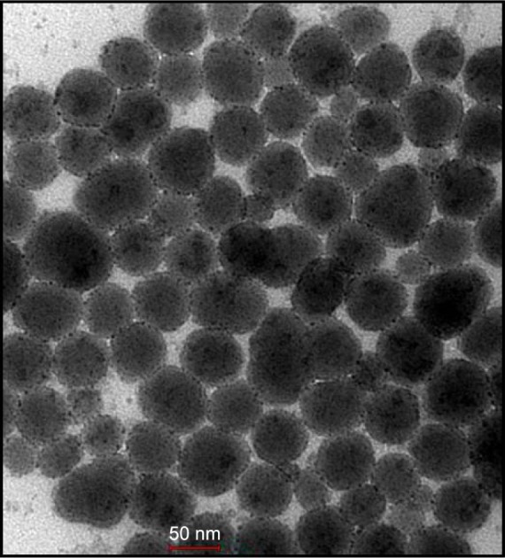
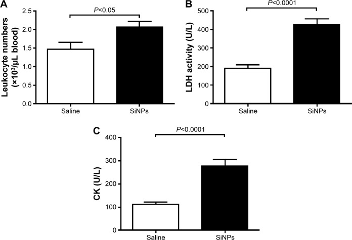
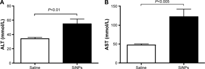
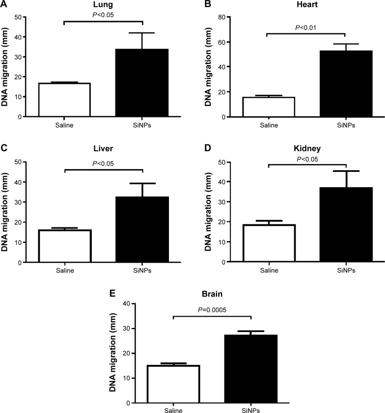
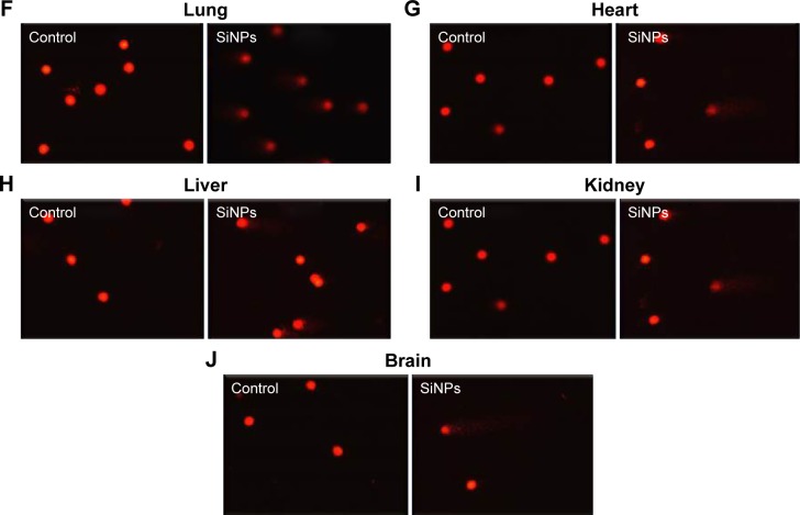
The use of amorphous silica (SiO2) in biopharmaceutical and industrial fields can lead to human exposure by injection, skin penetration, ingestion, or inhalation. However, the in vivo acute toxicity of amorphous SiO2 nanoparticles (SiNPs) on multiple organs and the mechanisms underlying these effects are not well understood. Presently, we investigated the acute (24 hours) effects of intraperitoneally administered 50 nm SiNPs (0.25 mg/kg) on systemic toxicity, oxidative stress, inflammation, and DNA damage in the lung, heart, liver, kidney, and brain of mice. Lipid peroxidation was significantly increased by SiNPs in the lung, liver, kidney, and brain, but was not changed in the heart. Similarly, superoxide dismutase and catalase activities were significantly affected by SiNPs in all organs studied. While the concentration of tumor necrosis factor α was insignificantly increased in the liver and brain, its increase was statistically significant in the lung, heart, and kidney. SiNPs induced a significant elevation in pulmonary and renal interleukin 6 and interleukin-1 beta in the lung, liver, and brain. Moreover, SiNPs caused a significant increase in DNA damage, assessed by comet assay, in all the organs studied. SiNPs caused leukocytosis and increased the plasma activities of lactate dehydrogenase, creatine kinase, alanine aminotranferase, and aspartate aminotransferase. These results indicate that acute systemic exposure to SiNPs causes oxidative stress, inflammation, and DNA damage in several major organs, and highlight the need for thorough evaluation of SiNPs before they can be safely used in human beings.
Keywords: DNA damage; amorphous silica nanoparticles; inflammation; organ toxicity; oxidative stress.
Figures
Similar articles
-
Cerium Oxide Nanoparticles in Lung Acutely Induce Oxidative Stress, Inflammation, and DNA Damage in Various Organs of Mice.Oxid Med Cell Longev. 2017;2017:9639035. doi: 10.1155/2017/9639035. Epub 2017 Mar 14. Oxid Med Cell Longev. 2017. PMID: 28392888 Free PMC article.
-
Amorphous silica nanoparticles impair vascular homeostasis and induce systemic inflammation.Int J Nanomedicine. 2014 Jun 2;9:2779-89. doi: 10.2147/IJN.S52818. eCollection 2014. Int J Nanomedicine. 2014. PMID: 24936130 Free PMC article.
-
Characterization of in vitro genotoxic, cytotoxic and transcriptomic responses following exposures to amorphous silica of different sizes.Mutat Res Genet Toxicol Environ Mutagen. 2016 Jan 15;796:8-22. doi: 10.1016/j.mrgentox.2015.11.011. Epub 2015 Nov 25. Mutat Res Genet Toxicol Environ Mutagen. 2016. PMID: 26778505
-
Toxicology of silica nanoparticles: an update.Arch Toxicol. 2017 Sep;91(9):2967-3010. doi: 10.1007/s00204-017-1993-y. Epub 2017 Jun 1. Arch Toxicol. 2017. PMID: 28573455 Free PMC article. Review.
-
The toxicity of silica nanoparticles to the immune system.Nanomedicine (Lond). 2018 Aug 1;13(15):1939-1962. doi: 10.2217/nnm-2018-0076. Epub 2018 Aug 28. Nanomedicine (Lond). 2018. PMID: 30152253 Review.
Cited by
-
SiNPs induce ferroptosis in HUVECs through p38 inhibiting NrF2 pathway.Front Public Health. 2023 Feb 8;11:1024130. doi: 10.3389/fpubh.2023.1024130. eCollection 2023. Front Public Health. 2023. PMID: 36844840 Free PMC article.
-
Cerium Oxide Nanoparticles in Lung Acutely Induce Oxidative Stress, Inflammation, and DNA Damage in Various Organs of Mice.Oxid Med Cell Longev. 2017;2017:9639035. doi: 10.1155/2017/9639035. Epub 2017 Mar 14. Oxid Med Cell Longev. 2017. PMID: 28392888 Free PMC article.
-
Nose-Only Water-Pipe Smoke Exposure in Mice Elicits Renal Histopathological Alterations, Inflammation, Oxidative Stress, DNA Damage, and Apoptosis.Front Physiol. 2020 Feb 11;11:46. doi: 10.3389/fphys.2020.00046. eCollection 2020. Front Physiol. 2020. PMID: 32116758 Free PMC article.
-
Magnesium Supplementation Alleviates the Toxic Effects of Silica Nanoparticles on the Kidneys, Liver, and Adrenal Glands in Rats.Toxics. 2023 Apr 17;11(4):381. doi: 10.3390/toxics11040381. Toxics. 2023. PMID: 37112608 Free PMC article.
-
Aortic Oxidative Stress, Inflammation and DNA Damage Following Pulmonary Exposure to Cerium Oxide Nanoparticles in a Rat Model of Vascular Injury.Biomolecules. 2019 Aug 17;9(8):376. doi: 10.3390/biom9080376. Biomolecules. 2019. PMID: 31426470 Free PMC article.
References
-
- Napierska D, Thomassen LC, Rabolli V, et al. Size-dependent cytotoxicity of monodisperse silica nanoparticles in human endothelial cells. Small. 2009;5:846–853. - PubMed
-
- Lison D, Thomassen LC, Rabolli V, et al. Nominal and effective dosimetry of silica nanoparticles in cytotoxicity assays. Toxicol Sci. 2008;104:155–162. - PubMed
Publication types
MeSH terms
Substances
LinkOut - more resources
Full Text Sources
Other Literature Sources
