

doi: 10.1155/2016/3608602.
Epub 2016 Feb 23.
Histological Features and Biocompatibility of Bone and Soft Tissue Substitutes in the Atrophic Alveolar Ridge Reconstruction
Affiliations
- PMID: 27022489
- PMCID: PMC4781934
- DOI: 10.1155/2016/3608602
Item in Clipboard
Histological Features and Biocompatibility of Bone and Soft Tissue Substitutes in the Atrophic Alveolar Ridge Reconstruction
Case Rep Dent.
2016.
Abstract
The reconstruction of the atrophic alveolar ridges for implant placement is today a common procedure in dentistry daily practice. The surgical reconstruction provides for the optimization of the supporting bone for the implants and a restoration of the amount of keratinized gingiva for esthetic and functional reasons. In the past, tissue regeneration has been performed with autogenous bone and free gingival or connective tissue grafts. Nowadays, bone substitutes and specific collagen matrix allow for a complete restoration of the atrophic ridge without invasive harvesting procedures. A maxillary reconstruction of an atrophic ridge by means of tissue substitutes and its histological features are then presented.
Figures
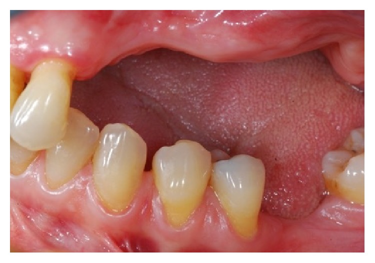
Clinical view of the left upper maxilla atrophic ridge.
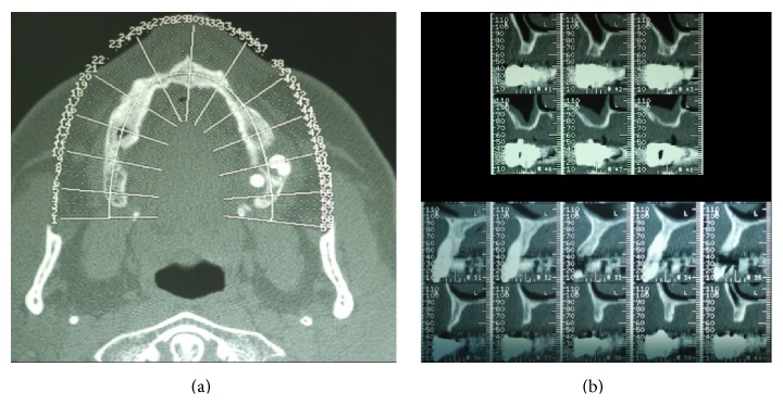
The CT dental scan confirms the clinical view showing atrophic maxillary ridge with important horizontal bone defect. (a) It is possible to underline all the atrophies of the upper maxilla; (b) a particular of the axial section of the area involved in the grafting procedures.
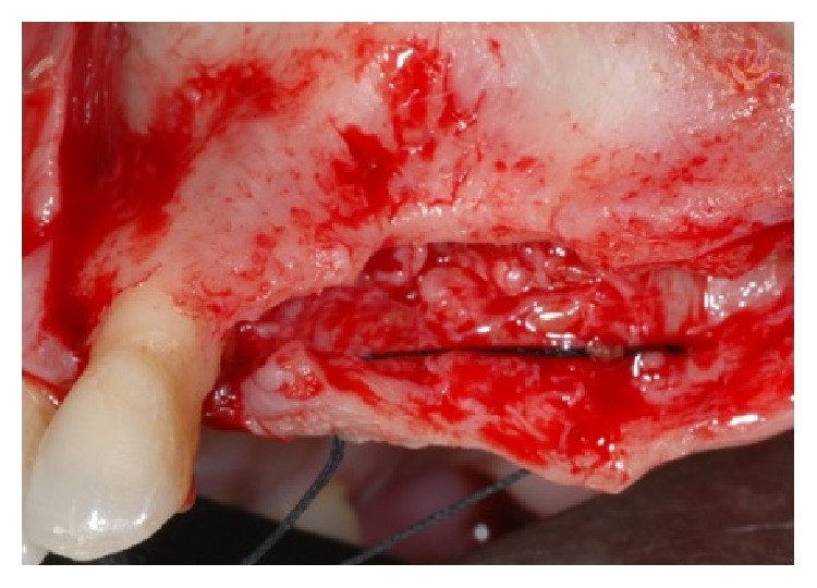
Clinical view of the bone after the elevation of the mucoperiosteal flap.
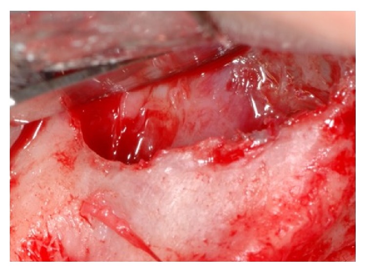
Sinus lift was performed in order to increase the volume of the bone height.
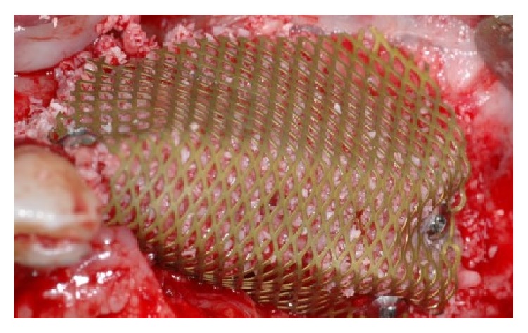
A titanium mesh has been placed for increasing the vertical and horizontal defect.
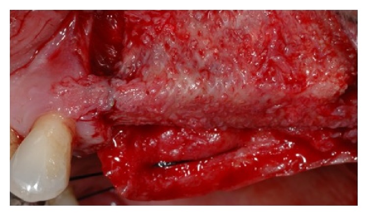
The time titanium mesh removal showed a good healing and the bone volume recovering.
Occlusal view of the increased bone after the bone regeneration procedure with the applied titanium mesh.
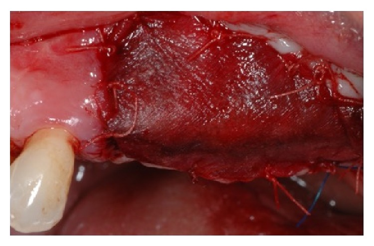
A collagen membrane has been placed in order to favor the soft tissue healing.
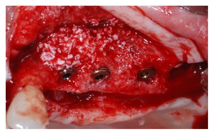
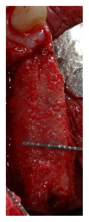
At the time of dental implants the healed bone has been removed and analyzed in order to perform the histologic analysis.
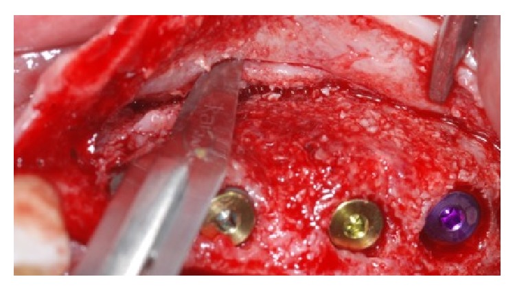
Dental implant was placed and periosteal incision has to be made in order to have a complete soft tissue covering of the dental implants placed.
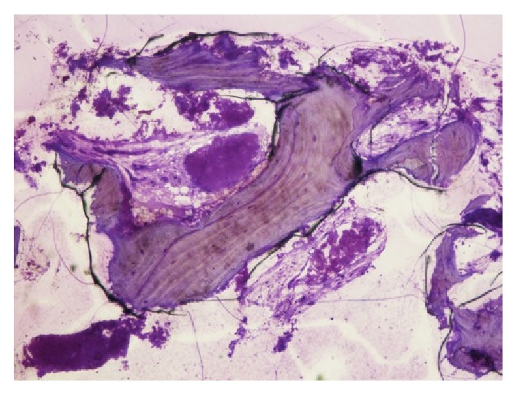
Histological image 40x magnification underlines the presence of new bone cells and some residual particles of the substitute material.
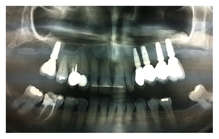
Two-year postoperative X-ray control evidences the integration of the dental implants for support of a fixed prosthetic rehabilitation.
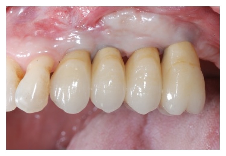
Two-year clinical analyses show a good healing of the soft tissue.
References
-
- Bouri A., Jr., Bissada N., Al-Zahrani M. S., Faddoul F., Nouneh I. Width of keratinized gingiva and the health status of the supporting tissues around dental implants. The International Journal of Oral & Maxillofacial Implants. 2008;23(2):323–326. - PubMed
-
- Cicciù M., Herford A. S., Stoffella E., Cervino G., Cicciù D. Protein-signaled guided bone regeneration using titanium mesh and Rh-BMP2 in oral surgery: a case report involving left mandibular reconstruction after tumor resection. The Open Dentistry Journal. 2012;6(1):51–55. doi: 10.2174/1874210601206010051. - DOI - PMC - PubMed
-
- Yamada S., Shima N., Kitamura H., Sugito H. Effect of porous xenographic bone graft with collagen barrier membrane on periodontal regeneration. International Journal of Periodontics and Restorative Dentistry. 2002;22(4):389–397. - PubMed
LinkOut - more resources
Full Text Sources
Other Literature Sources
Miscellaneous
