. 2016 May;157(5):2160-72.
doi: 10.1210/en.2015-2064. Epub 2016 Mar 29.

De Novo-Synthesized Retinoic Acid in Ovarian Antral Follicles Enhances FSH-Mediated Ovarian Follicular Cell Differentiation and Female Fertility

Affiliations

De Novo-Synthesized Retinoic Acid in Ovarian Antral Follicles Enhances FSH-Mediated Ovarian Follicular Cell Differentiation and Female Fertility

Tomoko Kawai et al. Endocrinology. 2016 May.

Abstract

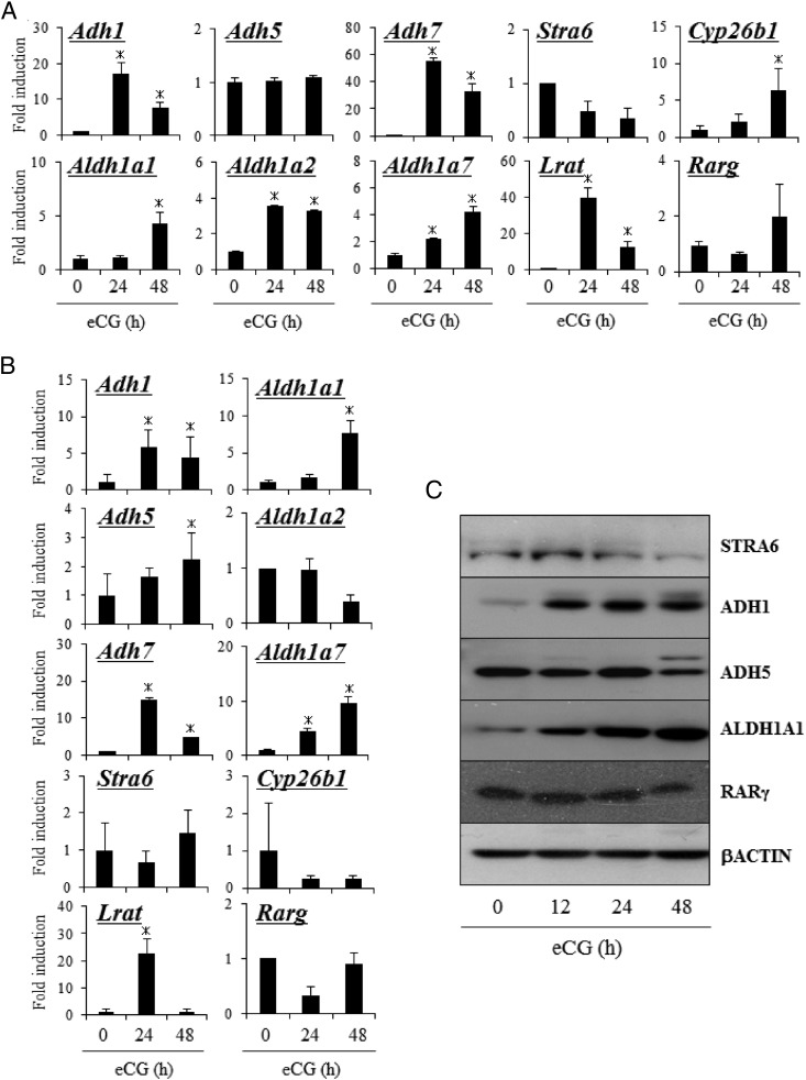
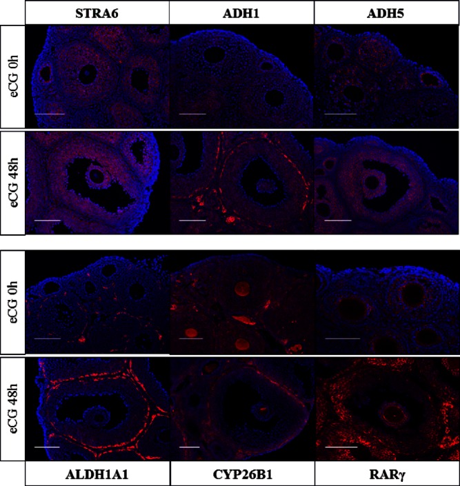
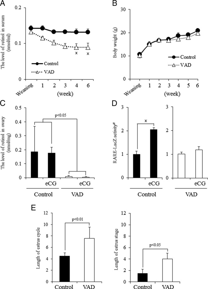
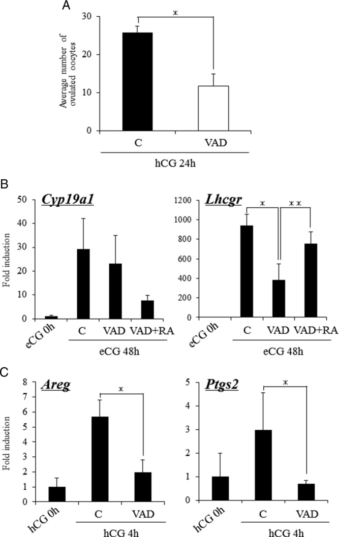
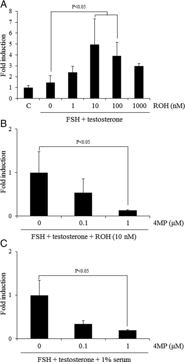
Retinoic acid (RA) is the active form of vitamin A and is synthesized from retinol by two key enzymes, alcohol dehydrogenase (ADH) and acetaldehyde dehydrogenase (ALDH). As the physiological precursor of RA, retinol impacts female reproductive functions and fertility. The expression of Adh1 and Adh5 as well as Aldh1a1 and Aldh1a7 are significantly increased in the ovaries of mice treated with equine chorionic gonadotropin/FSH. The RA receptor is expressed and localized in granulosa cells and is activated by endogenous RA as indicated by LacZ expression in granulosa cells of RA-responsive transgene-LacZ transgenic mice (RA reporter mice). Coinjection of the ADH inhibitor, 4-methylpyrazole, with equine chorionic gonadotropin significantly decreases the number and developmental competence of oocytes ovulated in response to human chorionic gonadotropin/LH as compared with controls. Injections of RA completely reverse the effects of the inhibitor of ovulation and oocyte development. When mice were fed a retinol-free, vitamin A-deficient diet that significantly reduced the serum levels of retinol, the expression of the LH receptor (Lhcgr) was significantly lower in the ovaries of the vitamin A-deficient mice, and injections of human chorionic gonadotropin failed to induce genes controlling ovulation. These results indicate that ovarian de novo biosynthesis of RA is required for the follicular expression of Lhcgr in granulosa cells and their ability to respond to the ovulatory LH surge.


Figures

Figure 1.
Figure 1.
The expression of genes related to RA synthesis in ovary of mice treated with or without eCG. A, Kinetic changes in the expression of Stra6, Adh1,5,7, Aldh1a1, Aldh1a2, Aldh1a7, Stra6, Lrat, Cyp26b1, and Rarg mRNA in ovaries of mice treated with or without eCG. For reference, the eCG 0 hour value in ovary is set as 1, and the data are presented as fold induction. Values are mean ± SEM of three replicates. *, Significant differences are observed between ovaries from control compared with those of eCG-primed mice (eCG 0 h) (P < .05). B, Kinetic changes in the expression of Stra6, Adh1, Aldh5, Aldh7, Aldh1a1, Aldh1a2, Aldh1a7, Stra6, Lrat, Cyp26b1, and Rarg mRNA in granulosa cells (GCs) of mice treated with or without eCG. For reference, the eCG 0 hour value in GCs is set as 1, and the data are presented as fold induction. Values are mean ± SEM of three replicates. *, Significant differences are observed between GCs from control mice compared with those of eCG-primed mice (eCG 0 h) (P < .05). C, The expression of STRA6, ADH1, ADH5, ALDH1A1, RARγ, and β-actin protein in whole-ovary samples detected by Western blot analysis.
Figure 2.
Figure 2.
The localization of factors related to RA synthesis and its functions in ovary of mice treated with or without eCG. Localizations of STRA6, ADH1, ADH5, ALDH1A1, CYP26B1, and RARγ in ovary were detected by fluorescent staining in the ovary of mice treated with or without eCG for 48 hours. Each protein was visualized with Cy3 and nuclear was stained with 4′,6′-diamino-2-phenylindole. Scale bur is 100 μm.
Figure 3.
Figure 3.
The detection of de novo synthesized RA in ovary using RARE reporter mice. A, Localization of LacZ-positive cells in the ovary of RARE reporter mice treated with eCG or without eCG (control). B, Activity of LacZ enzyme driven by RARE promoter in ovary (left panel) or granulosa cells (right panel) of RARE reporter mice treated with or without eCG. Values were absorbance in 595 nm after the protein samples were incubated with the substrate, CPRG, for 2 hours at 37°C. #, The RARE-LacZ activity is presented as fold induction. The control value in ovary (left panel) or granulosa cells (right panel) is set as 1. Values are mean ± SEM of three replicates. eCG treatment significantly increased the RARE-LacZ enzyme activity in ovaries (P < .01) or granulosa cells (P < .05) of mice as compared with those in mice without eCG treatment (control), respectively.
Figure 4.
Figure 4.
Characteristics of mice fed vitamin A (retinol)-deficient diets from weaning. A, Kinetic change of retinol levels in serum in mice fed a normal diet (control) or VAD. *, Significant differences are observed at each time interval as compared with control mice (P < .05). B, The body weight of control mice or VAD mice. C, Levels of retinol in the ovary of control mice or VAD mice with or without eCG treatment. eCG, 48 hours after eCG priming. Values are mean ± SEM of three replicates. Statistical analysis was done by a two-way ANOVA. Significant differences are observed between control and VAD mice (P < .05) but not between treatment groups with or without eCG priming. D, Activity of LacZ enzyme driven by RARE promoter in granulosa cells of control or VAD mice with or without eCG priming. eCG, 48 hours after eCG priming. Values are absorbance in 595 nm after incubation for 2 hours at 37°C from the addition of LacZ enzyme substrate, CPRG. #, The RARE-LacZ activity is presented as fold induction. The eCG 0 hours value in granulosa cells is set as 1. Values are mean ± SEM of three replicates. *, Significant induction is observed by eCG treatment in control mice (P < .05). E, Length of the estrus cycle and the length of the estrous stage of control mice or VAD mice. Values are mean ± SEM of five mice.
Figure 5.
Figure 5.
Ovarian function of mice fed a VAD diet. A, The effects of the VAD diet on the ovulation of hormone-treated mice. Mice fed normal diet (control) and VAD diet were injected with eCG. After 48 hours, these mice were further treated with hCG. For 24 hours after hCG injection, the ovulated oocytes were collected from oviducts and then the number of them was counted. Values are mean ± SEM of 4 mice. *, Significant differences are observed as compared with those in control mice (P < .05). B, The expression of genes known to be markers of follicular development, Cyp19a1 and Lhcgr, in granulosa cells of control mice and VAD mice injected with eCG and/or RA. For reference, the eCG 0 hours values are set as 1, and the data are presented as fold induction. *, Significant differences are observed between control (C) and VAD groups (P < .05); **, the addition of RA to VAD mice (RA+VAD) significantly increased the expression of Lhcgr in VAD mice (P < .05). Values are mean ± SEM of three replicates. RA, coinjection with eCG and RA (2.5 mg/kg) for 48 hours. C, The expression of genes known to be the target of the LH-LH receptor (LHR)-dependent pathway in granulosa cells of control mice and VAD mice injected with hCG for 4 hours. For reference, the hCG 0 hour values are set as 1, and the data are presented as fold induction. *, Significant differences are observed as compared with those in ovaries of mice fed normal diet between control and VAD mice. Values are mean ± SEM of three replicates.
Figure 6.
Figure 6.
The effects of ADH inhibitor on follicular development and/or ovulation process. A, The number of ovulated oocytes when the mice were coadministered with eCG and ADH inhibitor (4MP) followed by hCG injection or hCG and 4MP after eCG stimulation. Day 23 immature female mice were coinjected with eCG and 4MP (8 mg/kg). After 48 hours, these mice were treated with hCG. In other group, day 23 immature female mice treated with eCG for 48 hours were coinjected with hCG and 4MP. For 16 hours after hCG injection, the ovulated oocytes were collected from oviducts and counted. Values are mean ± SEM of three mice. *, Significant differences were observed as compared with that in control (P < .05). Control, immature female mice were treated with eCG followed by hCG stimulation. eCG+4MP; the mice were coinjected with eCG and 4MP (8 mg/kg) for 48 hours, and then hCG was injected into the mice. hCG+4MP, the mice were coinjected with hCG and 4MP (8 mg/kg) 48 hours after eCG injection. B, The expression of Lhcgr and Cyp19a1in granulosa cells of mice treated with eCG and 4MP for 48 hours or the expression of LH-LHR target genes, Areg and Ptgs2, in granulosa cells of mice injected with hCG for 4 hours after eCG and 4MP treatment. For reference, the values of eCG 0 hour are set as 1, and the data are presented as fold induction. *, 4MP treatment significantly suppressed the gene expressions as compared with those in control mice treated with eCG alone (P < .05). Values are mean ± SEM of three replicates. C, Activity of LacZ enzyme driven by RARE promoter in granulosa cells of eCG-stimulated mice with or without 4MP treatment for 48 hours. Values are absorbance in 595 nm after the protein samples are incubated for 2 hours at 37°C with CPRG, LacZ substrate. #, The RARE-LacZ activity is presented as fold induction. The eCG 0 hour value in granulosa cells is set as 1. Values are mean ± SEM of three replicates. *, 4MP treatment significantly decreased the level of LacZ enzyme activity after eCG stimulation (P < .05). D, Circular levels of estrogen in mice treated with eCG and/or 4MP, RA, or 17β-estradiol. *, The estrogen level was significantly increased by eCG injection (P < .05); however, there was no significant difference among the treatment group of eCG-primed mice (P < .05). Values are mean ± SEM of three replicates. 4MP, Mice were coinjected with eCG and 4MP (8 mg/kg) for 48 hours. E2, Mice were coinjected with eCG+4MP and 17β-estradiol (10 mg/kg) for 48 hours. RA; mice were coinjected with eCG+4MP and RA (2.5 mg/kg) for 48 hours. E, The addition of RA treatment overcomes 4MP-induced negative effect of Lhcgr expression in granulosa cells. #, The expression level of Lhcgr is presented as fold induction. The eCG 0 hour value is set as 1. *, 4MP treatment significantly decreased the expression of Lhcgr in granulosa cells of eCG-stimulated mice (P < .05); **, the addition of RA but not 17β-estradiol (E2) significantly increased the level of Lhcgr expression as compared with that in granulosa cells of mice cotreated with eCG and 4MP. Values are mean ± SEM of three replicates.
Figure 7.
Figure 7.
The effect of activation of the RA synthesis from retinol in the ovary on the Lhcgr expression. A, The effects of additional retinol (ROH) to FSH+T-containing medium on the expression of Lhcgr when the ovary was cultured in the absence of serum. For reference, the control (C) value was set as 1, and the data are presented as fold induction. The addition of 10 or 100 nM of retinol (ROH) to FSH and T-containing medium significantly increased the expression level of Lhcgr as compared with those in the ovaries cultured for 48 hours with FSH and T (P < .05). Values are mean ± SEM of three replicates. B, The conversion from retinol to RA is required for the induction of Lhcgr expression in vitro under serum-free condition. For reference, FSH+T+ROH (10 nM) value was set as 1, and the data are presented as fold induction. **, The treatment with 1 μM 4MP significantly decreased the expression level of Lhcgr as compared with that in ovary cultured with FSH and T with 10 nM of retinol (ROH) (P < .05). Values are mean ± SEM of three replicates. C, Ovaries of immature day 21 mice were embedded in alginate gels and then cultured for 48 hours without any hormones. The conversion from retinol to RA is required for the induction of Lhcgr expression in vitro under 1% serum condition. For reference, FSH+T value was set as 1, and the data are presented as fold induction. ***, The treatment with 1 μM 4MP significantly decreased the expression level of Lhcgr as compared with that in ovary cultured with FSH and T (P < .05). Values are mean ± SEM of three replicates.

References

    1. Richards JS. Hormonal control of gene expression in the ovary. Endocr Rev. 1994;15:725–751. - PubMed
    1. Elvin JA, Yan C, Matzuk MM. Oocyte-expressed TGF-β superfamily members in female fertility. Mol Cell Endocrinol. 2000;159:1–5. - PubMed
    1. Shimasaki S, Moore RK, Otsuka F, Erickson GF. The bone morphogenetic protein system in mammalian reproduction. Endocr Rev. 2004;25:72–101. - PubMed
    1. Barnett KR, Schilling C, Greenfeld CR, Tomic D, Flaws JA. Ovarian follicle development and transgenic mouse models. Hum Reprod. 2006;12:537–555. - PubMed
    1. Shimasaki S, Zachow RJ, Li D, et al. A functional bone morphogenetic protein system in the ovary. Proc Natl Acad Sci USA. 1999;96:7282–7287. - PMC - PubMed