Review
. 2016 Jun:28:50-6.
doi: 10.1016/j.coph.2016.03.001. Epub 2016 Mar 26.

Bone marrow adipose tissue: formation, function and regulation

Affiliations
Review

Bone marrow adipose tissue: formation, function and regulation

Karla J Suchacki et al. Curr Opin Pharmacol. 2016 Jun.

Abstract

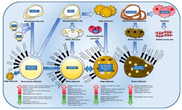
The human body requires an uninterrupted supply of energy to maintain metabolic homeostasis and energy balance. To sustain energy balance, excess consumed calories are stored as glycogen, triglycerides and protein, allowing the body to continue to function in states of starvation and increased energy expenditure. Adipose tissue provides the largest natural store of excess calories as triglycerides and plays an important role as an endocrine organ in energy homeostasis and beyond. This short review is intended to detail the current knowledge of the formation and role of bone marrow adipose tissue (MAT), a largely ignored adipose depot, focussing on the role of MAT as an endocrine organ and highlighting the pharmacological agents that regulate MAT.


Figures

Figure 1
Figure 1. Location of the principal white, brown, beige and marrow fat depots in mice and humans
In human adults and rodents the main white adipose tissue depots are subdivided into subcutaneous and visceral adipose tissue. Visceral adipose tissue surrounds the vital organs and is further subdivided into 1. Pericardial and epicardial in the thoracic cavity and 2. Retroperitoneal, 3. Omental, 4. Mesenteric and 5. Gonadal in the abdominal cavity. Subcutaneous white adipose tissue is located 6. Intramuscularly and out-with the abdominal and thoracic cavities, here exemplified by 7. gluteofemoral adipose tissue (human) and inguinal adipose tissue (mouse). Inguinal adipose tissue is composed of both white and beige adipocytes and can be highly influenced by environmental and dietary factors. Brown adipose tissue is located 8. Deep within the neck (humans) and in the interscapular region (rodent), with both of these depots containing classical brown adipocytes. Humans also contain some brown adipose tissue supercalivclularly (9.), although this may also contain white and beige adipocytes. 10. Marrow adipose tissue shows a similar distribution in rodents and humans, predominating in the arms and legs but being sparse (or non-existent) in the spine and more central skeleton. Adapted from [64,65].
Figure 2
Figure 2. Bone marrow, white, beige and brown fat developmental origins, functions and regulators
Originating from mesenchymal stem cells that express Pdgfrα, white adipocytes are enriched in several markers, including HoxC9 and Tcf21. White adipose tissue (WAT) is involved in physiological and metabolic processes such as glucose homeostasis and lipid metabolism [66]. White adipose tissue expansion and breakdown is induced by pathological, hormonal, therapeutic and environmental, cues including obesity, diabetes, anorexia nervosa and growth hormone. Beige adipocytes originate predominantly from Pdgfrα-positive, MYF5-negative mesoderm precursors, with a subset of beige adipocytes arising from MYH11-positive smooth muscle–like precursors [15], and from the transdifferentiation of differentiated white adipocytes. Beige adipocytes are enriched in markers such as Ucp1, Pgc1α, Cidea, Cited, Cd137, Tbx, Tmem26, Hoxc9, Cd137 and their differentiation is induced by cues such as cold exposure and exercise. Classical brown adipocytes originate from MYF5-positive dermomyotomes and express Ucp1, Pgc1α, Cidea, Zic1, Dio2 [13,16]. Proliferation and breakdown of beige and brown adipocytes is very similar, however irisin has been found to have selective actions in beige adipocytes only. The origin of bone marrow (BM) adipocytes remains unclear: it has been suggested that BM adipocytes differentiate from mesenchymal stem cells and upon formation may transdifferentiate into beige and white adipocytes. We leave the question open if regulated and constitutive marrow adipocytes have different origins (discussed further in text). BM adipocytes proliferate in conditions such as anorexia nervosa and type 1 diabetes. Indeed, therapeutic agents that increase MAT, including glucocorticoids and thiazolidinediones, also effect on bone formation. Adapted from [13,15,16,65,66].

References

    1. Feller DD, Feist E. Metabolism of adipose tissue. II. Incorporation of propionate carbon into lipides by slices of adipose tissue. J Biol Chem. 1957;228:275–284. - PubMed
    1. Fruhbeck G. Overview of adipose tissue and its role in obesity and metabolic disorders. Methods Mol Biol. 2008;456:1–22. - PubMed
    1. Chau Y-Y, Bandiera R, Serrels A, Martínez-Estrada OM, Qing W, Lee M, Slight J, Thornburn A, Berry R, McHaffie S, et al. Visceral and subcutaneous fat have different origins and evidence supports a mesothelial source. Nat Cell Biol. 2014;16:367–375. The autohors demonstrate that there is a major ontogenetic difference between visceral and subcutaneous white adipose tissue. - PMC - PubMed
    1. Shah RV, Murthy VL, Abbasi SA, Blankstein R, Kwong RY, Goldfine AB, Jerosch-Herold M, Lima JA, Ding J, Allison MA. Visceral adiposity and the risk of metabolic syndrome across body mass index: the MESA Study. JACC Cardiovasc Imaging. 2014;7:1221–1235. - PMC - PubMed
    1. Wajchenberg BL. Subcutaneous and visceral adipose tissue: their relation to the metabolic syndrome. Endocr Rev. 2000;21:697–738. - PubMed

Publication types