

Comparison of Whole-Body (18)F FDG PET/MR Imaging and Whole-Body (18)F FDG PET/CT in Terms of Lesion Detection and Radiation Dose in Patients with Breast Cancer
- PMID: 27023002
- PMCID: PMC5028256
- DOI: 10.1148/radiol.2016151155
Comparison of Whole-Body (18)F FDG PET/MR Imaging and Whole-Body (18)F FDG PET/CT in Terms of Lesion Detection and Radiation Dose in Patients with Breast Cancer
Abstract
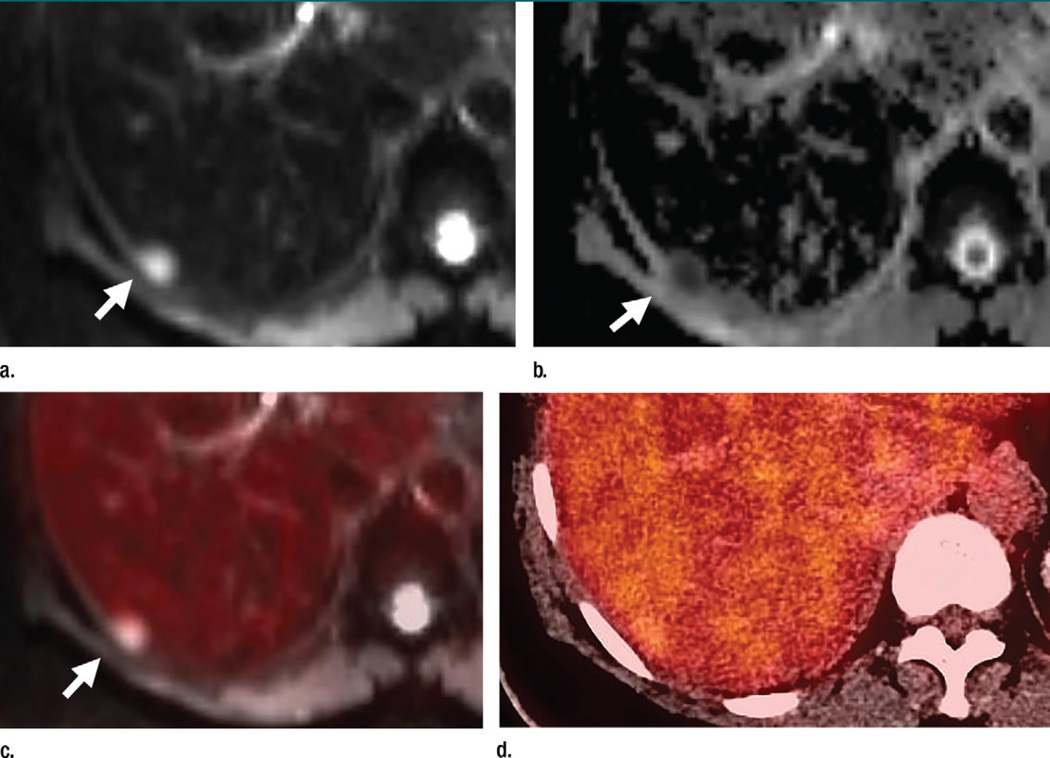
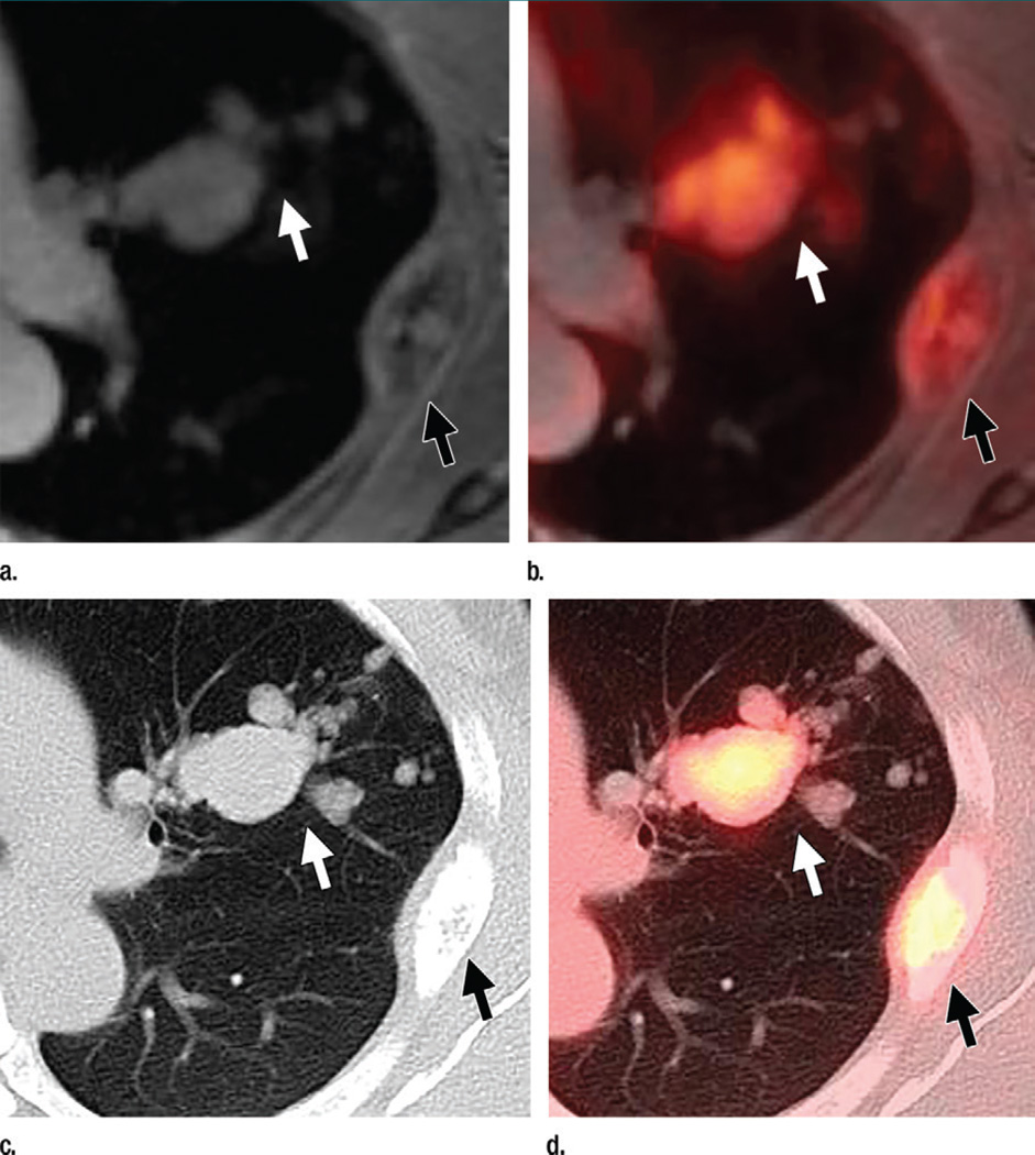
Purpose To compare fluorine 18 ((18)F) fluorodeoxyglucose (FDG) combined positron emission tomography (PET) and magnetic resonance (MR) imaging with (18)F FDG combined PET and computed tomography (CT) in terms of organ-specific metastatic lesion detection and radiation dose in patients with breast cancer. Materials and Methods From July 2012 to October 2013, this institutional review board-approved HIPAA-compliant prospective study included 51 patients with breast cancer (50 women; mean age, 56 years; range, 32-76 years; one man; aged 70 years) who completed PET/MR imaging with diffusion-weighted and contrast material-enhanced sequences after unenhanced PET/CT. Written informed consent for study participation was obtained. Two independent readers for each modality recorded site and number of lesions. Imaging and clinical follow-up, with consensus in two cases, served as the reference standard. Results There were 242 distant metastatic lesions in 30 patients, 18 breast cancers in 17 patients, and 19 positive axillary nodes in eight patients. On a per-patient basis, PET/MR imaging with diffusion-weighted and contrast-enhanced sequences depicted distant (30 of 30 [100%] for readers 1 and 2) and axillary (eight of eight [100%] for reader 1, seven of eight [88%] for reader 2) metastatic disease at rates similar to those of unenhanced PET/CT (distant metastatic disease: 28 of 29 [96%] for readers 3 and 4, P = .50; axillary metastatic disease: seven of eight [88%] for readers 3 and 4, P > .99) and outperformed PET/CT in the detection of breast cancer (17 of 17 [100%] for readers 1 and 2 vs 11 of 17 [65%] for reader 3 and 10 of 17 [59%] for reader 4; P < .001). PET/MR imaging showed increased sensitivity for liver (40 of 40 [100%] for reader 1 and 32 of 40 [80%] for reader 2 vs 30 of 40 [75%] for reader 3 and 28 of 40 [70%] for reader 4; P < .001) and bone (105 of 107 [98%] for reader 1 and 102 of 107 [95%] for reader 2 vs 106 of 107 [99%] for reader 3 and 93 of 107 [87%] for reader 4; P = .012) metastases and revealed brain metastases in five of 51 (10%) patients. PET/CT trended toward increased sensitivity for lung metastases (20 of 23 [87%] for reader 1 and 17 of 23 [74%] for reader 2 vs 23 of 23 [100%] for reader 3 and 22 of 23 [96%] for reader 4; P = .065). Dose reduction averaged 50% (P < .001). Conclusion In patients with breast cancer, PET/MR imaging may yield better sensitivity for liver and possibly bone metastases but not for pulmonary metastases, as compared with that attained with PET/CT, at about half the radiation dose. (©) RSNA, 2016 Online supplemental material is available for this article.
Conflict of interest statement
of Conflicts of Interest: A.N.M. disclosed no relevant relationships. R.A.R. disclosed no relevant relationships. A.C.P. disclosed no relevant relationships. F.D.P. disclosed no relevant relationships. K.M.P. disclosed no relevant relationships. K.J. disclosed no relevant relationships. J.S.B. disclosed no relevant relationships. E.E.S. disclosed no relevant relationships. S.G.K. disclosed no relevant relationships. L.A.M. disclosed no relevant relationships.
Figures
Similar articles
-
What is the diagnostic performance of 18-FDG-PET/MR compared to PET/CT for the N- and M- staging of breast cancer?Eur Radiol. 2019 Apr;29(4):1787-1798. doi: 10.1007/s00330-018-5720-8. Epub 2018 Sep 28. Eur Radiol. 2019. PMID: 30267154
-
Detection of occult primary tumors in patients with cervical metastases of unknown primary tumors: comparison of (18)F FDG PET/CT with contrast-enhanced CT or CT/MR imaging-prospective study.Radiology. 2015 Mar;274(3):764-71. doi: 10.1148/radiol.14141073. Epub 2014 Nov 17. Radiology. 2015. PMID: 25405771
-
Prospective Comparison of 99mTc-MDP Scintigraphy, Combined 18F-NaF and 18F-FDG PET/CT, and Whole-Body MRI in Patients with Breast and Prostate Cancer.J Nucl Med. 2015 Dec;56(12):1862-8. doi: 10.2967/jnumed.115.162610. Epub 2015 Sep 24. J Nucl Med. 2015. PMID: 26405167
-
Recurrent and metastatic breast cancer PET, PET/CT, PET/MRI: FDG and new biomarkers.Q J Nucl Med Mol Imaging. 2013 Dec;57(4):352-66. Q J Nucl Med Mol Imaging. 2013. PMID: 24322792 Review.
-
Comparison of [18F] FDG PET/CT and [18F]FDG PET/MRI in the Detection of Distant Metastases in Breast Cancer: A Meta-Analysis.Clin Breast Cancer. 2025 Feb;25(2):e113-e123.e4. doi: 10.1016/j.clbc.2024.09.015. Epub 2024 Sep 28. Clin Breast Cancer. 2025. PMID: 39438190 Review.
Cited by
-
What is the Diagnostic Performance of 18F-FDG-PET/MRI in the Detection of Bone Metastasis in Patients with Breast Cancer?Eur J Breast Health. 2019 Oct 1;15(4):213-216. doi: 10.5152/ejbh.2019.4885. eCollection 2019 Oct. Eur J Breast Health. 2019. PMID: 31620678 Free PMC article.
-
Lung Nodules Missed in Initial Staging of Breast Cancer Patients in PET/MRI-Clinically Relevant?Cancers (Basel). 2022 Jul 15;14(14):3454. doi: 10.3390/cancers14143454. Cancers (Basel). 2022. PMID: 35884513 Free PMC article.
-
Current and Emerging Magnetic Resonance-Based Techniques for Breast Cancer.Front Med (Lausanne). 2020 May 12;7:175. doi: 10.3389/fmed.2020.00175. eCollection 2020. Front Med (Lausanne). 2020. PMID: 32478083 Free PMC article. Review.
-
Clinical pediatric positron emission tomography/magnetic resonance program: a guide to successful implementation.Pediatr Radiol. 2020 May;50(5):607-617. doi: 10.1007/s00247-019-04578-z. Epub 2020 Feb 19. Pediatr Radiol. 2020. PMID: 32076750 Review.
-
Breast PET/MR Imaging.Radiol Clin North Am. 2017 May;55(3):579-589. doi: 10.1016/j.rcl.2016.12.011. Epub 2017 Feb 1. Radiol Clin North Am. 2017. PMID: 28411681 Free PMC article. Review.
References
-
- Kamel EM, Wyss MT, Fehr MK, von Schulthess GK, Goerres GW. [18F]-Fluorodeoxyglucose positron emission tomography in patients with suspected recurrence of breast cancer. J Cancer Res Clin Oncol. 2003;129(3):147–153. - PubMed
-
- Moon DH, Maddahi J, Silverman DH, Glaspy JA, Phelps ME, Hoh CK. Accuracy of whole-body fluorine-18-FDG PET for the detection of recurrent or metastatic breast carcinoma. J Nucl Med. 1998;39(3):431–435. - PubMed
-
- Huang B, Law MW, Khong PL. Whole-body PET/CT scanning: estimation of radiation dose and cancer risk. Radiology. 2009;251(1):166–174. - PubMed
-
- Brix G, Lechel U, Glatting G, et al. Radiation exposure of patients undergoing whole-body dual-modality 18F-FDG PET/CT examinations. J Nucl Med. 2005;46(4):608–613. - PubMed
Publication types
MeSH terms
Substances
Grants and funding
LinkOut - more resources
Full Text Sources
Other Literature Sources
Medical
