. 2016 Mar 29;11(3):e0151837.
doi: 10.1371/journal.pone.0151837. eCollection 2016.

TCR and IL-7 Signaling Are Altered in the Absence of Functional GTPase of the Immune Associated Nucleotide Binding Protein 5 (GIMAP5)

Affiliations

TCR and IL-7 Signaling Are Altered in the Absence of Functional GTPase of the Immune Associated Nucleotide Binding Protein 5 (GIMAP5)

Xi-Lin Chen et al. PLoS One. .

Abstract

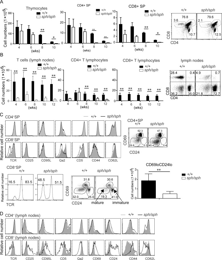
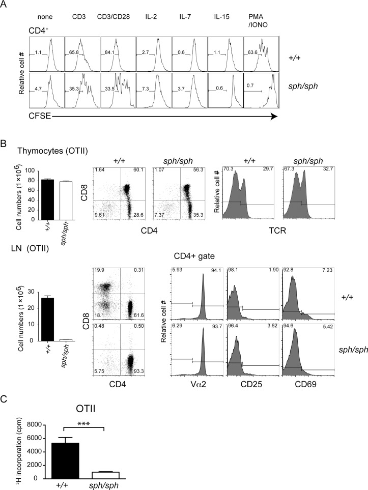
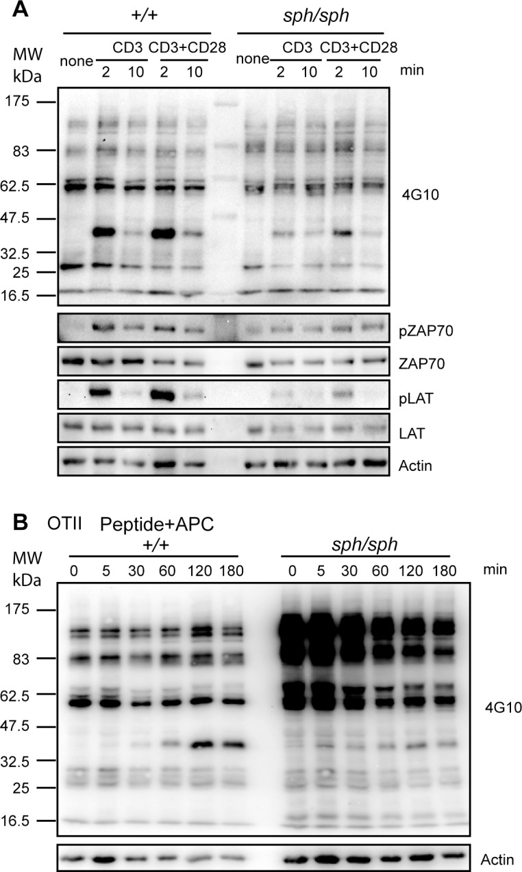
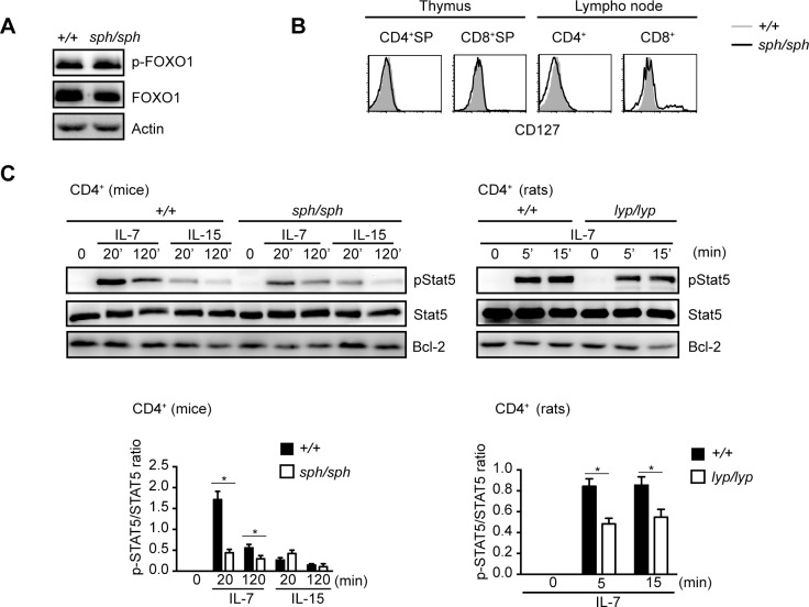
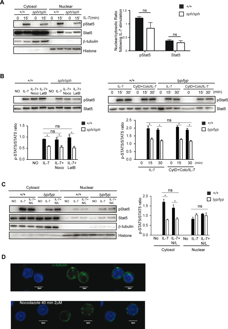
GTPase of the immune associated nucleotide binding protein (GIMAP) family of proteins are expressed essentially in cells of the hematopoietic system. Mutation in the founding member of this gene family, Gimap5, results in the lymphopenic phenotype in Bio-Breeding diabetes prone rats. In mice, deletion of functional Gimap5 gene affects the survival and renewal of hematopoietic stem cells in addition to the defects observed in T cells. Here we show that T cells from OTII TCR-transgenic Gimap5sph/sph mice do not proliferate in response to its cognate antigen. Furthermore, T cells from Gimap5 mutant rats and mice show decreased phosphorylation of STAT5 following stimulation with IL-7. Our results suggest that functional Gimap5 is required for optimal signaling through TCR and IL-7R in T cells.


Conflict of interest statement

Competing Interests: The authors have declared that no competing interests exist.

Figures

Fig 1
Fig 1. Gimap5 deficiency results in disrupted T cell development.
(A) Total number of thymocytes from wildtype and Gimap5sph/sph mice at different ages (in weeks) was counted by trypan blue staining (bar graphs). Flow cytometric analysis of thymocytes following staining with anti-CD4 and anti-CD8 antibodies in wild-type and Gimap5sph/sph mice. (B) Total number of T, CD4+ and CD8+ T cells in pooled lymph nodes (inguinal, axillary, brachial and superficial cervical) from wildtype and Gimap5sph/sph mice at different ages (in weeks, from the same groups of mice as in A) were counted by trypan blue staining. The absolute numbers of total T, CD4+ and CD8+ T cells were calculated by factoring the frequency of CD3+ T cells and the frequency of CD4+ and CD8+ cells within the CD3+ gate. (C) The phenotype of CD4+ or CD8+ SP thymocytes from wild-type and Gimap5sph/sph mice for the indicated markers is shown as histograms. The data for TCR expression shown in the enlarged histogram and the overlap histogram for CD8+SP are from different mice. (D) The phenotype of CD4+ or CD8+ T cells from wild-type and Gimap5sph/sph mice for the indicated markers is shown as histograms. Data shown are representative of 3 independent experiments. Each experiment consisted of analyzing 2 individual sex- and age-matched mice from each genotype.
Fig 2
Fig 2. Gimap5 deficiency results in the absence of proliferative response to cognate antigen in CD4+ T cells.
(A) CFSE-labeled polyclonal lymphocytes from wildtype and Gimap5sph/sph mice were cultured with anti-CD3 antibody, anti-CD3&CD28 antibodies, IL-2 (10 ng/ml), IL-7 (5 ng/ml), IL-15 (10 ng/ml) or PMA/Ionomycin for 3 to 5 days. Medium and cytokines were replenished on day 3. The representative histogram shows CFSE dye dilution, indicative of proliferation in the gated CD4+ T cells. Cultures stimulated with IL-2, IL-7 and IL-15 were analyzed on day 5. Unstimulated cultures and cultures stimulated with anti-CD3 or anti-CD3/CD28 antibodies and PMA/inomycin were analyzed on day 3. (B) Flow cytometric analysis of thymocytes and lymph node populations by staining with anti-TCR, anti-CD4 and anti-CD8 antibodies in wild-type and Gimap5sph/sph OTII TCR transgenic mice. Data shown are representative of 3 four-week old mice. (C) Purified CD4+ T cells from OT-II TCR-transgenic wild type and Gimap5sph/sph mice were activated with irradiated APCs in the presence of OVA peptide and cellular proliferation was measured by [3H]-thymidine incorporation. Data were pooled from 3 independent mice in each group. *** P value <0.005.
Fig 3
Fig 3. TCR-induced proximal signaling is decreased in Gimap5 deficient T cells.
(A) Purified CD4+ T cells from control and Gimap5sph/sph mice were left un-stimulated or stimulated with 5 μg/μL anti-CD3 or anti-CD3&CD28 antibodies for the indicated duration. Lysates were subjected to Western blot analysis. Representative data from one of 3 independent experiments are shown. (B) CD4+ T cells from OT-II TCR-transgenic control and from Gimap5sph/sph mice were co-cultured with irradiated APC pulsed with OVA peptide at a ratio of APC: CD4+ T cells (8:1) for different durations of time. Cell lysates were prepared and phosphorylation of the tyrosine recidue was detected using 4G10 antibody. Representative data from one of 3 independent experiments are shown.
Fig 4
Fig 4. IL-7-induced STAT5 phosphorylation is down regulated in Gimap5-deficient T cells.
(A) CD4+ T cells were purified from freshly isolated lymphocytes from wild type and Gimap5sph/sph mice. Equal amounts of protein lysates were subjected to Western blot analysis for the expression of FOXO1 and pFOXO1. Data shown are representative of 3 independent experiments. (B) Thymocytes and lymph node cells were stained with antibodies to CD4 and CD8. CD127 expression was assessed in gated CD4+ SP or CD8+ SP thymocytes and CD4+ or CD8+ T cell subsets by flow cytometry. Data shown are representative of 3 independent experiments. (C) Purified CD4+ T from Gimap5sph/sph mice or Gimap5lyp/lyp rats were stimulated with IL-7 or IL-15 at 37°C. At the indicated time points the cells were lysed and subjected to Western blot analysis for the detection of phosphorylated STAT5, total STAT5 or Bcl-2. Lower panel represents the densitometric data pooled from 3 independent experiments. * p value < 0.05.
Fig 5
Fig 5. Nuclear translocation of pSTAT5 is not dependent on microtubules in murine T cells.
Purified CD4+ T cells from Gimap5sph/sph mice (A, B) or Gimap5lyp/lyp rats (B, C) were cultured with IL-7 in the absence or presence of cytoskeleton inhibitors nocodazole (Noco, 2 μM) or latrunculin B (LatB, 15 μg/ml) or both together (N/L in C), Cytochalasin D (CytD, 20 μM) or/and Colchicine (Colc, 10 μM) for 15 min. Western blot analysis of whole-cell lysate (B upper two panels), cytosolic and nuclear fractions (A, C) was performed using anti-phospho-STAT5 and anti-STAT5 antibodies. Antibodies to alpha-tubulin and histone were used as markers for cytosolic and nuclear fractions, respectively. The data presented are representative of 3 independent experiments. Bar histogram represent the densitometric data pooled from 3 independent experiments. * p value < 0.05; ns = not significant. D) Confocal image of naïve CD4+ T cells from rats which were treated (or not) with Nocadazole (2 μM; to disrupt microtubules) for 40 min and stained with anti-alpha-tubulin (counterstained with DAPI for the nucleus) to visualize the disruption of microtubules.

Similar articles

Cited by

References

    1. Ciucci T, Bosselut R. Gimap and T cells: a matter of life or death. European journal of immunology. 2014;44(2):348–51. 10.1002/eji.201344375 - DOI - PMC - PubMed
    1. Heinonen MT, Laine AP, Soderhall C, Gruzieva O, Rautio S, Melen E, et al. GIMAP GTPase family genes: potential modifiers in autoimmune diabetes, asthma, and allergy. J Immunol. 2015;194(12):5885–94. 10.4049/jimmunol.1500016 - DOI - PMC - PubMed
    1. Schwefel D, Arasu BS, Marino SF, Lamprecht B, Kochert K, Rosenbaum E, et al. Structural insights into the mechanism of GTPase activation in the GIMAP family. Structure. 2013;21(4):550–9. 10.1016/j.str.2013.01.014 . - DOI - PubMed
    1. Hornum L, Romer J, Markholst H. The diabetes-prone BB rat carries a frameshift mutation in Ian4, a positional candidate of Iddm1. Diabetes. 2002;51(6):1972–9. . - PubMed
    1. MacMurray AJ, Moralejo DH, Kwitek AE, Rutledge EA, Van Yserloo B, Gohlke P, et al. Lymphopenia in the BB rat model of type 1 diabetes is due to a mutation in a novel immune-associated nucleotide (Ian)-related gene. Genome research. 2002;12(7):1029–39. . - PMC - PubMed

Publication types

MeSH terms