

Hemorrhage Caused by Snake Venom Metalloproteinases: A Journey of Discovery and Understanding
- PMID: 27023608
- PMCID: PMC4848620
- DOI: 10.3390/toxins8040093
Hemorrhage Caused by Snake Venom Metalloproteinases: A Journey of Discovery and Understanding
Abstract
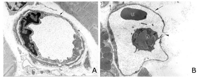
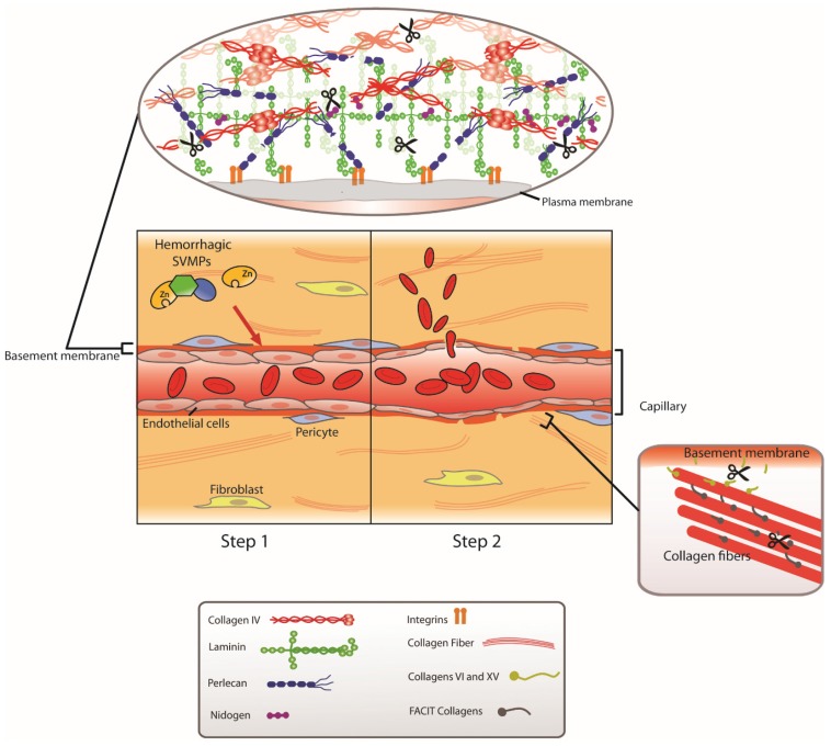
The historical development of discoveries and conceptual frames for understanding the hemorrhagic activity induced by viperid snake venoms and by hemorrhagic metalloproteinases (SVMPs) present in these venoms is reviewed. Histological and ultrastructural tools allowed the identification of the capillary network as the main site of action of SVMPs. After years of debate, biochemical developments demonstrated that all hemorrhagic toxins in viperid venoms are zinc-dependent metalloproteinases. Hemorrhagic SVMPs act by initially hydrolyzing key substrates at the basement membrane (BM) of capillaries. This degradation results in the weakening of the mechanical stability of the capillary wall, which becomes distended owing of the action of the hemodynamic biophysical forces operating in the circulation. As a consequence, the capillary wall is disrupted and extravasation occurs. SVMPs do not induce rapid toxicity to endothelial cells, and the pathological effects described in these cells in vivo result from the mechanical action of these hemodynamic forces. Experimental evidence suggests that degradation of type IV collagen, and perhaps also perlecan, is the key event in the onset of microvessel damage. It is necessary to study this phenomenon from a holistic, systemic perspective in which the action of other venom components is also taken into consideration.
Keywords: basement membrane; capillary vessels; hemorrhage; metalloproteinases; snake venom; type IV collagen; viperids.
Figures
Similar articles
-
Hemorrhage induced by snake venom metalloproteinases: biochemical and biophysical mechanisms involved in microvessel damage.Toxicon. 2005 Jun 15;45(8):997-1011. doi: 10.1016/j.toxicon.2005.02.029. Epub 2005 Apr 18. Toxicon. 2005. PMID: 15922771 Review.
-
Key events in microvascular damage induced by snake venom hemorrhagic metalloproteinases.J Proteomics. 2011 Aug 24;74(9):1781-94. doi: 10.1016/j.jprot.2011.03.026. Epub 2011 Apr 6. J Proteomics. 2011. PMID: 21447411 Review.
-
Role of collagens and perlecan in microvascular stability: exploring the mechanism of capillary vessel damage by snake venom metalloproteinases.PLoS One. 2011;6(12):e28017. doi: 10.1371/journal.pone.0028017. Epub 2011 Dec 8. PLoS One. 2011. PMID: 22174764 Free PMC article.
-
Mechanisms of vascular damage by hemorrhagic snake venom metalloproteinases: tissue distribution and in situ hydrolysis.PLoS Negl Trop Dis. 2010 Jun 29;4(6):e727. doi: 10.1371/journal.pntd.0000727. PLoS Negl Trop Dis. 2010. PMID: 20614020 Free PMC article.
-
Novel insights into capillary vessel basement membrane damage by snake venom hemorrhagic metalloproteinases: a biochemical and immunohistochemical study.Arch Biochem Biophys. 2006 Nov 15;455(2):144-53. doi: 10.1016/j.abb.2006.09.018. Epub 2006 Oct 6. Arch Biochem Biophys. 2006. PMID: 17055999
Cited by
-
Natural Inhibitors of Snake Venom Metalloendopeptidases: History and Current Challenges.Toxins (Basel). 2016 Aug 26;8(9):250. doi: 10.3390/toxins8090250. Toxins (Basel). 2016. PMID: 27571103 Free PMC article. Review.
-
Quantitative Characterization of the Hemorrhagic, Necrotic, Coagulation-Altering Properties and Edema-Forming Effects of Zebra Snake (Naja nigricincta nigricincta) Venom.J Toxicol. 2018 Oct 24;2018:6940798. doi: 10.1155/2018/6940798. eCollection 2018. J Toxicol. 2018. PMID: 30473709 Free PMC article.
-
Using organ-on-a-chip technology to study haemorrhagic activities of snake venoms on endothelial tubules.Sci Rep. 2024 Jun 4;14(1):11157. doi: 10.1038/s41598-024-60282-5. Sci Rep. 2024. PMID: 38834598 Free PMC article.
-
Plant-Derived Lapachol Analogs as Selective Metalloprotease Inhibitors Against Bothrops Venom: A Review.Int J Mol Sci. 2025 Apr 22;26(9):3950. doi: 10.3390/ijms26093950. Int J Mol Sci. 2025. PMID: 40362190 Free PMC article. Review.
-
Anti-ophidian activity of Bredemeyera floribunda Willd. (Polygalaceae) root extract on the local effects induced by Bothrops jararacussu venom.Braz J Med Biol Res. 2018 Dec 3;52(1):e7581. doi: 10.1590/1414-431X20187581. Braz J Med Biol Res. 2018. PMID: 30517287 Free PMC article.
References
-
- Kasturiratne A., Wickremasinghe A.R., de Silva N., Gunawardena N.K., Pathmeswaran A., Premaratna R., Savioli L., Lalloo D.G., de Silva H.J. The global burden of snakebite: A literature analysis and modelling based on regional estimates of envenoming and deaths. PLoS Med. 2008;5 doi: 10.1371/journal.pmed.0050218. - DOI - PMC - PubMed
Publication types
MeSH terms
Substances
Grants and funding
LinkOut - more resources
Full Text Sources
Other Literature Sources
Medical
Research Materials
