. 2016 Mar 29;12(3):e1005917.
doi: 10.1371/journal.pgen.1005917. eCollection 2016 Mar.

Stabilization of Microtubule-Unbound Tau via Tau Phosphorylation at Ser262/356 by Par-1/MARK Contributes to Augmentation of AD-Related Phosphorylation and Aβ42-Induced Tau Toxicity

Affiliations

Stabilization of Microtubule-Unbound Tau via Tau Phosphorylation at Ser262/356 by Par-1/MARK Contributes to Augmentation of AD-Related Phosphorylation and Aβ42-Induced Tau Toxicity

Kanae Ando et al. PLoS Genet. .

Abstract

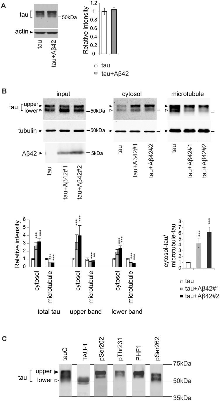
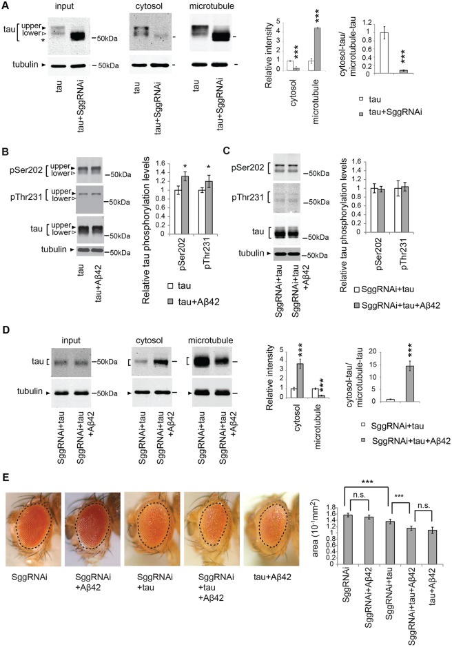
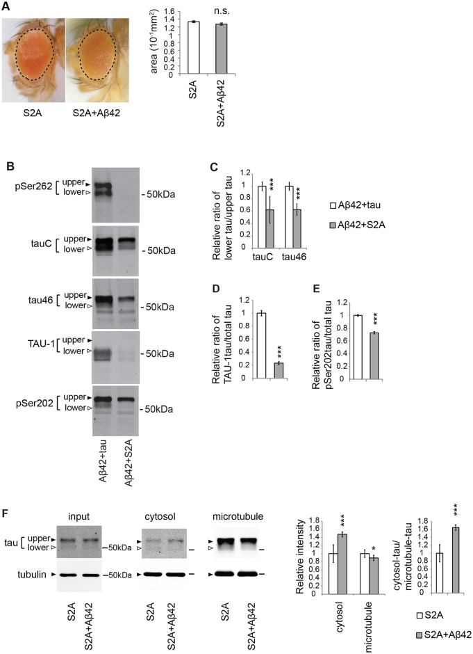
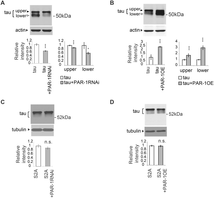
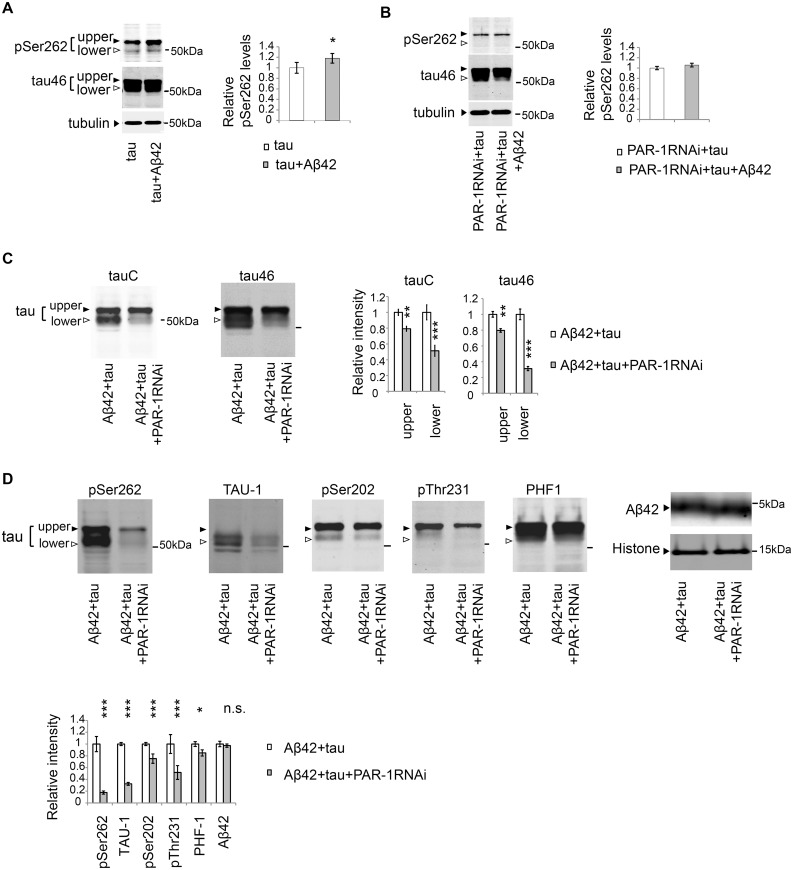
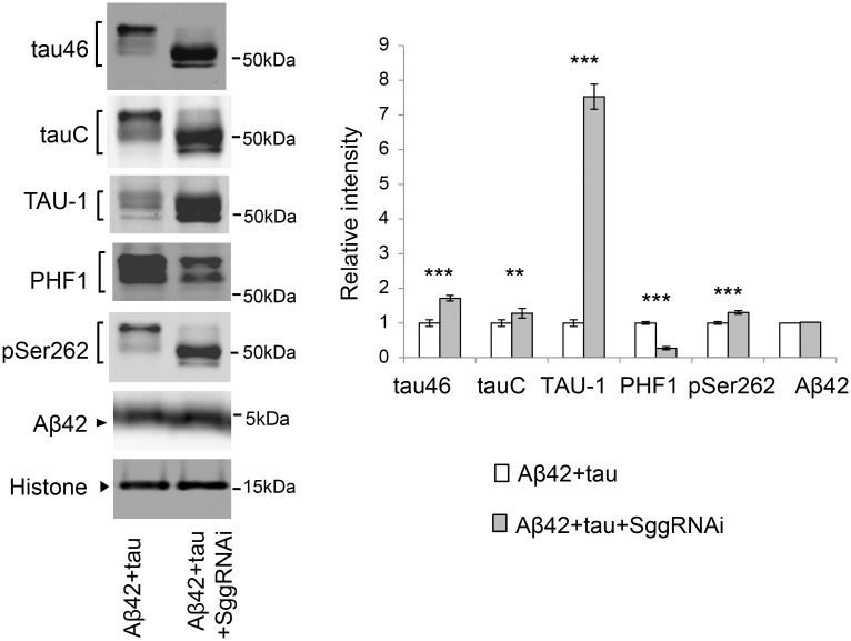
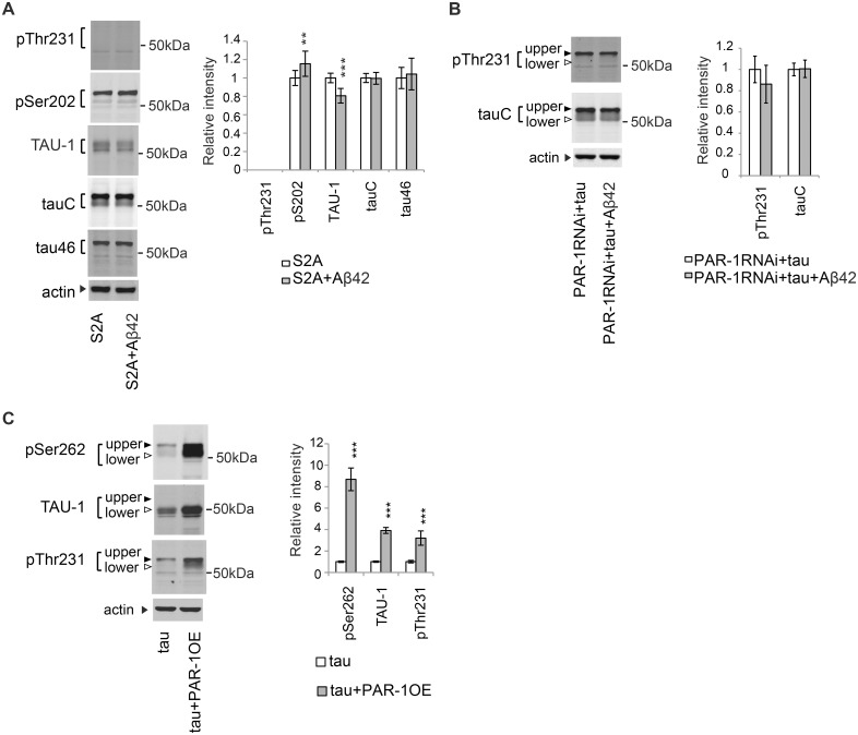
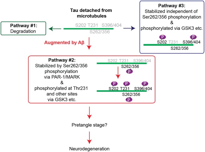
Abnormal accumulation of the microtubule-interacting protein tau is associated with neurodegenerative diseases including Alzheimer's disease (AD). β-amyloid (Aβ) lies upstream of abnormal tau behavior, including detachment from microtubules, phosphorylation at several disease-specific sites, and self-aggregation into toxic tau species in AD brains. To prevent the cascade of events leading to neurodegeneration in AD, it is essential to elucidate the mechanisms underlying the initial events of tau mismetabolism. Currently, however, these mechanisms remain unclear. In this study, using transgenic Drosophila co-expressing human tau and Aβ, we found that tau phosphorylation at AD-related Ser262/356 stabilized microtubule-unbound tau in the early phase of tau mismetabolism, leading to neurodegeneration. Aβ increased the level of tau detached from microtubules, independent of the phosphorylation status at GSK3-targeted SP/TP sites. Such mislocalized tau proteins, especially the less phosphorylated species, were stabilized by phosphorylation at Ser262/356 via PAR-1/MARK. Levels of Ser262 phosphorylation were increased by Aβ42, and blocking this stabilization of tau suppressed Aβ42-mediated augmentation of tau toxicity and an increase in the levels of tau phosphorylation at the SP/TP site Thr231, suggesting that this process may be involved in AD pathogenesis. In contrast to PAR-1/MARK, blocking tau phosphorylation at SP/TP sites by knockdown of Sgg/GSK3 did not reduce tau levels, suppress tau mislocalization to the cytosol, or diminish Aβ-mediated augmentation of tau toxicity. These results suggest that stabilization of microtubule-unbound tau by phosphorylation at Ser262/356 via the PAR-1/MARK may act in the initial steps of tau mismetabolism in AD pathogenesis, and that such tau species may represent a potential therapeutic target for AD.


Conflict of interest statement

The authors have declared that no competing interests exist.

Figures

Fig 1
Fig 1. Aβ42 increases the level of tau in the cytosol and decreases the level of microtubule-bound tau.
(A) Aβ42 does not change total tau levels. Western blot of heads of flies expressing tau alone (tau) or that co-expressing tau and Aβ42 (tau+Aβ42) driven by gmr-GAL4 with anti-tau antibody. Actin was used as a loading control. Mean ± SD, n = 5; no significant difference was found by Student's t-test (p>0.05). Representative blots are shown. (B) Co-expression of Aβ42 increases the levels of tau free from microtubules and reduces the levels of tau bound to microtubules. The levels of tau and tubulin in the lysate of fly heads expressing tau alone (tau) or co-expressing tau and Aβ42 (tau+Aβ42#1 and tau+Aβ42#2) before sedimentation (input), in the supernatant (cytosol) and in the pellet containing microtubules (microtubule) were analyzed by western blotting by using anti-tau antibody. The same amount of proteins from each genotype was loaded. Expression of Aβ42 was confirmed by western blot with anti-Aβ antibody (Aβ42). Two independent transgenic fly lines expressing Aβ42 at different expression levels (Aβ42#1 and Aβ42#2) yielded similar results, and the fly line with higher Aβ42 (Aβ42#2) expression exhibited a larger effect. Transgene expression was driven by gmr-GAL4. Mean ± SD, n = 4; **, p<0.01, ***, p<0.005 compared to tau by one-way ANOVA with Tukey's post hoc test. Representative blots are shown. (C) Two major bands detected by western blotting of fly heads expressing tau with a pan-tau antibody (tauC) differ in their phosphorylation patterns. Western blots with TAU-1 antibody, which specifically recognizes tau protein without phosphorylation at several AD-related sites (Ser194, Ser195, Ser198, and Ser202) (TAU-1), anti-phospho-Ser202, anti-phospho-Thr231, anti-phospho-Ser396/404 (PHF1), or anti-phospho-Ser262 antibody (pSer262) are shown. Transgene expression was driven by gmr-GAL4.
Fig 2
Fig 2. Knockdown of GSK3β/Sgg is not sufficient to suppress either mislocalization of tau to the cytosol or Aβ42-induced augmentation of tau toxicity.
(A) GSK3β/Sgg negatively regulates tau binding to microtubules. RNAi-mediated knockdown of GSK3/Sgg shifts all tau species to lower apparent molecular weights, and the resultant species migrate faster than the original taulower species (indicated as asterisk). The levels of tau and tubulin in the lysate before sedimentation (input), in the supernatant (cytosol) and in the pellet containing microtubules (microtubule) were analyzed by western blotting by using anti-tau antibody. The same amount of proteins from each genotype was loaded. Mean ± SD, n = 4; **, p < 0.01, ***, p < 0.005 by Student's t-test. Representative blots are shown. (B) Aβ42 increased the levels of tau phosphorylated at Ser202 and those phosphorylated at Thr231. Western blots of fly heads expressing tau alone (tau) or that co-expressing tau and Aβ42 (tau+Aβ42) with anti-phospho-Ser202 antibody (pSer202), anti-phospho-Thr231 antibody (pThr231), and anti-tau antibody. Tubulin was used as a loading control. Mean ± SD, n = 5; *, p < 0.05 by Student's t-test. Representative blots are shown. (C) Expression of Aβ42 did not increase tau phosphorylation at either of Ser202 and Thr231 in the Sgg knockdown background. Western blots of fly heads expressing Sgg RNAi tau and (SggRNAi+tau) or that co-expressing Sgg RNAi, tau and Aβ42 (SggRNAi+tau+Aβ42) with anti-phospho-Ser202 antibody (pSer202), anti-phospho-Thr231 antibody (pThr231), and anti-tau antibody. Tubulin was used as a loading control. Mean ± SD, n = 5; no significant difference by Student's t-test (p > 0.05). Representative blots are shown. (D) Aβ42 causes an increase in tau levels in the cytosol fraction and reduction in tau levels in the microtubule fraction in the Sgg knockdown background. The levels of tau and tubulin in fly heads expressing Sgg RNAi and tau (SggRNAi+tau) or that co-expressing Sgg RNAi, tau and Aβ42 (SggRNAi+tau+Aβ42) before sedimentation (input), in the supernatant (cytosol) and in the pellet containing microtubules (microtubule) were analyzed by western blotting with anti-tau and anti-tubulin. Mean ± SD, n = 4; ***, p < 0.005 by Student's t-test. Representative blots are shown. (E) Aβ42 enhances tau-induced retinal degeneration in the Sgg knockdown background (compare SggRNAi+tau and SggRNAi+tau+Aβ42). Mean ± SE, N = 6–8, asterisks indicate significant differences in the surface area of the external eye (***, p < 0.005, n.s., not significant (p > 0.05) by one-way ANOVA with Tukey's post hoc test). Transgene expression was driven by gmr-GAL4.
Fig 3
Fig 3. Blocking tau phosphorylation at Ser262/356 with unphosphorylatable alanine substitutions (S2A) preferentially reduces the levels of taulower and partially suppresses the Aβ42-mediated increase in tau mislocalization from the microtubule to the cytosol.
(A) Expression of either S2Atau (S2A) alone or co-expression of S2Atau and Aβ42 (S2A+Aβ42) does not cause eye degeneration. No significant difference in the surface area of the external eyes between S2A and S2A+ Aβ42 (mean ± SE, n = 6–8, N.S., not significant (p > 0.05) by one-way ANOVA). Transgene expression was driven by gmr-GAL4. (B) S2Atau shows different phosphorylation profiles compared to wild-type tau. Wild-type tau or S2Atau were co-expressed with Aβ42 (tau+Aβ42 and Aβ42+S2A, respectively) and subjected to western blotting with pan-tau antibody (tauC and tau46) or antibodies that recognize phosphorylation status of tau at the specific sites (pSer262, TAU-1 and pSer202). (C) The ratio of signal intensities of taulower to tauupper detected by tauC. (D) The ratio of signal intensities of TAU-1 blot to tauC blot. Mean ± SD, n = 5; ***, p < 0.005 by Student's t-test. (E) The ratio of signal intensities of pSer202 blot to tauC blot. Mean ± SD, n = 5; ***, p < 0.005 by Student's t-test. (F) Aβ42 increased the level of S2Atau in the cytosol and decreased those in the microtubule fraction, while the effects were smaller than those in wild-type tau. Mean ± SD, n = 4; *, p < 0.05, ***, p < 0.005 compared by Student's t-test. Representative blots are shown. Transgene expression was driven by gmr-GAL4.
Fig 4
Fig 4. PAR-1 stabilizes less phosphorylated forms of tau (taulower) through phosphorylation at Ser262 and Ser356.
(A) PAR-1 knockdown reduces the levels of tau with more prominent effect on taulower. Western blots of fly heads expressing tau (tau) or that co-expressing tau and PAR-1RNAi (tau+PAR-1RNAi) with anti-tau antibody. (B) PAR-1 overexpression increases the levels of tau with more prominent effect on taulower. Western blots of fly heads expressing tau (tau) or that co-expressing tau and PAR-1 (tau+PAR-1OE) with anti-tau antibody. (C) PAR-1 knockdown does not affect S2Atau levels. Western blots of fly heads expressing S2Atau (S2A) or that co-expressing S2Atau and PAR-1RNAi (S2A+PAR-1RNAi) with anti-tau antibody. (D) PAR-1 overexpression does not affect S2Atau levels. PAR-1 knockdown does not affect S2Atau levels. Western blots of fly heads expressing S2Atau (S2A) or that co-expressing S2Atau and PAR-1 (S2A+PAR-1OE) with anti-tau antibody. Tubulin or actin was used as loading control. Mean ± SD, n = 4–5; *, p < 0.05, ***, p < 0.005, n.s., not significant (p > 0.05) by Student's t-test. Representative blots are shown. Transgene expression was driven by gmr-GAL4.
Fig 5
Fig 5. PAR-1 mediates the increase in the level of tau phosphorylated at Ser262 caused by Aβ42, and knockdown of Par-1/MARK markedly decreases the levels of taulower.
(A) Aβ42 increased the levels of tau phosphorylated at Ser262. Western blots of fly heads expressing tau alone (tau) or that co-expressing tau and Aβ42 (tau+Aβ42) with anti-phospho-Ser262 antibody (pSer262) or anti-tau (tau46). Tubulin was used as a loading control. Mean ± SD, n = 5; *, p < 0.05 by Student's t-test. Representative blots are shown. (B) Expression of Aβ42 did not increase tau phosphorylation at Ser262 in the PAR-1 knockdown background. Western blots of fly heads expressing PAR-1 RNAi and tau (PAR-1RNAi+tau) or that co-expressing PAR-1RNAi, tau and Aβ42 (PAR-1RNAi+tau+Aβ42) with anti-phospho-Ser262 antibody (pSer262) and anti-tau antibody (tau46). Tubulin was used as a loading control. Mean ± SD, n = 5; no significant difference by Student's t-test (p > 0.05). Representative blots are shown. (C-D) PAR-1 knockdown reduces the levels of tau with more prominent effect on the levels of taulower than those of tauupper. Western blots of fly heads expressing Aβ42 and tau (Aβ42+tau) or that co-expressing Aβ42, tau and PAR-1 RNAi (Aβ42+tau+PAR-1RNAi) with pan-tau antibody (tauC and tau46), antibodies that recognize phosphorylation status of tau at the specific sites (pSer262, TAU-1, pSer202, pThr231, PHF-1) or anti-Aβ antibody (Aβ42). Histone was used as loading control. Mean ± SD, n = 5; *, p < 0.05, **, p < 0.01, ***, p<0.005. Representative blots are shown. Transgene expression was driven by gmr-GAL4.
Fig 6
Fig 6. Blocking tau phosphorylation at SP/TP sites by Sgg knockdown does not reduce the levels of tau.
Western blots of fly heads expressing Aβ42 and tau (Aβ42+tau) or that co-expressing Aβ42, tau and Sgg RNAi (Aβ42+tau+SggRNAi) with pan-tau antibody (tauC and tau46), antibodies that recognize phosphorylation status of tau at the specific sites (TAU-1, PHF-1 and pSer262) or anti-Aβ antibody (Aβ42). Histone was used as loading control. Mean ± SD, n = 5; **, p < 0.01, ***, p < 0.005. Representative blots are shown. Transgene expression was driven by gmr-GAL4.
Fig 7
Fig 7. Stabilization of tau through phosphorylation at Ser262 and Ser356 contributes to the Aβ42-induced increase in the level of tau phosphorylated at Thr231.
(A) Aβ42 does not increase the levels of S2Atau phosphorylated at Thr231. Western blots of fly heads expressing S2Atau (S2A) or that co-expressing S2Atau and Aβ42 (S2A+Aβ42) with antibodies that recognize phosphorylation status of tau at the specific sites (pThr231, pSer202, and TAU-1) or pan-tau antibodies (tauC and tau46). (B) Aβ42 does not increase the levels of tau phosphorylated at Thr231 in the PAR-1 knockdown background. Western blots of fly heads expressing PAR-1RNAi and tau (PAR-1RNAi+tau) or that co-expressing PAR-1RNAi, tau and Aβ42 (PAR-1RNAi+tau+Aβ42) with anti-phospho-Thr231 antibody (pThr231) or pan-tau antibody (tauC). (C) PAR-1 overexpression increases the levels of tau phosphorylated at Thr231. Western blots of fly heads expressing tau (tau) or that co-expressing PAR-1 and tau (tau+PAR-1OE) with anti-phospho-Ser262 antibody (pSer262), TAU-1 or anti-phospho-Thr231 antibody (pThr231). Actin was used as loading control. Mean ± SD, n = 5; **, p < 0.01, ***, p < 0.005. Representative blots are shown. Transgene expression was driven by gmr-GAL4.
Fig 8
Fig 8. Working model of the early phase of tau mismetabolism.

Similar articles

Cited by

References

    1. Hardy J, Selkoe DJ. The amyloid hypothesis of Alzheimer's disease: progress and problems on the road to therapeutics. Science. 2002;297(5580):353–6. - PubMed
    1. Jin M, Shepardson N, Yang T, Chen G, Walsh D, Selkoe DJ. Soluble amyloid beta-protein dimers isolated from Alzheimer cortex directly induce Tau hyperphosphorylation and neuritic degeneration. Proc Natl Acad Sci U S A. 2011;108(14):5819–24. 10.1073/pnas.1017033108 - DOI - PMC - PubMed
    1. Lewis J, Dickson DW, Lin WL, Chisholm L, Corral A, Jones G, et al. Enhanced neurofibrillary degeneration in transgenic mice expressing mutant tau and APP. Science. 2001;293(5534):1487–91. - PubMed
    1. Gotz J, Chen F, van Dorpe J, Nitsch RM. Formation of neurofibrillary tangles in P301l tau transgenic mice induced by Abeta 42 fibrils. Science. 2001;293(5534):1491–5. - PubMed
    1. Oddo S, Billings L, Kesslak JP, Cribbs DH, LaFerla FM. Abeta immunotherapy leads to clearance of early, but not late, hyperphosphorylated tau aggregates via the proteasome. Neuron. 2004;43(3):321–32. - PubMed

Publication types

MeSH terms