. 2016 Jan 28;5(1):10.
doi: 10.3390/antibiotics5010010.

Identification of a Fragment-Based Scaffold that Inhibits the Glycosyltransferase WaaG from Escherichia coli

Affiliations

Identification of a Fragment-Based Scaffold that Inhibits the Glycosyltransferase WaaG from Escherichia coli

Claudio Muheim et al. Antibiotics (Basel). .

Abstract

WaaG is a glycosyltransferase that is involved in the biosynthesis of lipopolysaccharide in Gram-negative bacteria. Inhibitors of WaaG are highly sought after as they could be used to inhibit the biosynthesis of the core region of lipopolysaccharide, which would improve the uptake of antibiotics. Herein, we establish an activity assay for WaaG using (14)C-labeled UDP-glucose and LPS purified from a ∆waaG strain of Escherichia coli. We noted that addition of the lipids phosphatidylglycerol (PG) and cardiolipin (CL), as well as the detergent 3-[(3-cholamidopropyl)dimethylammonio]-1-propanesulfonate (CHAPS) increased activity. We then use the assay to determine if three molecular scaffolds, which bind to WaaG, could inhibit its activity in vitro. We show that 4-(2-amino-1,3-thiazol-4-yl)phenol inhibits WaaG (IC50 1.0 mM), but that the other scaffolds do not. This study represents an important step towards an inhibitor of WaaG by fragment-based lead discovery.

Keywords: Gram-negative bacteria; fragment-based lead discovery; glucosyltransferase; lipopolysaccharide; scaffold.


Figures

Figure 1
Figure 1
Schematic of the action of the glycosyltransferase WaaG transferring a d-glucose residue (blue ball) from UDP-glucose to heptose-II of the inner core of an LPS molecule.
Figure 2
Figure 2
His-WaaG activity under different lipid compositions. (A) Mixed micelles containing CHAPS and/or various lipids were incubated with LPSTRUNC (0.5 µg·mL−1), UDP-Glc* and His-WaaG (0.1 mg·mL−1) to monitor LPS-glycosylation. After 1 h, the reaction was stopped by the addition of Laemmli buffer, and the products were separated by SDS-PAGE and detected by digital autoradiography. Lane 1: no added lipids or detergents; Lane 2: 20 mM CHAPS; Lane 3: 10 mM PG; Lane 4: 5 mM CL; Lane 5: 20 mM CHAPS + 10 mM PG; Lane 6: 20 mM CHAPS + 10 mM PG + 1 mM CL; Lane 7: 20 mM CHAPS + 10 mM PG + 5 mM CL; Lane 8: 3% DHPC. Quantification of LPSTRUNC-Glc* and UDP-Glc* was performed with ImageJ and the specific activity calculated (see the Experimental Section). Specific activities plotted as the mean ± SD; n = 2. (B) Nitrocellulose membranes containing immobilized lipids (100 pmol/spot) were incubated with purified His-WaaG (3 µg·mL−1). After washing to remove unbound protein, His-WaaG that bound to different lipids was detected by immunoblotting with antisera to the His-tag. (C) His-WaaG activity was monitored in vitro with different concentrations of MgCl2.
Figure 3
Figure 3
Compound L1 inhibits His-WaaG. (A) Structures of ligands: L1, 4-(2-amino-1,3-thiazol-4-yl)phenol; L2, 4-(1H-pyrrol-1-yl)benzoic acid; L3, 2-(1H-pyrrol-1-ylmethyl)pyridine. (B) Mixed micelles containing 20 mM CHAPS, 10 mM PG and 1 mM CL were mixed with LPSTRUNC (0.5 µg·mL−1) and UDP-Glc*. The reaction was started by adding His-WaaG (0.1 mg·mL−1) and incubated either with 2.5% DMSO (v/v) or 25 mM of compound L1, L2 or L3. Samples were collected after different time points, inactivated by adding Laemmli buffer, separated by SDS-PAGE and detected by autoradiography. (C) Quantification of the activity of His-WaaG in the presence of either DMSO or 25 mM L1, L2 or L3. Gels in (B) were quantified by densitometry, and the activity was calculated as described in the Experimental Section.
Figure 3
Figure 3
Compound L1 inhibits His-WaaG. (A) Structures of ligands: L1, 4-(2-amino-1,3-thiazol-4-yl)phenol; L2, 4-(1H-pyrrol-1-yl)benzoic acid; L3, 2-(1H-pyrrol-1-ylmethyl)pyridine. (B) Mixed micelles containing 20 mM CHAPS, 10 mM PG and 1 mM CL were mixed with LPSTRUNC (0.5 µg·mL−1) and UDP-Glc*. The reaction was started by adding His-WaaG (0.1 mg·mL−1) and incubated either with 2.5% DMSO (v/v) or 25 mM of compound L1, L2 or L3. Samples were collected after different time points, inactivated by adding Laemmli buffer, separated by SDS-PAGE and detected by autoradiography. (C) Quantification of the activity of His-WaaG in the presence of either DMSO or 25 mM L1, L2 or L3. Gels in (B) were quantified by densitometry, and the activity was calculated as described in the Experimental Section.
Figure 4
Figure 4
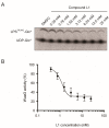
Dose-response curve for compound L1. (A) To evaluate the IC50, the activity of His-WaaG was assayed in the presence of 20 mM CHAPS, 10 mM PG, 1 mM CL and different concentrations of L1. Samples were collected after 1 h and inactivated by adding Laemmli buffer. The products were separated by SDS-PAGE, detected by digital autoradiography and quantified by densitometry. The IC50 was calculated as the midpoint between the DMSO control (which was assumed to be 100% activity) and the activity at 25 mM of L1 (which was assumed to be 0% activity). (B) His-WaaG activity is shown as a function of L1 concentration. The resulting IC50 value for compound L1 was calculated to be 1.0 mM (n = 2).

References

    1. Amor K., Heinrichs D.E., Frirdich E., Ziebell K., Johnson R.P., Whitfield C. Distribution of core oligosaccharide types in lipopolysaccharides from Escherichia coli. Infect. Immun. 2000;68:1116–1124. doi: 10.1128/IAI.68.3.1116-1124.2000. - DOI - PMC - PubMed
    1. Stenutz R., Weintraub A., Widmalm G. The structures of Escherichia coli O-polysaccharide antigens. FEMS Microbiol. Rev. 2006;30:382–403. doi: 10.1111/j.1574-6976.2006.00016.x. - DOI - PubMed
    1. Nikaido H. Molecular basis of bacterial outer membrane permeability revisited. Microbiol. Mol. Biol. Rev. 2003;67:593–656. doi: 10.1128/MMBR.67.4.593-656.2003. - DOI - PMC - PubMed
    1. Liu A., Tran L., Becket E., Lee K., Chinn L., Park E., Tran K., Miller J.H. Antibiotic sensitivity profiles determined with an Escherichia coli gene knockout collection: Generating an antibiotic bar code. Antimicrob. Agents Chemother. 2010;54:1393–1403. doi: 10.1128/AAC.00906-09. - DOI - PMC - PubMed
    1. Laxminarayan R., Duse A., Wattal C., Zaidi A.K.M., Wertheim H.F.L., Sumpradit N., Vlieghe E., Hara G.L., Gould I.M., Goossens H., et al. Antibiotic resistance—The need for global solutions. Lancet Infect. Dis. 2013;13:1057–1098. doi: 10.1016/S1473-3099(13)70318-9. - DOI - PubMed

LinkOut - more resources