Case Reports
. 2016 Jun;263(6):1083-91.
doi: 10.1007/s00415-016-8083-6. Epub 2016 Mar 30.

N-methyl-D-aspartate receptor encephalitis mediates loss of intrinsic activity measured by functional MRI

Affiliations
Case Reports

N-methyl-D-aspartate receptor encephalitis mediates loss of intrinsic activity measured by functional MRI

Matthew R Brier et al. J Neurol. 2016 Jun.

Abstract

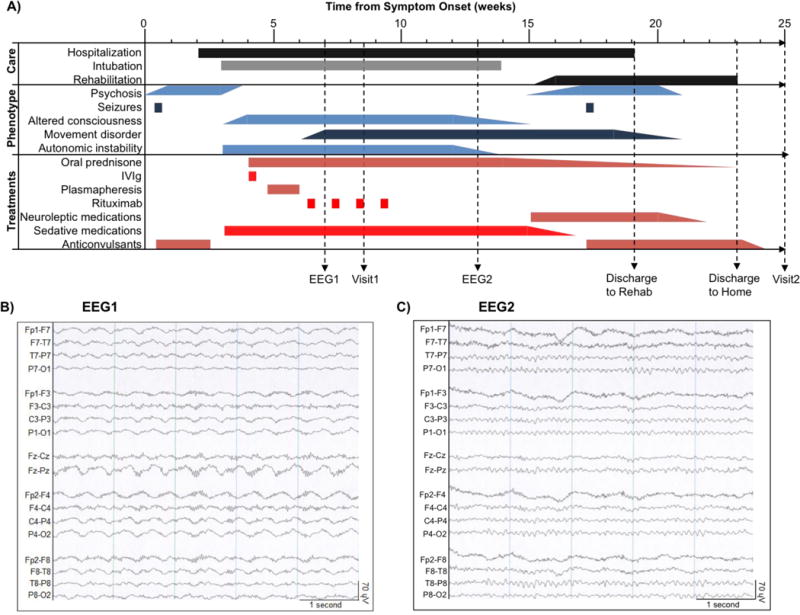
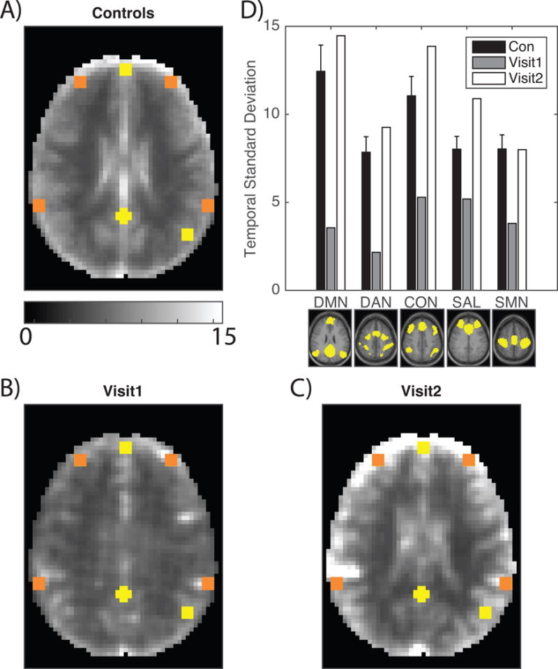
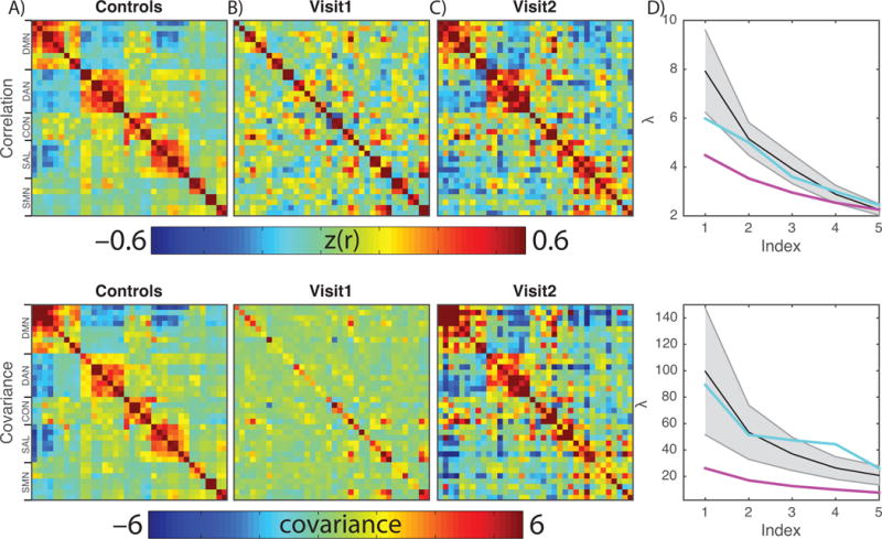
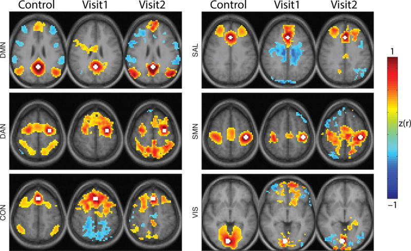
Spontaneous brain activity is required for the development and maintenance of normal brain function. Many disease processes disrupt the organization of intrinsic brain activity, but few pervasively reduce the amplitude of resting state blood oxygen level dependent (BOLD) fMRI fluctuations. We report the case of a female with anti-N-methyl-D-aspartate receptor (NMDAR) encephalitis, longitudinally studied during the course of her illness to determine the contribution of NMDAR signaling to spontaneous brain activity. Resting state BOLD fMRI was measured at the height of her illness and 18 weeks following discharge from hospital. Conventional resting state networks were defined using established methods. Correlation and covariance matrices were calculated by extracting the BOLD time series from regions of interest and calculating either the correlation or covariance quantity. The intrinsic activity was compared between visits, and to expected activity from 45 similarly aged healthy individuals. Near the height of the illness, the patient exhibited profound loss of consciousness, high-amplitude slowing of the electroencephalogram, and a severe reduction in the amplitude of spontaneous BOLD fMRI fluctuations. The patient's neurological status and measures of intrinsic activity improved following treatment. We conclude that NMDAR-mediated signaling plays a critical role in the mechanisms that give rise to organized spontaneous brain activity. Loss of intrinsic activity is associated with profound disruptions of consciousness and cognition.

Keywords: Functional connectivity; NMDA receptor encephalitis; Resting-state; fMRI.


Conflict of interest statement

Conflict of Interest: The authors have no conflict of interest. GSD is the clinical director of the Anti-NMDA Receptor Encephalitis Foundation; the Foundation is supported by private donations.

Figures

Figure 1
Figure 1
Clinical course and interventions. A: Line diagram (from time of symptom onset) depicting landmarks in clinical care, emergence of symptoms and signs (phenotype), treatments and the timing of electroencephalograms (EEG1 and EEG2) and structural / functional neuroimaging (Visit1 and Visit2). B: Abnormal EEG showing diffuse, rhythmic, delta activity at 1–3 Hz with superimposed bursts of rhythmic 20–30 Hz beta frequency activity riding on each delta wave (“extreme delta brush”[48]). C: Normal EEG with characteristic posterior-dominant alpha rhythm (8–13 Hz).
Figure 2
Figure 2
BOLD signal amplitude is diminished in a patient with NMDAR encephalitis. A: Mean BOLD temporal standard deviation derived from 45 age-matched healthy individuals. Yellow boxes correspond to DMN ROIs and orange boxes correspond to non-DMN ROIs. The control group demonstrates strong grey versus white matter contrast. B: Temporal standard deviation at Visit1 in the same style as A. Grey versus white matter contrast is markedly diminished and the overall amplitude is reduced. C: Temporal standard deviation at Visit2 in the same style as A. Grey versus white matter contrast is restored and the overall amplitude is restored. D: The temporal standard deviation of the BOLD signal measured in ROIs from each of 5 RSNs shown below the corresponding set of bars. Black bars indicate healthy individuals (±1 standard deviation); grey bars correspond to Visit1; white bars correspond to Visit2.
Figure 3
Figure 3
BOLD organization and amplitude is disrupted in a patient with NMDAR encephalitis. Top row shows BOLD correlation matrices; the bottom row shows BOLD covariance matrices. The salient difference in these results is that the former is normalized with respect to signal amplitude. A: BOLD correlation and covariance matrices derived from 45 age-matched healthy individuals. Rows and columns index individual regions of interest; the color indicates the correlation/covariance value of the BOLD signal extracted from ROIs. The prominent blocks represent RSNs. B: RSN structure is absent at Visit1. The covariance matrix emphasizes that the signal is both disorganized and has low amplitude. C: RSN structure and amplitude is restored at Visit2. D: Quantification of this observation is achieved by comparing the five largest eigenvalues of the matrices in healthy individuals (black line, grey area is +/− 1SD), Visit1 (magenta) and Visit2 (teal). The Visit1 result is abnormally flattened while the Visit2 result is within the normal range.
Figure 4
Figure 4
RSN topography is disrupted and recovers during NMDAR encephalitis. Seed-based functional connectivity maps for healthy individuals (first column), Visit1 and Visit2. Seed regions for each network were posterior cingulate cortex (DMN), left frontal eye field (DAN), dorsal medial prefrontal cortex (CON), anterior cingulate (SAL), left primary motor cortex (SMN) and right primary visual cortex (VIS) indicated in white. Conventional functional connectivity was calculated between the seed region and all other voxels. Resulting correlations were Fisher z-transformed. z(r) maps were thresholded voxel-wise |z(r)|>0.2 and only suprathreshold clusters consisting of more than 100 voxels are shown. Healthy individuals demonstrate focal and defined RSN topographies that are absent at Visit1 and restored at Visit2. Images are presented in radiologic convention.

Similar articles

Cited by

References

    1. Abikoff H, Alvir J, Hong G, Sukoff R, Orazio J, Solomon S, Saravay S. Logical memory subtest of the Wechsler Memory Scale: age and education norms and alternate-form reliability of two scoring systems. Journal of clinical and experimental neuropsychology. 1987;9:435–448. - PubMed
    1. Akeju O, Westover MB, Pavone KJ, Sampson AL, Hartnack KE, Brown EN, Purdon PL. Effects of sevoflurane and propofol on frontal electroencephalogram power and coherence. Anesthesiology. 2014;121:990–998. - PMC - PubMed
    1. Attwell D, Buchan AM, Charpak S, Lauritzen M, Macvicar BA, Newman EA. Glial and neuronal control of brain blood flow. Nature. 2010;468:232–243. - PMC - PubMed
    1. Bartlett TE, Wang YT. The intersections of NMDAR-dependent synaptic plasticity and cell survival. Neuropharmacology. 2013;74:59–68. - PubMed
    1. Benjamini Y, Hochberg Y. Controlling the False Discovery Rate: A Practical and Powerful Approach to Multiple Testing. Journal of the Royal Statistical Society Series B (Methodological) 1995;57(1):289–300.

Publication types

MeSH terms

LinkOut - more resources