. 2016 Jul;30(7):2557-69.
doi: 10.1096/fj.201600102R. Epub 2016 Mar 29.

Neuronal Wiskott-Aldrich syndrome protein regulates TGF-β1-mediated lung vascular permeability

Affiliations

Neuronal Wiskott-Aldrich syndrome protein regulates TGF-β1-mediated lung vascular permeability

Brant M Wagener et al. FASEB J. 2016 Jul.

Abstract

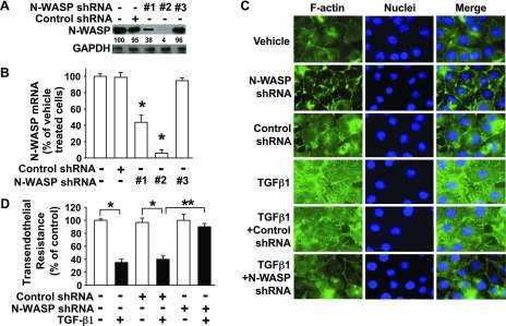
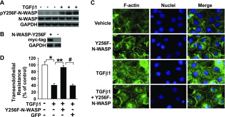
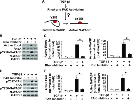
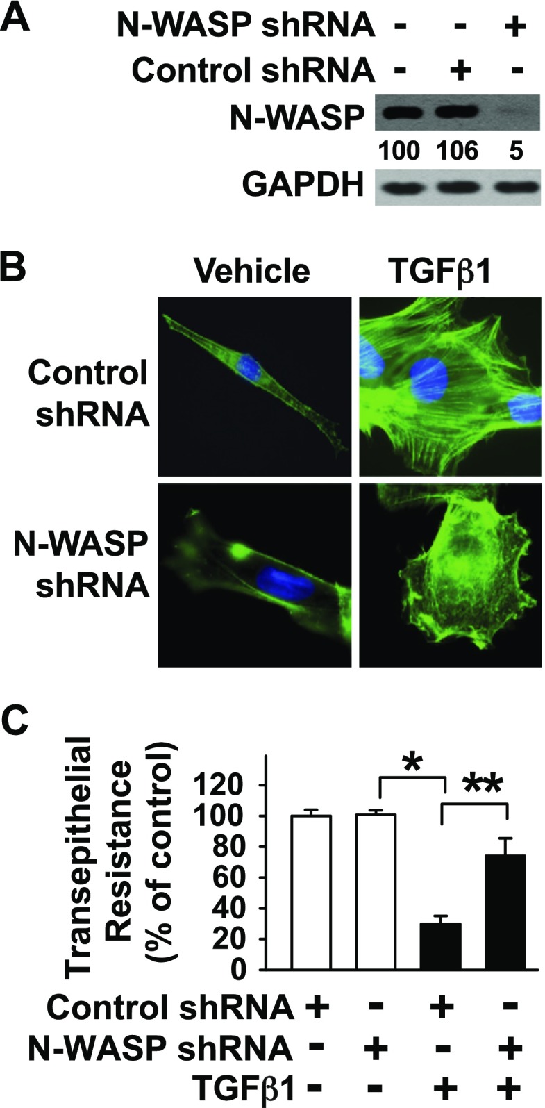
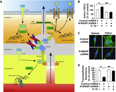
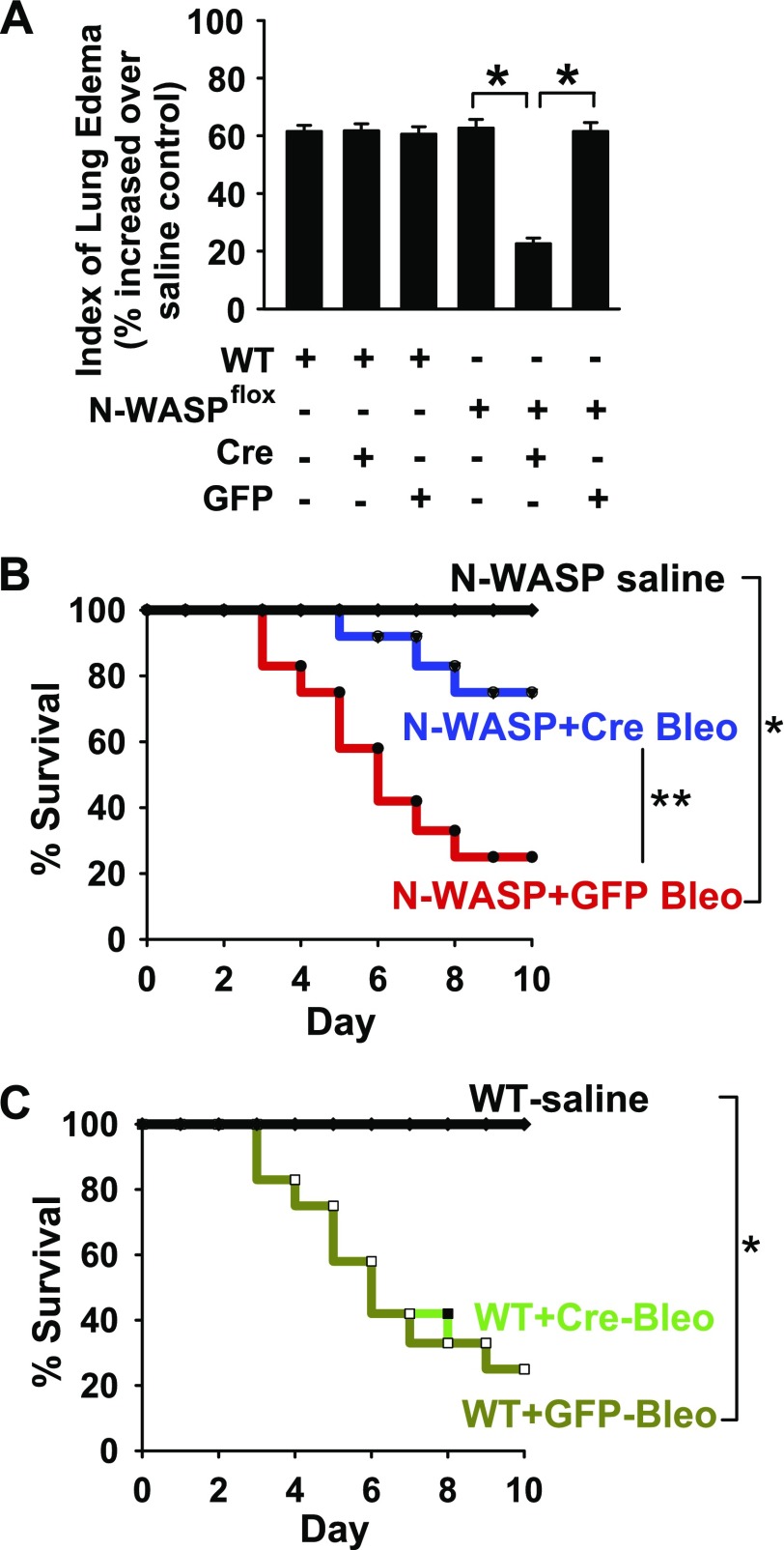
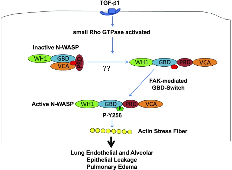
TGF-β1 induces an increase in paracellular permeability and actin stress fiber formation in lung microvascular endothelial and alveolar epithelial cells via small Rho GTPase. The molecular mechanism involved is not fully understood. Neuronal Wiskott-Aldrich syndrome protein (N-WASP) has an essential role in actin structure dynamics. We hypothesized that N-WASP plays a critical role in these TGF-β1-induced responses. In these cell monolayers, we demonstrated that N-WASP down-regulation by short hairpin RNA prevented TGF-β1-mediated disruption of the cortical actin structure, actin stress filament formation, and increased permeability. Furthermore, N-WASP down-regulation blocked TGF-β1 activation mediated by IL-1β in alveolar epithelial cells, which requires actin stress fiber formation. Control short hairpin RNA had no effect on these TGF-β1-induced responses. TGF-β1-induced phosphorylation of Y256 of N-WASP via activation of small Rho GTPase and focal adhesion kinase mediates TGF-β1-induced paracellular permeability and actin cytoskeleton dynamics. In vivo, compared with controls, N-WASP down-regulation increases survival and prevents lung edema in mice induced by bleomycin exposure-a lung injury model in which TGF-β1 plays a critical role. Our data indicate that N-WASP plays a crucial role in the development of TGF-β1-mediated acute lung injury by promoting pulmonary edema via regulation of actin cytoskeleton dynamics.-Wagener, B. M., Hu, M., Zheng, A., Zhao, X., Che, P., Brandon, A., Anjum, N., Snapper, S., Creighton, J., Guan, J.-L., Han, Q., Cai, G.-Q., Han, X., Pittet, J.-F., Ding, Q. Neuronal Wiskott-Aldrich syndrome protein regulates TGF-β1-mediated lung vascular permeability.

Keywords: FAK; IL-1β; acute lung injury; cytoskeletal dynamics; small Rho GTPases.


Figures

Figure 1.
Figure 1.
N-WASP plays a critical role in TGF-β1–induced de novo actin filament formation and paracellular permeability in RMVECs. A) N-WASP shRNA effectively decreases N-WASP protein levels in RMVECs. Cells were infected with lentiviral vectors that contained N-WASP shRNA #1 (or #2, or #3) or control nontargeting shRNA. Equivalent amounts of whole-cell detergent lysates were Western blotted with antibodies against N-WASP or GAPDH. Densitometry was used to determine down-regulation of N-WASP relative to control cells, and the relative percentage was shown between top and bottom panels. B) N-WASP shRNA effectively decreases N-WASP mRNA levels. Cells were treated as in panel A. N-WASP mRNA level was examined by quantitative real-time RT-PCR using NWASP-specific primers. Data are presented as percent of mRNA in vehicle-treated cells; means ± sem; n = 3. P < 0.001. C) N-WASP knockdown ameliorates TGF-β1–mediated actin filament formation. RMVECs were cultured in serum-free media with 1% bovine serum albumin, treated as in panel A, and stimulated by TGF-β1 (5 ng/ml) for 24 h. Cells were then fixed, stained with Alexa Fluor 488-phalloidin, counterstained with nuclear staining dye (Hoechst), and imaged via confocal fluorescence microscopy. Representative images are shown. D) N-WASP knockdown inhibits TGF-β1–mediated increases in endothelial paracellular permeability. Cells were infected with N-WASP shRNA #2 or control shRNA as in panel A, treated with TGF-β1 (5 ng/ml, or vehicle), and transendothelial resistance was measured via electric cell-substrate impedance sensing. Data are presented as percent of control in vehicle-treated cells; n = 8. P < 0.001. Single asterisk indicates significant difference compared with controls.; Double asterisk indicates significant difference compared with TGF-β1 and control shRNA. Original magnification, ×600.
Figure 2.
Figure 2.
TGF-β1 induces phosphorylation of Y256 of N-WASP. N-WASP Y256 plays a critical role in TGF-β1–induced paracellular permeability and actin stress fiber formation in RMVECs. A) TGF-β1 induces phosphorylation of Y256 of N-WASP in RMVECs. Phosphorylation of Y256 of N-WASP was examined in lung microvascular endothelial cells that were treated with TGF-β1 (5 ng/ml) by Western blotting. B) Validation of N-WASP Y256F mutant overexpression. Cells were infected with adenoviral vectors that contained myc-tagged Y256F-NWASP mutant. Mutant expression was confirmed by Western blotting against myc. C) Y256F-NWASP mutant overexpression in endothelial cells prevents TGF-β1–mediated actin filament formation. Endothelial cells overexpressing (or not) NWASP-Y256F mutant were fixed, stained with Alexa Fluor 488-phalloidin, counterstained with nuclear staining dye (Hoechst), and imaged via confocal fluorescence microscopy. Representative images are shown. D) Y256F-NWASP mutant overexpression inhibits TGF-β1–mediated increases in endothelial paracellular permeability. Transendothelial resistance was measured in endothelial cells overexpressing (or not) Y256F-NWASP mutant to evaluate paracellular permeability after exposure to TGF-β1 (5 ng/ml, or vehicle) via electric cell-substrate impedance sensing. Data are presented as percent of control in vehicle-treated cells; n = 8. P < 0.001. Single asterisk indicates significant difference compared with control. Double asterisk indicates significant difference compared with TGF-β1 alone. Pound symbol indicatesignificant difference compared with TGF-β1 with overexpressed N-WASP mutant. Original magnification, ×600.
Figure 3.
Figure 3.
Activation of Rho and FAK is necessary for Y256 phosphorylation of N-WASP induced by TGF-β1 in RMVECs. A) Proposed mechanism of N-WASP activation by TGF-β1. Previous data indicate that Rho GTPase activation unlocks N-WASP from an autoinhibitory, folded structure to an open structure primed for activation; however, the role of TGF-β1–induced RhoA activation in phosphorylation of Y256 of N-WASP is unknown. FAK is also activated by TGF-β1 and we hypothesize that FAK is critical in N-WASP activation (phosphorylation of Y256) in endothelial cells treated with TGF-β1. B, C) Rho inhibition prevents TGF-β1–mediated phosphorylation of Y256 of N-WASP. B) RMVECs were pretreated with a pan Rho inhibitor (1 µg/ml Rho inhibitor I) or vehicle, followed by TGF-β1 treatment (5 ng/ml), and cell lysates were collected. Rho GTPase activation and phosphorylation of Y256 of N-WASP were determined by Western blot by using indicated antibodies. C) Normalized active RhoA/total RhoA or active N-WASP/total N-WASP ratios determined by densitometry analysis from bands in panel B; n = 3. D, E) FAK inhibition prevents TGF-β1–mediated phosphorylation of Y256 of N-WASP. D) Endothelial cells were pretreated with FAK inhibitor (1 µM PF573228) or vehicle, followed by TGF-β1 treatment (5 ng/ml), and cell lysates were collected. FAK and N-WASP phosphorylation was determined by Western blot using indicated antibodies. E) Normalized active FAK/total FAK or active N-WASP/total N-WASP ratios determined by densitometry analysis from bands in panel B; n = 3. For all experiments, the results are presented as means ± sem. *P < 0.05 from TGF-β1 alone.
Figure 4.
Figure 4.
N-WASP plays a critical role in TGF-β1–induced actin stress fiber formation and paracellular permeability in alveolar epithelial cells. A) N-WASP shRNA effectively decreases N-WASP protein levels in L2 (rat lung alveolar epithelial) cells. L2 cells were infected with lentiviral vectors that contained N-WASP shRNA #2 or control nontargeting shRNA. Equivalent amounts of whole-cell detergent lysates were Western blotted with indicated antibodies. Densitometry was used to determine down-regulation of N-WASP relative to control cells, and the relative percentage was shown between top and bottom panels. B) N-WASP knockdown ameliorates TGF-β1–mediated actin filament formation. L2 cells were treated as in panel A, followed by TGF-β1 (5 ng/ml) or vehicle for 24 h. Cells were then fixed, stained with Alexa Fluor 488-phalloidin, counterstained with nuclear staining dye (Hoechst), and imaged via confocal fluorescence microscopy. Representative images are shown. C) N-WASP knockdown inhibits TGF-β1–mediated increases in epithelial paracellular permeability. L2 cells were treated as in panel B and transepithelial resistance was measured via electric cell-stimulated impedance sensing. Data are presented as percent of control in vehicle-treated cells; n = 8. P < 0.01. Single asterisk indicates significant difference compared with control. Double asterisk indicates significant difference compared with TGF-β1 and control shRNA. Original magnification, ×600.
Figure 5.
Figure 5.
N-WASP is required for IL-1β–induced release of active TGF-β1 in alveolar epithelial cells. A) Schematic of epithelial paracellular permeability mediated by IL-1β, TGF-β1, and RhoA (11). The role of N-WASP in this critical signaling cascade is unknown. B) N-WASP knockdown inhibits IL-1β–induced release of active TGF-β1 in alveolar epithelial cells. N-WASP was knocked down in L2 cells via infection with lentiviral N-WASP shRNA #2 or control nontargeting shRNA and followed by treatment with IL-1β (10 ng/ml). Levels of active TGF-β1 released into culture medium were measured by ELISA at 6 h after IL-1β treatment. Bar 1 represents 1300 ± 72 pg/ml active TGF-β1 levels. Data are presented as percent of control in vehicle-treated cells; n = 8. P < 0.01. C) N-WASP knockdown ameliorates IL-1β–mediated actin filament formation in epithelial cells. L2 cells were treated as in panel B, followed by IL-1β (10 ng/ml) or vehicle for 24 h. Cells were then fixed, stained with Alexa Fluor 488-phalloidin, counterstained with nuclear staining dye (Hoechst), and imaged via confocal fluorescence microscopy. Representative images are shown. D) N-WASP knockdown inhibits IL-1β–mediated increases in epithelial paracellular permeability. L2 cells were treated as in panel B and transepithelial resistance was measured via electric cell-stimulated impedance sensing. Data are presented as percent of control in vehicle treated cells; n = 8; P < 0.01. CFTR, cystic fibrosis transmembrane conductance regulator; LAP, latency-associted peptide. Single asterisk indicates significant difference compared with control. Double asterisk indicates sgnificant difference compared with IL-1β and control shRNA. Original magnification, ×600.
Figure 6.
Figure 6.
N-WASP down-regulation significantly decreases pulmonary edema and increases survival in bleomycin-challenged mice. A) N-WASP knockdown in vivo prevents bleomycin-induced lung edema. N-WASP floxed (N-WASPflox/flox) and WT mice were intratracheally instilled with adenoviral vectors that contained Cre recombinase (Cre) or GFP control protein (GFP; 1 × 10⁸ pfu), then challenged with bleomycin (4 U/kg body weight) or saline. Wet/dry ratio was examined at d 6. Data are presented as percent of wet/dry ratio in bleomycin-challenged mice over the wet/dry ratio in same genotype of mice treated with saline. Median wet/dry ratio value for WT mice treated with saline was 3.29 and for NWASP-flox mice treated with saline was 3.23; n = 12 for N-WASPflox/flox with Ad-Cre or Ad-GFP; n = 12–13 for the rest groups. P < 0.01. B, C) N-WASP knockdown in vivo improves bleomycin-induced survival. N-WASPflox/flox (B) and WT (C) mice were treated as in panel A, and survival was evaluated for 10 d; n = 16 per group. P < 0.01. Single asterisk indicates significant difference when compared to control. Double asterisk indicates significant difference when compared to Ad-GFP.
Figure 7.
Figure 7.
Proposed schematics of N-WASP–mediated actin cytoskeletal dynamics and permeability derangements during lung injury. ALI mediator TGF-β1 causes RhoA activation. We speculate that N-WASP is unlocked by active RhoA GTPase from an autoinhibited conformation to an open molecule structure ready for next step during activation. Data suggest that phosphorylation of Y256 of N-WASP by FAK is a critical switch that activates N-WASP and enhances the ability of N-WASP to promote actin stress fiber formation, actin cytoskeletal dynamics, and increase paracellular permeability during lung injury. PRD, proline-rich domain; WH1, WASH homology domain 1.

Similar articles

Cited by

References

    1. Ware L. B., Matthay M. A. (2000) The acute respiratory distress syndrome. N. Engl. J. Med. 342, 1334–1349 - PubMed
    1. Rubenfeld G. D., Caldwell E., Peabody E., Weaver J., Martin D. P., Neff M., Stern E. J., Hudson L. D. (2005) Incidence and outcomes of acute lung injury. N. Engl. J. Med. 353, 1685–1693 - PubMed
    1. Wagener B. M., Roux J., Carles M., Pittet J. F. (2015) Synergistic inhibition of β2-adrenergic receptor-mediated alveolar epithelial fluid transport by interleukin-8 and transforming growth factor-β. Anesthesiology 122, 1084–1092 - PubMed
    1. Lewis J. F., Jobe A. H. (1993) Surfactant and the adult respiratory distress syndrome. Am. Rev. Respir. Dis. 147, 218–233 - PubMed
    1. Pittet J. F., Koh H., Fang X., Iles K., Christiaans S., Anjun N., Wagener B. M., Park D. W., Zmijewski J. W., Matthay M. A., Roux J. (2013) HMGB1 accelerates alveolar epithelial repair via an IL-1β- and αvβ6 integrin-dependent activation of TGF-β1. PLoS One 8, e63907 - PMC - PubMed

Publication types

MeSH terms

Substances

LinkOut - more resources