. 2016 Mar 30:7:11063.
doi: 10.1038/ncomms11063.

Essential role of the TFIID subunit TAF4 in murine embryogenesis and embryonic stem cell differentiation

Affiliations

Essential role of the TFIID subunit TAF4 in murine embryogenesis and embryonic stem cell differentiation

Diana Langer et al. Nat Commun. .

Abstract

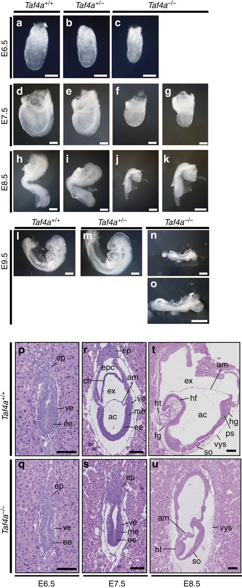
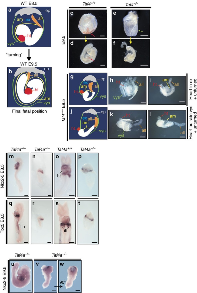
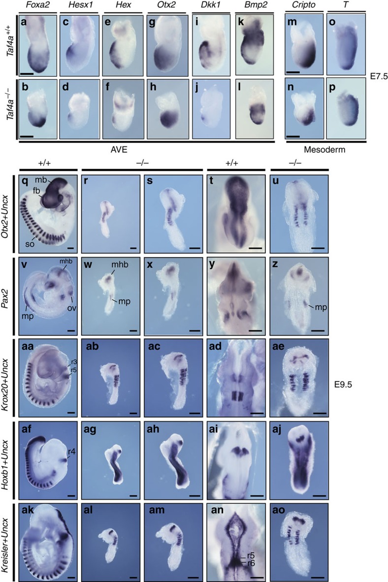
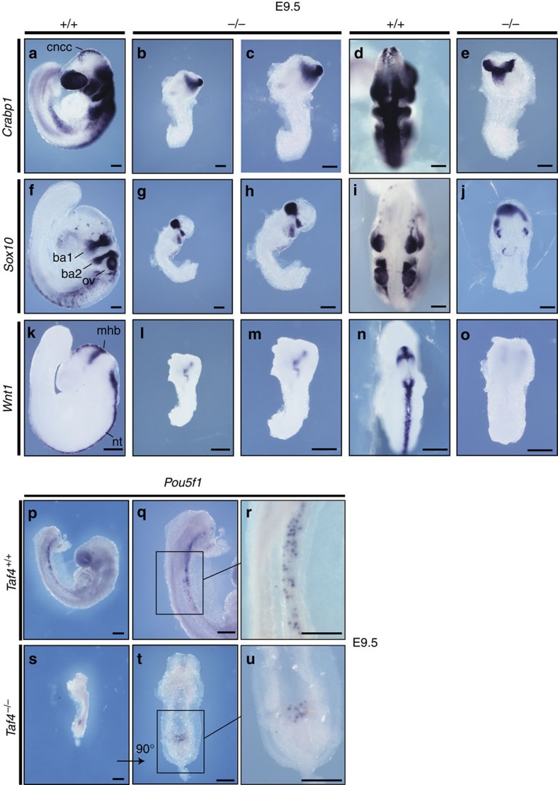
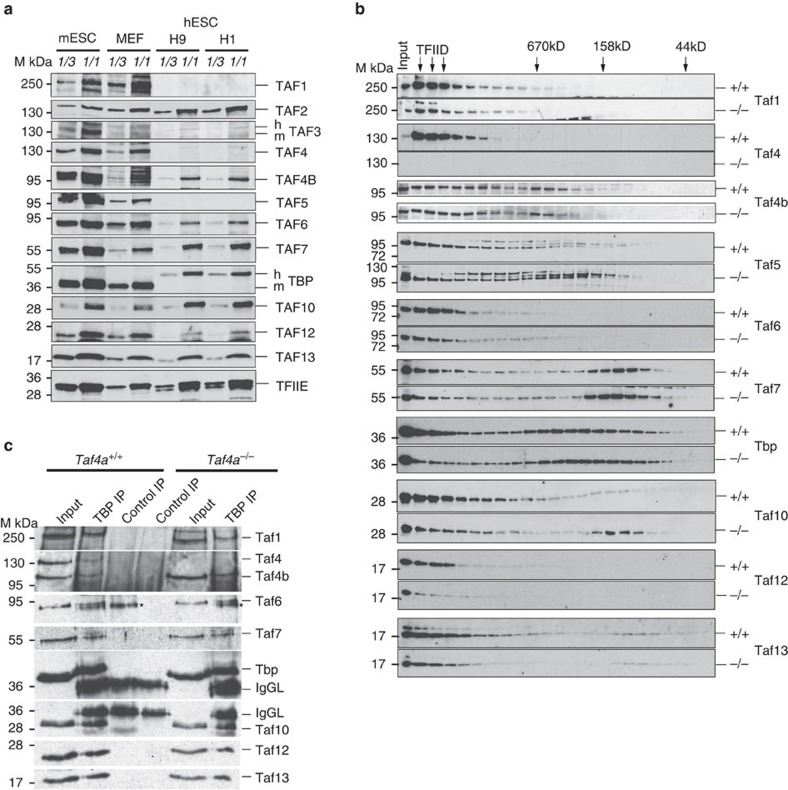
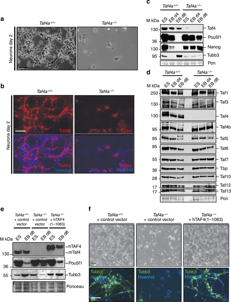
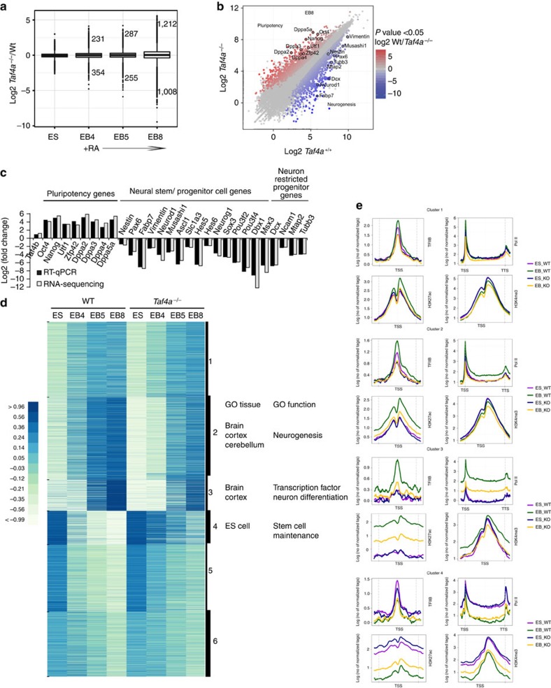
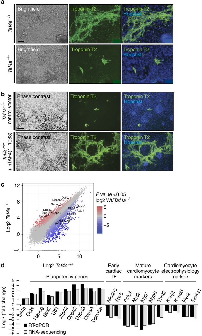
TAF4 (TATA-binding protein-associated factor 4) and its paralogue TAF4b are components of the TFIID core module. We inactivated the murine Taf4a gene to address Taf4 function during embryogenesis. Here we show that Taf4a(-/-) embryos survive until E9.5 where primary germ layers and many embryonic structures are identified showing Taf4 is dispensable for their specification. In contrast, Taf4 is required for correct patterning of the trunk and anterior structures, ventral morphogenesis and proper heart positioning. Overlapping expression of Taf4a and Taf4b during embryogenesis suggests their redundancy at early stages. In agreement with this, Taf4a(-/-) embryonic stem cells (ESCs) are viable and comprise Taf4b-containing TFIID. Nevertheless, Taf4a(-/-) ESCs do not complete differentiation into glutamatergic neurons and cardiomyocytes in vitro due to impaired preinitiation complex formation at the promoters of critical differentiation genes. We define an essential role of a core TFIID TAF in differentiation events during mammalian embryogenesis.


Figures

Figure 1
Figure 1. Taf4 is required during mid-gestation.
(ac) E6.5 stage embryos of the indicated genotypes. (dg). E7.5-stage embryos of the indicated genotypes, g shows a blow up of f. Scale bar, 100 μm. (hk). E8.5-stage embryos of the indicated genotypes, k shows a blow up of j. (lo) E9.5-stage embryos of the indicated genotypes, o shows a blow up of n. Scale bar, 200 μm. (pu) Haematoxylin and eosin-stained sections through WT or mutant embryos at the indicated stages. Scale bar, 100 μm. ep, ectoplacental cone; ve, visceral endoderm; ee, embryonic ectoderm; me, mesoderm;, epc, ectoplacental cavity; ch, chorion; ex, exocoelomic cavity; am, amnion; ac, amniotic cavity; hf, headfold; ht, heart; fg, foregut; hg, hindgut; so, somites; vys, visceral yolk sac; ps, primitive streak.
Figure 2
Figure 2. Taf4 is required for correct positioning of head and heart.
(a,b). Schematic representations illustrate normal development at E8.5 and E9.5 highlighting positions of head and heart during embryo turning. (cf) WT and mutant embryos at E9.5 before and after dissection from the yolk sac highlighting the blood-filled yolk sac vascular plexus in control embryos (red arrow in c) and the embryo protruding partially out of the yolk sac (green arrow in e). WT embryos show a strong accumulation of blood in the atrium of the heart (red arrow in d), which was absent in Taf4a−/− embryos (f). (gi) Schematic illustration of observed mutant phenotypes at E9.5. All mutant embryos fail to undergo turning at E9.5 and fusion of the allantois to the chorion. In addition to this, mutant embryos develop heart structures either in the exocoelomic cavity (hi) or outside of the visceral yolk sac (kl). (mw) In situ hybridization of wild-type and Taf4a−/− embryos for the cardiac transcription factors Nkx2-5 at E8.5 (mp) or Tbx5 at E8.5 (qt) and E9.5 (uw). ep, ectoplacental cone; all, allantois; ht, heart; flp, forelimb precursors; ift, inflow tract. Scale bar, 500 μm (cf), 300 μm (h-i), 200 μm (mw).
Figure 3
Figure 3. Abnormal patterning of Taf4a−/− embryos.
(ao) (upper row). In situ hybridization with the indicated probes for anterior visceral endoderm (AVE) and mesoderm markers on E7.5 WT embryos. (bp) (lower row). In situ hybridization with the indicated probes on E7.5 Taf4−/− embryos. (qao). In situ hybridization with neuronal markers in E9.5 WT or Taf4−/− embryos (lateral views (qak); ventral view R dorsal views T-AN) and (lateral views RP-AL, higher magnifications of same embryos sam; ventral view U; dorsal views zao). Scale bar, 200 μm in all panels.
Figure 4
Figure 4. Neural crest cell markers and primordial germ cells in Taf4 mutant embryos.
(ao) In situ hybridizations for neural crest markers Sox10, Crabp1 and Wnt1 in E9.5 control (lateral views a,f,k dorsal views d,i,n) and Taf4a−/− embryos (lateral views b,g,l higher magnifications of the same embryos c,h,m dorsal views E, J, O ). (pu). In situ hybridizations at E9.5 with Pou5f1 (Oct4) as a marker for primordial germ cells (PGCs). ba1, first branchial arch; ba2, second branchial arch; ov, otic vesicle; cncc, cranial neural crest cells; mhb, midbrain–hindbrain boundary; nt, neural tube. Scale bar, 200 μm in all panels.
Figure 5
Figure 5. Biochemical analysis of TFIID in Taf4a−/− ES cells.
(a). Immunoblots showing expression of TFIID subunits in ES cells, MEFs and two human ES cell lines, H1 and H9. (b). Immmunoblots on gel filtration fractions of nuclear extracts from WT and mutant ES cells. (c). Immunoblots of Tbp IPs from the pooled TFIID fractions indicated by arrows in b. * indicates a non-specific band migrating just below Taf6.
Figure 6
Figure 6. Taf4 is required for RA-driven neuronal ES cell differentiation.
(a) Phase-contrast microscopy of RA-treated EBs differentiated into neurons 2 days after plating. (b) Tubb3 labelling of RA-treated EBs differentiated into neurons 2 days after plating. (c,d). Immunoblots on extracts from differenting ES cells and EBs with the indicated antibodies. The membranes were stained with Ponceau as loading controls. (e). Immunoblots on extracts from ES cell lines engineered to re-express recombinant Taf4. (f). Phase-contrast microscopy and Tubb3 labelling of RA-treated EBs differentiated into neurons 2 days after plating. All images were taken at × 20 magnification. Scale bars 100 μm.
Figure 7
Figure 7. Taf4 is required for activation of neurogenic genes in differentiating ES cells.
(a). Gene expression changes at different stages of differentiation in Taf4a−/− compared with WT ES cells. The numbers of genes with altered expression and their fold changes at each stage are indicated. (b). Global comparison of RNA-seq data in Taf4a−/− compared to WT at the EB8 stage. The expression of several neurogenic and pluripotency genes are highlighted. (c). Comparison of the changes in expression of selected marker genes measured by RNA-seq and by RT–qPCR. (d). Kinetics of gene expression and their alterations during differentiation are represented as a clustered heatmap. The ontology terms associated with the genes in clusters 2–4 that are most affected by Taf4 loss are indicated. (e). Metaprofiles for the indicated ChIP-seq data sets corresponding to genes in clusters 2–4 are shown. For TFIIB, H3K4me3 and H3K27ac the profiles are centred on the TSS. For Pol II the profiles are show over the gene from the transcription start site (TSS) to the transcription termination site (TTS).
Figure 8
Figure 8. Taf4 is required for cardiomyocyte differentiation.
(a) Phase-contrast microscopy and Tnnt2 labelling of EBs differentiated into cardiomyocytes 9 days after plating. (b) Phase-contrast microscopy and Tnnt2 labelling of ES cells engineered to re-express exogenous Taf4 differentiated into cardiomyocytes 9 days after plating. All images were taken at × 20 magnification. (c) Global comparison of RNA-seq data in Taf4a−/− compared with WT at the EB8 stage. The expression of several cardiomyocyte markers and pluripotency genes are highlighted. (d) Comparison of the changes in expression of selected marker genes measured by RNA-seq and by RR–qPCR. Scale bars 50 μm.

References

    1. Wright K. J., Marr M. T. 2nd & Tjian R. TAF4 nucleates a core subcomplex of TFIID and mediates activated transcription from a TATA-less promoter. Proc. Natl Acad. Sci. USA 103, 12347–12352 (2006) . - PMC - PubMed
    1. Cler E., Papai G., Schultz P. & Davidson I. Recent advances in understanding the structure and function of general transcription factor TFIID. Cell Mol. Life Sci. 66, 2123–2134 (2009) . - PMC - PubMed
    1. Bieniossek C. et al.. The architecture of human general transcription factor TFIID core complex. Nature 493, 699–702 (2013) . - PubMed
    1. Zhou H. et al.. Dual functions of TAF7L in adipocyte differentiation. eLife 2, e00170 (2013) . - PMC - PubMed
    1. Cheng Y. et al.. Abnormal sperm in mice lacking the taf7l gene. Mol. Cell. Biol. 27, 2582–2589 (2007) . - PMC - PubMed

Publication types

MeSH terms