. 2016 Aug:107:305-316.
doi: 10.1016/j.neuropharm.2016.03.044. Epub 2016 Mar 26.

Effects of lack of microRNA-34 on the neural circuitry underlying the stress response and anxiety

Affiliations

Effects of lack of microRNA-34 on the neural circuitry underlying the stress response and anxiety

Diego Andolina et al. Neuropharmacology. 2016 Aug.

Abstract

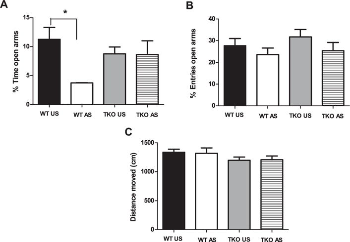
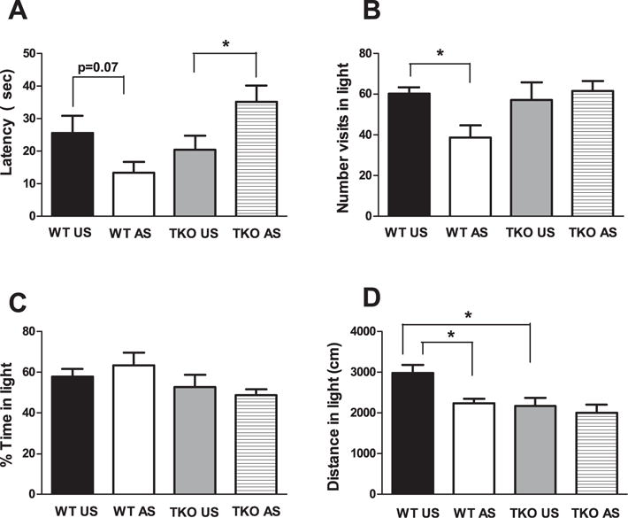
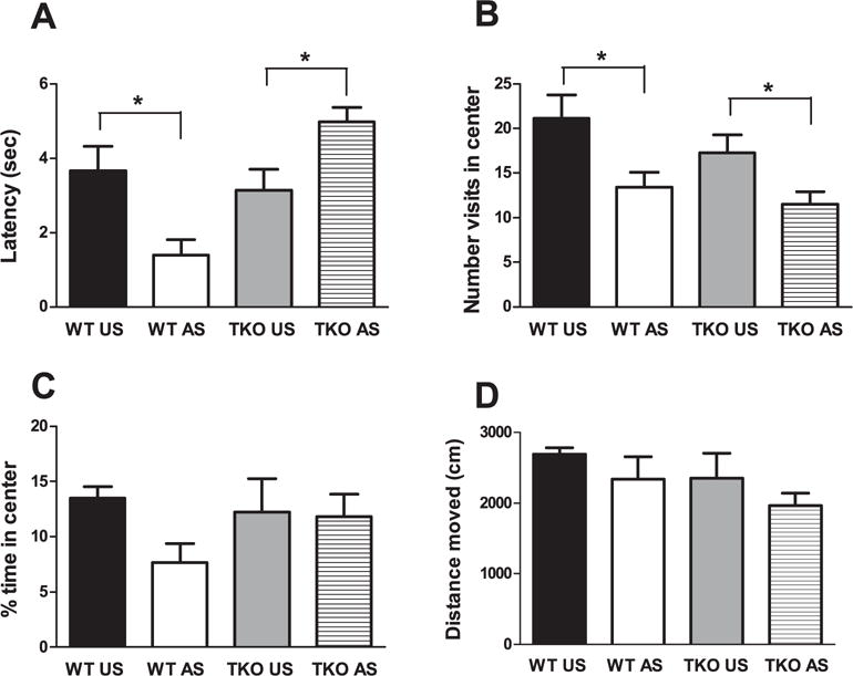
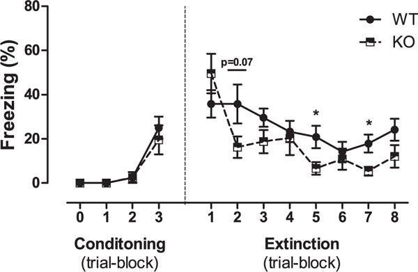
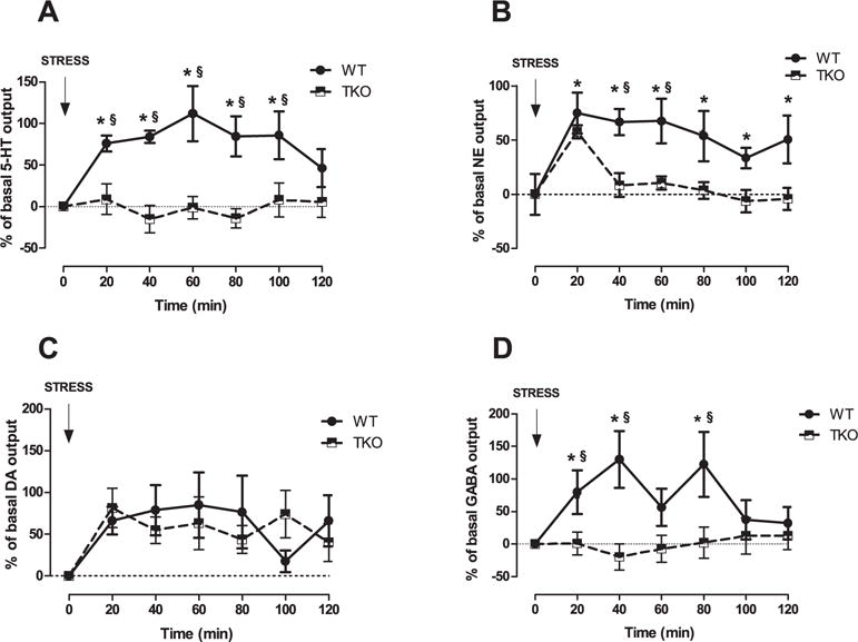
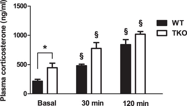
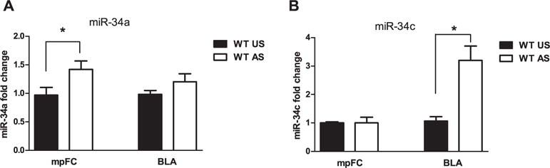
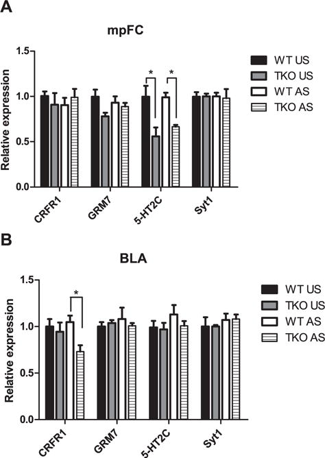
Stress-related psychiatric disorders, including anxiety, are complex diseases that have genetic, and environmental causes. Stressful experiences increase the release of prefrontal amygdala neurotransmitters, a response that is relevant to cognitive, emotional, and behavioral coping. Moreover, exposure to stress elicits anxiety-like behavior and dendritic remodeling in the amygdala. Members of the miR-34 family have been suggested to regulate synaptic plasticity and neurotransmission processes, which mediate stress-related disorders. Using mice that harbored targeted deletions of all 3 members of the miR-34-family (miR-34-TKO), we evaluated acute stress-induced basolateral amygdala (BLA)-GABAergic and medial prefrontal cortex (mpFC) aminergic outflow by intracerebral in vivo microdialysis. Moreover, we also examined fear conditioning/extinction, stress-induced anxiety, and dendritic remodeling in the BLA of stress-exposed TKO mice. We found that TKO mice showed resilience to stress-induced anxiety and facilitation in fear extinction. Accordingly, no significant increase was evident in aminergic prefrontal or amygdala GABA release, and no significant acute stress-induced amygdalar dendritic remodeling was observed in TKO mice. Differential GRM7, 5-HT2C, and CRFR1 mRNA expression was noted in the mpFC and BLA between TKO and WT mice. Our data demonstrate that the miR-34 has a critical function in regulating the behavioral and neurochemical response to acute stress and in inducing stress-related amygdala neuroplasticity.

Keywords: Amygdala; Anxiety; Prefrontal cortex; Stress; miR-34.


Figures

Fig. 1
Fig. 1
Effects of stress exposure (30 min of restraint) on (A) the percentage of time spent in the open arms (B) the percentage of entries in the open arms, (C) the traveled distance in the apparatus in elevated plus maze in WT (WT Unstressed (US) n = 8; WT stressed (WT AS) n = 8) and TKO (TKO US n = 8; TKO AS n = 8) mice. Data are expressed as mean ± SE. *P < 0.05.
Fig. 2
Fig. 2
Effects of stress exposure (30 min of restraint) on (A) the latency to entry in the dark compartment, (B) the number of visits in the lighted compartment, (C) the percentage of time spent in the lighted compartment and (D) the distance covered in the lighted compartment in the dark/light test in WT (WT Unstressed (US) n = 7; WTstressed (WT AS) n = 7) and TKO (TKO US n = 7; TKO AS n = 8) mice. Data are expressed as mean = SE. *P < 0.05.
Fig. 3
Fig. 3
Effects of stress exposure (30 min of restraint) on (A) the latency to exit from the center, (B) the number of visit in the center, (C) the percentage of time spent in the center and (D) the traveled distance in the apparatus in WT (WT Unstressed (US) n = 6; WT stressed (WT AS) n = 7) and TKO (TKO US n = 7; TKO AS n = 8) mice. Data are expressed as mean ± SE. *P < 0.05.
Fig. 4
Fig. 4
Effects of the genotype (WT, n = 8; TKO, n = 8) on fear memory extinction following contextual and cued fear conditioning training. Data are expressed as mean ± SE of percent freezing. *P < 0.05.
Fig. 5
Fig. 5
Effects of stress exposure (120 min of restraint) on (A) Serotonin (5-HT), (B) Norepinephrine (NE), (C) Dopamine (DA) outflow in the medial prefrontal cortex (mpFC) and (D) GABA outflow in the Basolateral Amygdala (BLA) of WT (NE, n = 9; DA, n = 8; 5-HT, n = 7; GABA, n = 9) and TKO (NE, n = 9; DA, n = 7; 5-HT, n = 12; GABA, n = 9) mice. Results are expressed as percent changes (mean ± SE) from basal values. Statistical analyses were performed on raw data. § P < 0.05 in comparison with the corresponding time point of miR-34s TKO group. *P < 0.05 from basal values.
Fig. 6
Fig. 6
CORT levels in basal condition and after 30 or 120 min of acute restraint stress exposure in WT (WT basal, n = 8; WT stressed (AS) 30 min, n = 8; WT AS 120 min, n = 6) and TKO mice (TKO basal, n = 8; TKO stressed (AS) 30 min, n = 7; TKO AS 120 min, n = 6 mice). Data are expressed as mean ± SE. *P < 0.05. § P < 0.05 from basal values.
Fig. 7
Fig. 7
Effects of stress exposure (30 min of restraint) on (A) miR-34a and (B) miR-34c expression in the medial prefrontal cortex (mpFC) and Basolateral Amygdala (BLA) of WT (WT Unstressed (US), mpFC miR-34a, n = 9; mpFC miR-34c, n = 8; BLA miR-34a, n = 8; BLA miR-34c, n = 8); WTstressed (AS), mpFC miR-34a, n = 8; mpFC miR-34c, n = 6; BLA miR-34a, n = 9; BLA miR-34c, n = 9)) mice. Data are expressed as mean ± SE. *P < 0.05.
Fig. 8
Fig. 8
Effect of the stress exposure (30 min of restraint) on genes mRNA expression in the (A) medial prefrontal cortex (mpFC) and (B) Basolateral amygdala (BLA) of WT (WT Unstressed (US), mpFC n = 4, BLA n = 4; WT stressed (AS), mpFC n = 4, BLA n = 5) and TKO (TKO US, mpFC n = 4, BLA n = 4; TKO AS, mpFC n = 4, BLA n = 4) mice. Data are expressed as mean ± SE. *P < 0.05.
Fig. 9
Fig. 9
Effect of the stress exposure (30 min of restraint) on (A) spines density, (B) numbers of branch points, and (C) dendritic length in the Basolateral Amygdala (BLA) of WT (WT Unstressed (US), n = 5/neurons = 26; WT stressed (AS), n = 5/neurons = 26) and TKO (TKO Unstressed (US), n = 5/neurons = 25; TKO stressed (AS), n = 5/neurons = 27) mice. (D) High-power photomicrographs of the representative dendritic segment in the BLA of WT and TKO mice. (E) Representative camera lucida drawing the neurons of WT and miR-34 TKO mice. Data are expressed as mean ± SE. *P < 0.05.

Similar articles

Cited by

References

    1. Abbas MG, Shoji H, Soya S, Hondo M, Miyakawa T, Sakurai T. Comprehensive behavioral analysis of male Ox1r (−/−) mice showed implication of orexin receptor-1 in mood, anxiety, and social behavior. Front Behav Neurosci. 2015;9:324. - PMC - PubMed
    1. Agostini M, Tucci P, Steinert JR, Shalom-Feuerstein R, Rouleau M, Aberdam D, Forsythe ID, Young KW, Ventura A, Concepcion CP, Han YC, Candi E, Knight RA, Mak TW, Melino G. microRNA-34a regulates neurite outgrowth, spinal morphology, and function. Proc Natl Acad Sci U S A. 2011;108:21099–21104. - PMC - PubMed
    1. Akirav I, Maroun M. The role of the medial prefrontal cortex amygdala circuit in stress effects on the extinction of fear. Neural Plast. 2007:30873. - PMC - PubMed
    1. Andolina D, Conversi D, Cabib S, Trabalza A, Ventura R, Puglisi-Allegra S, Pascucci T. 5-Hydroxytryptophan during critical postnatal period improves cognitive performances and promotes dendritic spine maturation in genetic mouse model of phenylketonuria. Int J Neuropsychopharmacol. 2011;14:479–489. - PMC - PubMed
    1. Andolina D, Maran D, Valzania A, Conversi D, Puglisi-Allegra S. Prefrontal/amy gdalar system determines stress coping behavior through 5-HT/ GABA connection. Neuropsychopharmacology. 2013;38:2057–2067. - PMC - PubMed