. 2016 Mar 30:7:11069.
doi: 10.1038/ncomms11069.

A developmentally regulated switch from stem cells to dedifferentiation for limb muscle regeneration in newts

Affiliations

A developmentally regulated switch from stem cells to dedifferentiation for limb muscle regeneration in newts

Hibiki Vincent Tanaka et al. Nat Commun. .

Abstract

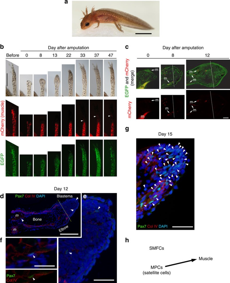
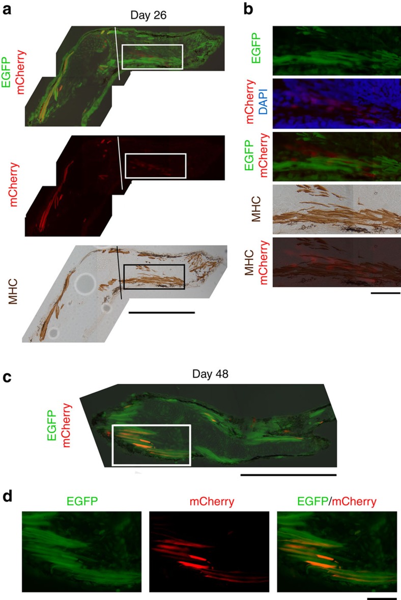
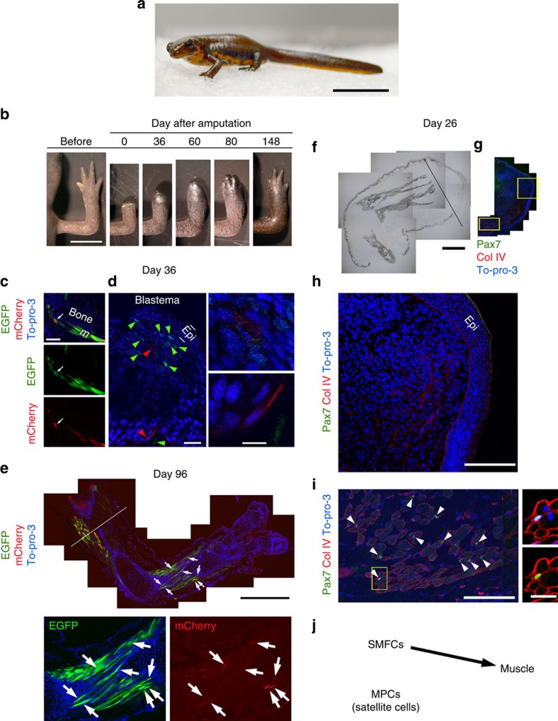
The newt, a urodele amphibian, is able to repeatedly regenerate its limbs throughout its lifespan, whereas other amphibians deteriorate or lose their ability to regenerate limbs after metamorphosis. It remains to be determined whether such an exceptional ability of the newt is either attributed to a strategy, which controls regeneration in larvae, or on a novel one invented by the newt after metamorphosis. Here we report that the newt switches the cellular mechanism for limb regeneration from a stem/progenitor-based mechanism (larval mode) to a dedifferentiation-based one (adult mode) as it transits beyond metamorphosis. We demonstrate that larval newts use stem/progenitor cells such as satellite cells for new muscle in a regenerated limb, whereas metamorphosed newts recruit muscle fibre cells in the stump for the same purpose. We conclude that the newt has evolved novel strategies to secure its regenerative ability of the limbs after metamorphosis.


Figures

Figure 1
Figure 1. SMFC tracking in larval newt limb regeneration.
(a) Larva (3 months old). It has four limbs, as well as the gills and tail fin. Scale bar, 4 mm. (b) Monitoring of SMFCs (mCherry+) during limb regeneration (n=6). mCherry was not detected in the regenerating part of the limb until ∼30 days when the amputated limb had almost been recovered (see Supplementary Movie 1). Arrowheads: flexor muscle for digits (see Fig. 2). Scale bar, 1 mm. (c) Sections of regenerating limbs (n=3 for each stage). SMFC-derived mCherry+ cells were not observed in the blastema. Lines: amputation site. m: muscle. Scale bar, 100 μm. (df) Pax7 immunolabelling of regenerating limbs on day 12 (n=3) and (g) day 15 (n=3) after amputation. (d) On day 12, a few Pax7+ nuclei (arrowheads) were detected in blastema cells and in satellite cells along the muscle fibres. Col IV, collagen type IV immunoreactivity. DAPI (4,6-diamidino-2-phenylindole), nuclei. Scale bar, 300 μm. The Pax7+ nuclei pointed by arrowheads were enlarged in e and f, respectively. Scale bars, 100 μm. (g) On day 15 when the regenerating part of the limb grew more distally, the number of Pax7+ nuclei (arrowheads) in the blastema was dramatically increased. Scale bar, 100 μm. (h) Summary. In larval newts, MPCs, potentially satellite cells, were recruited for new muscle during limb regeneration, whereas SMFCs were not.
Figure 2
Figure 2. Appearance of mCherry in a late stage of larval limb regeneration.
(a) A section of the limb on day 26 after amputation (n=3). mCherry was detected in mononucleated cells, which had occupied the same area as the differentiating flexor muscle for digits (rectangle). Lines: amputation site. MHC, myosin heavy chain immunoreactivity. Scale bar, 500 μm. (b) Enlargement of the area enclosed by the rectangle in a. At this stage, mCherry did not overlap with the flexor muscle. Scale bar, 100 μm. (c) A section of the forearm on day 48 after amputation (n=3). Scale bar, 500 μm. (d) Enlargement of the area enclosed by a rectangle in c. The flexor muscle for digits at this stage exhibited mCherry. Scale bar, 100 μm.
Figure 3
Figure 3. SMFC tracking in metamorphosed newt limb regeneration.
(a) Juvenile (16 months old). Scale bar, 15 mm. (b) Limb regeneration. Scale bar, 5 mm. (ce) Tracking of SMFCs (mCherry+) (n=2). This animal was a mosaic expressing EGFP in muscle only. mCherry+ fibres in the forearm were ∼25% of total EGFP+ fibres. (c) On day 36 after amputation, fragments of muscle fibres (arrows) were observed in distal regions adjacent to the blastema. Scale bar, 100 μm. (d) mCherry+ mononucleated cells (red arrowheads; enlarged in right-hand panels) and EGFP+ cells (green arrowheads) were observed in the blastema. Epi, epidermis. To-pro-3: nuclei. Scale bars, 50 μm (left), 10 μm (right-hand panels). (e) In the same limb, at day 96 after the second amputation in the upper arm (line), mCherry (arrows) and EGFP were observed only in muscle fibres. Scale bars, 1 mm (upper panel), 500 μm (lower panels). (fi) Pax7 immunolabelling of a regenerating limb on day 26 after amputation (n=4). Pax7 immunoreactivity was not detected in the blastema. (f) Translucent image. Line: amputation site. (g) Merged fluorescence image. Col IV, collagen type IV immunoreactivity. To-pro-3: nuclei. Scale bar, 1 mm. (h) Enlargement of a region in the blastema and (i) a region proximal to the amputation site, enclosed by boxes in g. Scale bars, 250 μm. Arrowheads in (i) Pax7+ nuclei. An example satellite cell (box) is enlarged in the right-hand panels (upper: Col VI/To-pro-3; lower: Col IV/Pax7). Scale bar, 50 μm. (j) Summary. In metamorphosed newts, SMFCs were recruited for new muscle during limb regeneration, whereas MPCs such as satellite cells were not.
Figure 4
Figure 4. Lineage tracing in adult newt limb regeneration by tissue transplantation.
(a,b) Tracing of skin cells by allograft of mCherry+ skin (n=3). (a) mCherry+ cells were distributed along the epidermis and dermis, as well as around the cartilage in a regenerating part of the limb (day 40). c, cartilage. Asterisk, mucous gland. Scale bar, 200 μm. (b) In a growing tip of new digit, mCherry was sometimes observed in chondrocytes (arrows) in the cartilage and in interstitial cells surrounding the cartilage. Epi: epidermis. Scale bars, 100 μm. (c,d) Tracing of epidermal cells by transplantation of mCherry+ ectoderm at embryonic stage (n=2). mCherry was observed in epidermal cells such as those in multilayered epithelium (arrowheads in c) and mucous glands (asterisks in d), as well as in interstitial cells around the cartilage (arrowheads in d) in a regenerating part of the limb (day 148). Scale bars, 200 μm. (e) Tracing of bone cells by allograft of mCherry+ bone (n=4). mCherry was observed in chondrocytes (arrows) in new cartilage of a regenerating limb (day 81). Asterisk: mucous gland. Scale bar, 100 μm. (f) Tracing of muscle cells by allograft of mCherry+ muscle (n=3). mCherry was observed in muscle fibres in a regenerating part of the limb (day 81). MHC, myosin heavy chain immunoreactivity. Scale bar, 50 μm. (g) Tracing of Schwann cells by implantation of a fragment of mCherry+ nerve (n=3). mCherry was observed in Schwann cells along regenerated nerve fibres in a regenerating part of the limb (day 133). Arrowhead: myelin sheath covering a regenerated nerve fibre; a-tubulin, acetylated tubulin immunoreactivity. Scale bar, 20 μm. Blue: nuclei stained by To-pro-3 (bd,f,g) and DAPI (4,6-diamidino-2-phenylindole) (e). (h) Summary. The skin, bone, muscle and nerve (Schwann cell) regenerated themselves. Exceptionally, dermis contributed to new bone/cartilage.

References

    1. Tsonis P. A. Limb Regeneration Cambridge Univ. Press (1996) .
    1. Sandoval-Guzmán T. et al. Fundamental differences in dedifferentiation and stem cell recruitment during skeletal muscle regeneration in two salamander species. Cell Stem Cell 14, 174–187 (2014) . - PubMed
    1. Casco-Robles M. M. et al. Expressing exogenous genes in newts by transgenesis. Nat. Protoc. 6, 600–608 (2011) . - PubMed
    1. Holder N. Organization of connective tissue patterns by dermal fibroblasts in the regenerating axolotl limb. Development 105, 585–593 (1989) . - PubMed
    1. Feil R., Wagner J., Metzger D. & Chambon P. Regulation of Cre recombinase activity by mutated estrogen receptor ligand-binding domains. Biochem. Biophys. Res. Commun. 237, 752–757 (1997) . - PubMed

Publication types

Substances