. 2016 Dec;27(12):3639-3652.
doi: 10.1681/ASN.2015111227. Epub 2016 Mar 29.

TWEAK-Fn14 Signaling Activates Myofibroblasts to Drive Progression of Fibrotic Kidney Disease

Affiliations

TWEAK-Fn14 Signaling Activates Myofibroblasts to Drive Progression of Fibrotic Kidney Disease

Ivan G Gomez et al. J Am Soc Nephrol. 2016 Dec.

Abstract

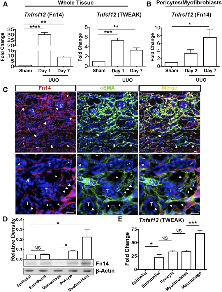
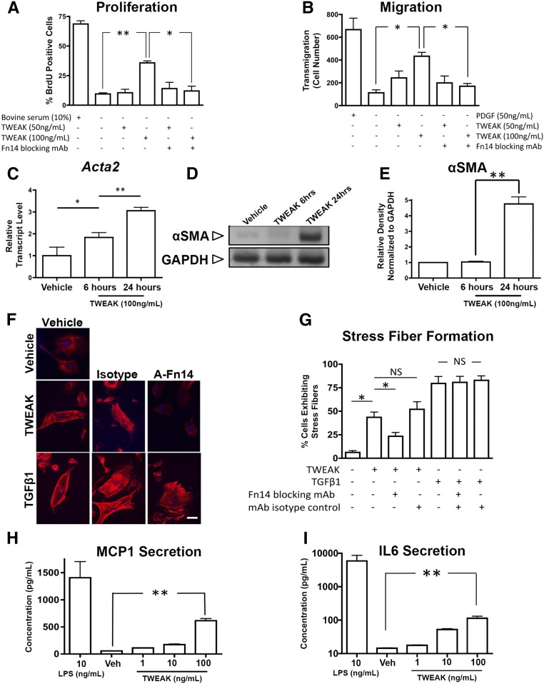
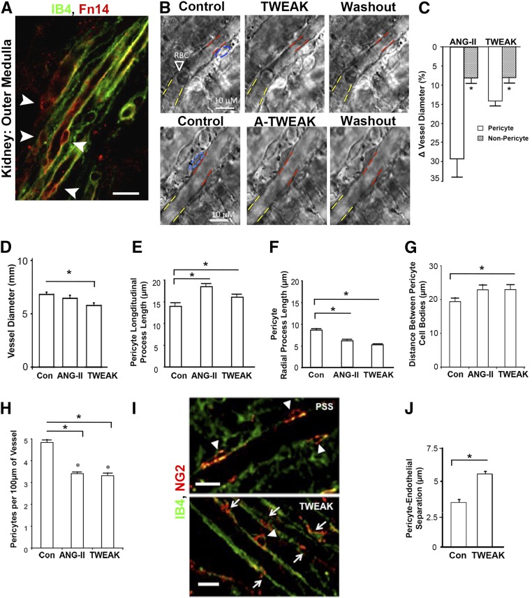
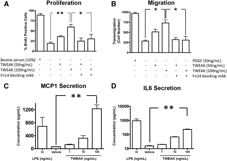
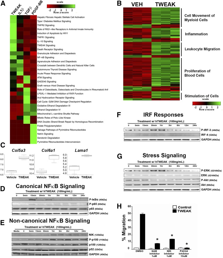
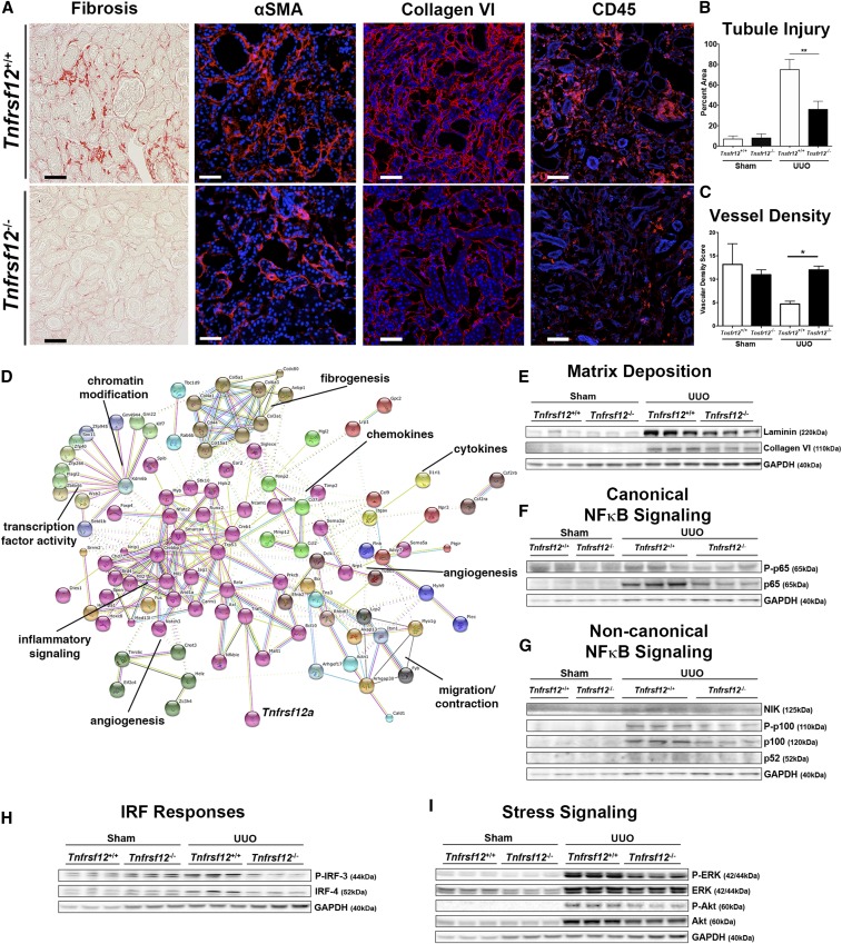
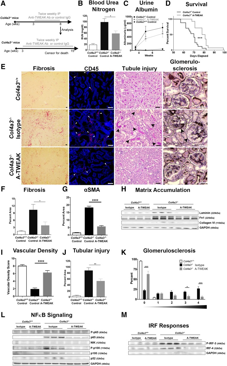
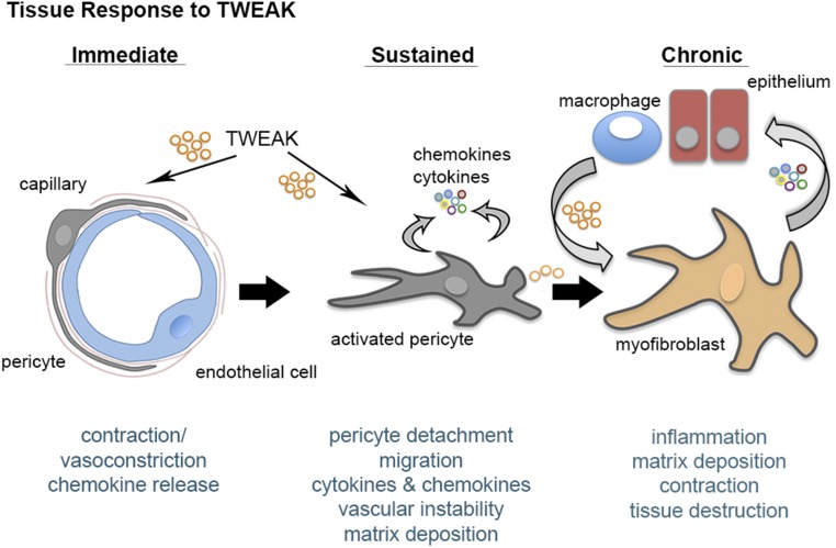
The identification of the cellular origins of myofibroblasts has led to the discovery of novel pathways that potentially drive myofibroblast perpetuation in disease. Here, we further investigated the role of innate immune signaling pathways in this process. In mice, renal injury-induced activation of pericytes, which are myofibroblast precursors attached to endothelial cells, led to upregulated expression of TNF receptor superfamily member 12a, also known as fibroblast growth factor-inducible 14 (Fn14), by these cells. In live rat kidney slices, administration of the Fn14 ligand, TNF-related weak inducer of apoptosis (TWEAK), promoted pericyte-dependent vasoconstriction followed by pericyte detachment from capillaries. In vitro, administration of TWEAK activated and differentiated pericytes into cytokine-producing myofibroblasts, and further activated established myofibroblasts in a manner requiring canonical and noncanonical NF-κB signaling pathways. Deficiency of Fn14 protected mouse kidneys from fibrogenesis, inflammation, and associated vascular instability after in vivo injury, and was associated with loss of NF-κB signaling. In a genetic model of spontaneous CKD, therapeutic delivery of anti-TWEAK blocking antibodies attenuated disease progression, preserved organ function, and increased survival. These results identify the TWEAK-Fn14 signaling pathway as an important factor in myofibroblast perpetuation, fibrogenesis, and chronic disease progression.

Keywords: Alport nephropathy; chronic; kidney disease; myofibroblast; pericyte; vascular biology.


Figures

Figure 1.
Figure 1.
Fn14 is highly expressed by kidney myofibroblasts and TWEAK is highly expressed by macrophages. (A) Quantitative PCR (qPCR) results showing changes in Tnfrsf12 (Fn14) and Tnfsf12 (TWEAK) transcript levels in whole tissue from the UUO model of kidney disease in mice versus sham. (B) qPCR showing changes in Tnfrsf12 transcript levels in FACS-sorted collagen I-producing cells in the UUO model of kidney disease in mice versus sham. (C) Immunofluorescence images showing the localization of Fn14 in the UUO kidney disease model at day 10. Note a predominantly interstitial pattern of expression and predominant colocalization with a subpopulation of myofibroblasts, particularly in areas of intense interstitial cell accumulation (arrowheads), whereas some myofibroblasts do not coexpress Fn14. Weak expression in the mesangium (*) of glomeruli (g) can be seen. Expression of Fn14 on endothelium is also detected (thick arrows) and while almost all tubular epithelium (T) does not express Fn14, a minority of injured cells can be seen to express Fn14 (thin arrows) (a, arteriole). (D) Western blot showing Fn14 protein expression in primary cultures of cells. Quantitation is based on three independent experiments. (E) qPCR showing relative expression of Tnfsf12 transcripts in primary cell cultures. Bar, 25 μm; *P<0.05; **P<0.01; ***P<0.001; n=5/group unless otherwise stated.
Figure 2.
Figure 2.
Kidney pericyte cultures are activated by TWEAK. (A, B) Graphs showing the (A) proliferative response and (B) migratory response of pericytes across a transwell barrier in response to TWEAK. (C–E) The effect of TWEAK on pericyte expression of Acta2 transcript levels and its protein, the myofibroblast marker αSMA (C) quantitative PCR, (D) Western blot, and (E) normalized densitometry of Western blots. (F) Fluorescence images and (G) quantification of stress fiber formation as detected by phalloidin-Cy3 in pericytes in response (24 hours) to TWEAK or TGFβ1. Note, anti-Fn14 (A-Fn14) blocking antibodies inhibit TWEAK-mediated stress fiber formation, but not TGFβ-mediated stress fibers. (H, I) Graphs showing release of (H) MCP1 or (I) IL6 into the supernatant by primary pericyte cultures. Note, at higher concentrations pericytes release substantial amounts of these cytokines. Bar, 25 μm; *P<0.05; **P<0.01; n=4/group.
Figure 3.
Figure 3.
TWEAK stimulates acute microvascular vasoconstriction mediated by pericytes in situ, followed by pericyte detachment. (A) Fluorescence micrograph showing isolectin (IB4) labeled endothelium and immunodetection of Fn14 in rat kidney slice. Note prominent expression of Fn14 by perivascular cells (arrowheads) (bar, 25 µm). (B) Phase contrast images of medullary capillaries (vasa recta) in live kidney slice showing the effect of TWEAK on capillary diameter after 270 seconds of exposure (blue: pericyte body, red: diameter at pericyte body, yellow: diameter at nonpericyte area). Note washout of TWEAK (after 6 minutes) results in relaxation. Application of anti-TWEAK antibodies has no effect on diameter. (C) Quantification of change in capillary diameter 300 seconds after application of AngII or TWEAK in pericyte or nonpericyte areas of the capillary (n=8/group). (D) Graph of capillary diameter after treatment of kidney slice with TWEAK or AngII for 4 hours. (E, F) Graphs showing the length of processes of NG2-labeled pericytes where (E) longitudinal processes increase in length but (F) radial processes retract. (G, H) Morphometric measurements of pericyte density long the vasa recta capillary 4 hours after treatment with TWEAK or AngII compared with control. (I) Fluorescent images of pericytes labeled with NG2 and capillary lumen labeled with IB4. In control settings (PSS), long pericyte processes remain longitudinal with the capillary and associated with cell bodies (shown by arrowheads), whereas after 4 hours of TWEAK, pericyte processes without associated cell bodies are seen (arrows) indicative of separation, and in other instances cell bodies show enlargement and spreading (arrowhead) (bar, 10 µm). (J) Morphometry of pericyte endothelial distance between pericyte basal membrane and capillary wall. *P<0.05; n=12/group. PSS, physiological saline solution; RBC, red blood cell.
Figure 4.
Figure 4.
Established kidney myofibroblasts are highly sensitive to TWEAK mediated activation. (A, B) Graphs showing the proliferative response at (A) 16 hours and (B) migratory response (24 hours) across a transwell barrier of myofibroblasts in response to TWEAK. (C, D) Graphs showing release of (C) MCP1 or (D) IL6 into the supernatant by myofibroblast cultures. Note, at higher concentrations myofibroblasts release large amounts of these cytokines. *P<0.05; **P<0.01; n=4/group.
Figure 5.
Figure 5.
Transcriptional analysis of cytokine-stimulated myofibroblasts identifies TWEAK-induced fibrogenic signaling via canonical and noncanonical NF-κB, IRF, and ERK. (A) Ingenuity IPA software was used to identify pathways that are enriched in myofibroblasts treated with TWEAK, IL1β, TGFβ, or PDGF-BB compared with vehicle after 16 hours. The top ten pathways for each cytokine based on P values were pooled, and the heatmap shows z-scores for these pathways across all stimuli. (B) The heatmap shows the intensity of genes differentially expressed with TWEAK treatment at 16 hours for each of the biologic processes shown. Vehicle and TWEAK treatments were performed in triplicate. (C) Normalized gene expression for matrix proteins upregulated by TWEAK treatment (log base 2). (D) Blots showing canonical NF-κB activation by the increase in P-IκBα, P-p65 (RELA) with time in myofibroblasts in response to TWEAK. (E) Blots showing noncanonical NF-κB activation by accumulation of NIK and activation of P-p100 to generate the cleaved and active p52 subunit. (F) Blots showing the effect of TWEAK on active P-IRF-3 accumulation and IRF-4 accumulation. (G) The effect of TWEAK on active P-ERK and P-AKT signaling adaptors. (H) The effect of canonical NF-κB inhibitor (IκKβ inhibitor), noncanonical NF-κB inhibitor (NIK inhibitor), and ERK inhibitor on TWEAK stimulated myofibroblast migration. *P<0.05.
Figure 6.
Figure 6.
Fn14 deficiency (Tnfrsf12−/−) reduces fibrogenesis in the UUO model of kidney disease. (A) Photomicrographic images showing the extent of fibrosis (day 10), αSMA+ myofibroblasts (day 7), collagen type VI (day 7), and CD45+ leukocytes (day 7) in the UUO model. (B, C) Graphs quantifying tubular injury and vascular density. (D) String analysis showing known and predicted protein-protein interactions of 200 genes most enriched as a result of loss of Fn14 in the diseased kidney. Different types of evidence denoted by the color of the connecting lines, where green is neighborhood, red is gene fusion, blue is co-occurrence, black is coexpression, purple is experiments, cyan is databases, yellow is text mining, and lilac is homology. Proteins are colored according to nodes of interaction. (E) Blots showing the effect of Fn14 deficiency on matrix protein accumulation in whole tissue. (F, G) Blots showing the effect of disease and Fn14 deficiency on activation of factors in the (F) canonical and (G) noncanonical NF-κB signaling pathways. (H) Blots showing the effect of disease and Fn14 deficiency on activation of P-IRF-3 and IRF-4. (I) Blots showing the effect of Fn14 deficiency on active P-ERK and P-AKT. *P<0.05; **P<0.01; n=7/group; bar, 50 μm.
Figure 7.
Figure 7.
Anti-TWEAK antibodies block fibrogenesis and the progression of CKD in Col4a3−/− mice. (A) Schema showing the experimental approach. (B) BUN levels at 8 weeks showing the effect of anti-TWEAK in Col4a3−/− mice. (C) Time course of albuminuria in Col4a3−/− mice (D) Kaplein–Meier survival curve showing the effect of anti-TWEAK in Col4a3−/− mice. (E) Photomicrographs showing the extent of interstitial fibrosis (Sirius red stain), CD45+ leukocytes, tubular epithelial injury (periodic acid–Schiff stain), and glomerular scarring (silver stain) at 8 weeks of age in Col4a3−/− kidneys. (F–H) Quantification of Sirius red+ fibrosis, αSMA+ myofibroblasts, and matrix proteins. (I, J) Quantification of vascular density and tubular epithelial injury. (K) Glomerulosclerosis scores showing percentage of glomeruli with each of the disease scores (0=no sclerosis, 1=1%–25% sclerosis, 2=25%–50% sclerosis 3=50%–75% sclerosis, 4=75%–100% sclerosis). (L) Representative blots showing the effect of anti-TWEAK antibodies on canonical and noncanonical NF-κB signaling factors. (M) Representative blots showing the effect of anti-TWEAK antibodies on P-IRF-3 and IRF-4. *P<0.05; ** P<0.01; n=14/group; bar, 50 μm.
Figure 8.
Figure 8.
Tweak triggers vasoconstriction, pericyte activation and myofibroblast persistence in the kidney. Schema summarizing the effect of TWEAK on the fibroblast lineage in kidney disease.

Comment in

  • 3502–3504

References

    1. Duffield JS: Cellular and molecular mechanisms in kidney fibrosis. J Clin Invest 124: 2299–2306, 2014 - PMC - PubMed
    1. Lin SL, Kisseleva T, Brenner DA, Duffield JS: Pericytes and perivascular fibroblasts are the primary source of collagen-producing cells in obstructive fibrosis of the kidney. Am J Pathol 173: 1617–1627, 2008 - PMC - PubMed
    1. Schrimpf C, Xin C, Campanholle G, Gill SE, Stallcup W, Lin SL, Davis GE, Gharib SA, Humphreys BD, Duffield JS: Pericyte TIMP3 and ADAMTS1 modulate vascular stability after kidney injury. J Am Soc Nephrol 23: 868–883, 2012 - PMC - PubMed
    1. Grgic I, Krautzberger AM, Hofmeister A, Lalli M, DiRocco DP, Fleig SV, Liu J, Duffield JS, McMahon AP, Aronow B, Humphreys BD: Translational profiles of medullary myofibroblasts during kidney fibrosis. J Am Soc Nephrol 25: 1979–1990, 2014 - PMC - PubMed
    1. Lemos DR, Babaeijandaghi F, Low M, Chang CK, Lee ST, Fiore D, Zhang RH, Natarajan A, Nedospasov SA, Rossi FM: Nilotinib reduces muscle fibrosis in chronic muscle injury by promoting TNF-mediated apoptosis of fibro/adipogenic progenitors. Nat Med 21: 786–794, 2015 - PubMed