

Ivy Sign in Moyamoya Disease
- PMID: 27026766
- PMCID: PMC4792498
- DOI: 10.5152/eurasianjmed.2015.14142
Ivy Sign in Moyamoya Disease
Abstract
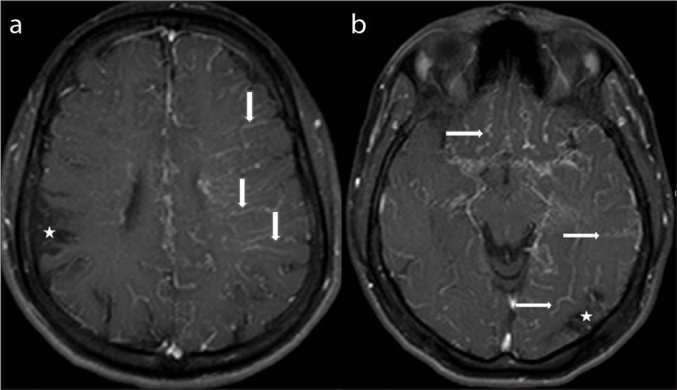
Moyamoya disease is an idiopathic disease characterized by the progressive stenosis and collateral development of the distal internal carotid arteries. In this disease, several collateral vascular structures develop following stenosis and occlusion. The ivy sign is a characteristic Magnetic rezonance imaging (MRI) finding frequently encountered in patients with moyamoya. It can be observed both in post contrast T1-weighted images and Fluid attenuated inversion recovery (FLAIR) images. While this sign manifests in the form of contrasting on the cortical surfaces due to the formation of leptomeningeal collateral development and increased numbers of pial vascular webs on post contrast images, in FLAIR images it originates from the slow arterial flow in the leptomeningeal collateral vascular structures. In this case, we presented the Digital subtraction angiography (DSA) signs of moyamoya disease and "ivy sign" in MRI and its development mechanism in a 16 years old female patient.
Moyamoya hastalığı distal internal karotid arterlerin ilerleyici stenozu ve kollateral gelişimi ile karakterize idiyopatik bir hastalıktır. Bu hastalıkta, stenoz ve oklüzyonu takiben kollateral vasküler yapılar gelişir. Ivy bulgusu sıklıkla moyamoyalı hastalarda izlenen karakteristik Magnetik Rezonans Görüntüleme (MRG) bulgusudur. Bu bulgu kontrastlı T1-ağırlıklı ve Fluid attenuated inversion recovery (FLAIR) görüntülerde gözlemlenebilir. Ivy bugusu postkontrast görüntülerde leptomeningeal kolaterallerin gelişimi ve artan pial vasküler ağ nedeniyle oluşan kollateral yüzeylerde kontrastlanma şeklinde izlenirken, FLAIR görüntülerde leptomeningeal kollateral vasküler yapılarda yavaş arteriyel akım nedeniyle oluşur. Bu olguda, moyamoya hastalığının Digital subtraction angiography (DSA) bulgularını, MRG’de Ivy bulgusunu ve 16 yaşında kadın hastada bu bulgunun gelişim mekanizmasını sunduk.
Keywords: DSA; Ivy Sign; MRI; Moyamoya Disease.
Figures


References
-
- Marshall S, Hawley JS, Nyquist PA, DeGraba T. The “Ivy sign” of adult Moyamoya Disease. Neurologist. 2009;6:367-8.v. - PubMed
-
- Baba T, Houkin K, Kuroda S. Novel Epidemiological features of moyamoya disease. J Neurol Neurosurg Psychiatry. 2008;79:900–4. http://dx.doi.org/10.1136/jnnp.2007.130666. - DOI - PubMed
-
- Yonekawa Y, Ogata N, Kaku Y, Taub E, Imhof HG. Moyamoya disease in Europe, past and present status. Clin Neurol Neurosurg. 1997;99:58–60. http://dx.doi.org/10.1016/S0303-8467(97)00042-5. - DOI - PubMed
-
- Wakai K, Tamakoshi A, Ikezaki K, et al. Epidemiological features of moyamoya disease in Japan: finding from a nationwide survey. Clin Neurol Neurosurg. 1997;99:1–5. http://dx.doi.org/10.1016/S0303-8467(97)00031-0. - DOI - PubMed
-
- Yamauchi T, Houkin K, Tada M, Abe H. (1997) Familial occurrence of moyamoya disease. Clin Neurosurg. 1997;99:162–7. http://dx.doi.org/10.1016/S0303-8467(97)00054-1. - DOI - PubMed
Publication types
LinkOut - more resources
Full Text Sources
Other Literature Sources