

Primary Pleomorphic Rhabdomyosarcoma of Thyroid Gland in an Adult Patient: A Case Report
- PMID: 27026769
- PMCID: PMC4792502
- DOI: 10.5152/eurasianjmed.2015.96
Primary Pleomorphic Rhabdomyosarcoma of Thyroid Gland in an Adult Patient: A Case Report
Abstract
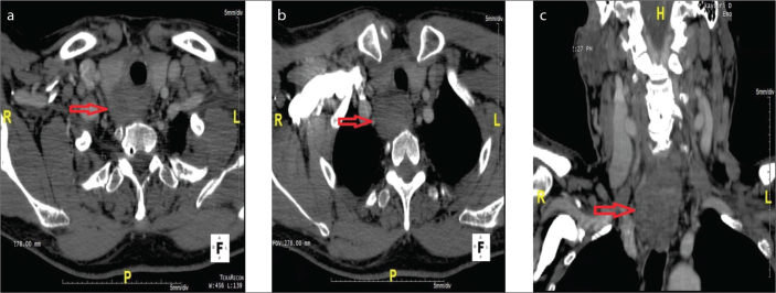
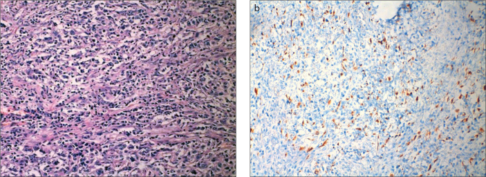
Thyroid sarcoma is a very rare entity, accounting for less than 1% of all malignant thyroid tumours. Rhabdomyosarcoma (RMS) is a sarcoma subtype, which is more common in children and adolescents. In this case, a 68-year old man, presented with hoarseness and diagnosed with pleomorphic RMS, was explored. No study of primary thyroid pure RMS has been reported in the literature, with the exception of the case reports of differentiated RMS.
Tiroid sarkomu, tüm tiroid malignitelerinin %1’inden azını oluşturan oldukça nadir bir klinik antitedir. Rabdomyosarkom (RMS) coçuk ve adölesanlarda daha yaygın görülen sarkom subtiptidir. Diferansiye RMS vaka sunumları hariç, primer saf tiroid RMS vakası rapor edilmemiştir. Biz ses kısıklığı ile şikayeti ile başvuran ve pleomorfik RMS tanısı konulan 68 yaşında erkek hastayı sunduk.
Keywords: Pleomorphic rhabdomyosarcoma; adult; thyroid cancer.
Figures
References
-
- Carling T, Udelsman R. Cancer of the Endocrine System. In: DeVita VT, Lawrence TS, Rosenberg SA, DePinho RA, Weinberg RA, editors. DeVita, Hellman And Rosenberg’s Cancer Principles Practice of Oncology. 8th edition. Lippincott Williams Wilkins; 2008. pp. 1669–70.
-
- Melmed S. Nontoxic diffuse and nodular goiter and thyroid neoplasia. In: Schlumberger JM, Filetti S, Hay DL, editors. Williams textbook of endocrinology. 12th edition. Saunders Elsevier; Philadelphia: 2011. pp. 440–75.
-
- Kraus DH, Saenz NC, Gollamudi S, et al. Pediatric rhabdomyosarcoma of the head and neck. Am J Surg. 1997;174:556–60. http://dx.doi.org/10.1016/S0002-9610(97)00171-2. - DOI - PubMed
-
- Davies CE, Davies AM, Kindblom LG, James SL. Soft tissue tumors with muscle differentiation. Semin Musculoskelet Radiol. 2010;14:245–56. http://dx.doi.org/10.1055/s-0030-1253165. - DOI - PubMed
-
- Furlong MA, Mentzel T, Fanburg-Smith JC. Pleomorphic rhabdomyosarcoma in adults: a clinicopathologic study of 38 cases with emphasis on morphologic variants and recent skeletal muscle-specific markers. Mod Pathol. 2001;14:595–603. http://dx.doi.org/10.1038/modpathol.3880357. - DOI - PubMed
Publication types
LinkOut - more resources
Full Text Sources
Other Literature Sources